

Chlamydia Serine Protease Inhibitor, targeting HtrA, as a New Treatment for Koala Chlamydia infection
- PMID: 27530689
- PMCID: PMC4987629
- DOI: 10.1038/srep31466
Chlamydia Serine Protease Inhibitor, targeting HtrA, as a New Treatment for Koala Chlamydia infection
Abstract
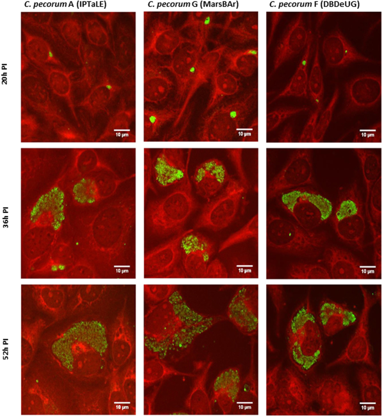
The koala, an iconic marsupial native to Australia, is a threatened species in many parts of the country. One major factor in the decline is disease caused by infection with Chlamydia. Current therapeutic strategies to treat chlamydiosis in the koala are limited. This study examines the effectiveness of an inhibitor, JO146, which targets the HtrA serine protease for treatment of C. pecorum and C. pneumoniae in vitro and ex vivo with the aim of developing a novel therapeutic for koala Chlamydia infections. Clinical isolates from koalas were examined for their susceptibility to JO146. In vitro studies demonstrated that treatment with JO146 during the mid-replicative phase of C. pecorum or C. pneumoniae infections resulted in a significant loss of infectious progeny. Ex vivo primary koala tissue cultures were used to demonstrate the efficacy of JO146 and the non-toxic nature of this compound on peripheral blood mononuclear cells and primary cell lines established from koala tissues collected at necropsy. Our results suggest that inhibition of the serine protease HtrA could be a novel treatment strategy for chlamydiosis in koalas.
Figures


Similar articles
-
Optimization of peptide-based inhibitors targeting the HtrA serine protease in Chlamydia: Design, synthesis and biological evaluation of pyridone-based and N-Capping group-modified analogues.Eur J Med Chem. 2021 Nov 15;224:113692. doi: 10.1016/j.ejmech.2021.113692. Epub 2021 Jul 7. Eur J Med Chem. 2021. PMID: 34265463
-
Preliminary characterisation of tumor necrosis factor alpha and interleukin-10 responses to Chlamydia pecorum infection in the koala (Phascolarctos cinereus).PLoS One. 2013;8(3):e59958. doi: 10.1371/journal.pone.0059958. Epub 2013 Mar 19. PLoS One. 2013. PMID: 23527290 Free PMC article.
-
Recent advances in understanding the biology, epidemiology and control of chlamydial infections in koalas.Vet Microbiol. 2013 Aug 30;165(3-4):214-23. doi: 10.1016/j.vetmic.2013.02.026. Epub 2013 Mar 1. Vet Microbiol. 2013. PMID: 23523170 Review.
-
Genetic diversity of Chlamydia pecorum strains in wild koala locations across Australia and the implications for a recombinant C. pecorum major outer membrane protein based vaccine.Vet Microbiol. 2013 Dec 27;167(3-4):513-22. doi: 10.1016/j.vetmic.2013.08.009. Epub 2013 Aug 18. Vet Microbiol. 2013. PMID: 24012135
-
Koala immunology and infectious diseases: How much can the koala bear?Dev Comp Immunol. 2018 May;82:177-185. doi: 10.1016/j.dci.2018.01.017. Epub 2018 Jan 31. Dev Comp Immunol. 2018. PMID: 29382557 Review.
Cited by
-
A 29-year retrospective analysis of koala rescues in New South Wales, Australia.PLoS One. 2020 Oct 28;15(10):e0239182. doi: 10.1371/journal.pone.0239182. eCollection 2020. PLoS One. 2020. PMID: 33112860 Free PMC article.
-
Evaluation of a biosecurity survey approach for contamination by Chlamydia pecorum in koala rehabilitation, field capture, and captive settings.PeerJ. 2023 Aug 15;11:e15842. doi: 10.7717/peerj.15842. eCollection 2023. PeerJ. 2023. PMID: 37601255 Free PMC article.
-
Epidemiology, Transmission Mode, and Pathogenesis of Chlamydia pecorum Infection in Koalas (Phascolarctos cinereus): An Overview.Animals (Basel). 2024 Sep 15;14(18):2686. doi: 10.3390/ani14182686. Animals (Basel). 2024. PMID: 39335275 Free PMC article. Review.
-
Molecular pathogenesis of Chlamydia trachomatis.Front Cell Infect Microbiol. 2023 Oct 18;13:1281823. doi: 10.3389/fcimb.2023.1281823. eCollection 2023. Front Cell Infect Microbiol. 2023. PMID: 37920447 Free PMC article. Review.
-
Characterization of shifts of koala (Phascolarctos cinereus) intestinal microbial communities associated with antibiotic treatment.PeerJ. 2018 Mar 12;6:e4452. doi: 10.7717/peerj.4452. eCollection 2018. PeerJ. 2018. PMID: 29576947 Free PMC article.
References
-
- Melzer A., Carrick F., Menkhorst P., Lunney D. & John B. Overview, Critical Assessment, and Conservation Implications of Koala Distribution and Abundance. Conservation Biology 14, 619–628 (2000).
-
- Dique D. S. et al. Koala mortality on roads in south-east Queensland: the koala speed-zone trial. Wildlife Research 30, 419–426 (2003).
-
- Lunney D., Gresser S., O’Neil L. E., Matthews A. & Rhodes J. R. The impact of fire and dogs on Koalas at Port Stephens, New South Wales, using population viability analysis. Pacific Conservation Biology 13, 189–201 (2007).
-
- Polkinghorne A., Hanger J. & Timms P. Recent advances in understanding the biology, epidemiology and control of chlamydial infections in koalas. Veterinary Microbiology 165, 214–223 (2013). - PubMed
-
- Jackson M., White N., Giffard P. & Timms P. Epizootiology of Chlamydia infections in two free-range koala populations. Veterinary Microbiology 65, 255–264 (1999). - PubMed
Publication types
MeSH terms
Substances
LinkOut - more resources
Full Text Sources
Other Literature Sources
Medical
