. 2016 Aug 17:6:31754.
doi: 10.1038/srep31754.

Pathophysiology of the inner ear after blast injury caused by laser-induced shock wave

Affiliations

Pathophysiology of the inner ear after blast injury caused by laser-induced shock wave

Katsuki Niwa et al. Sci Rep. .

Abstract

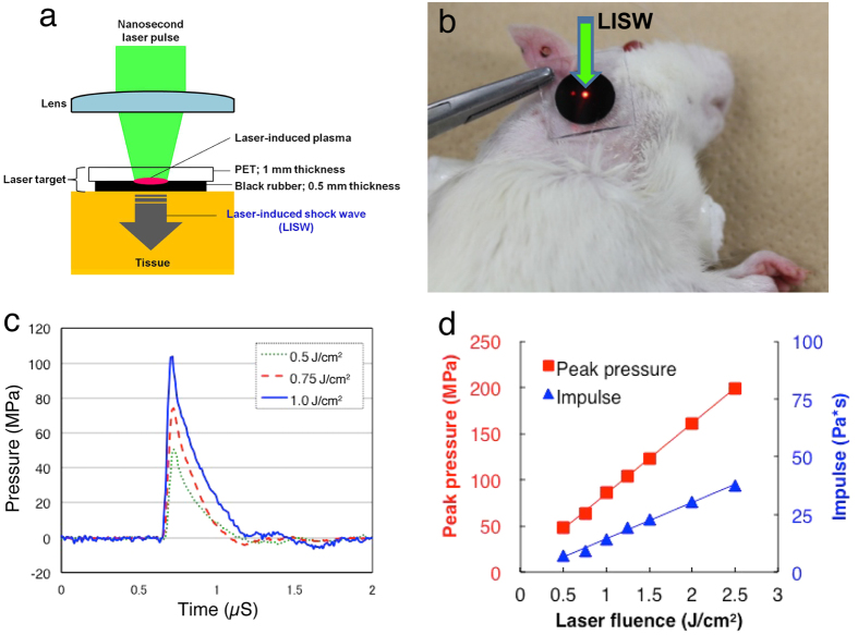
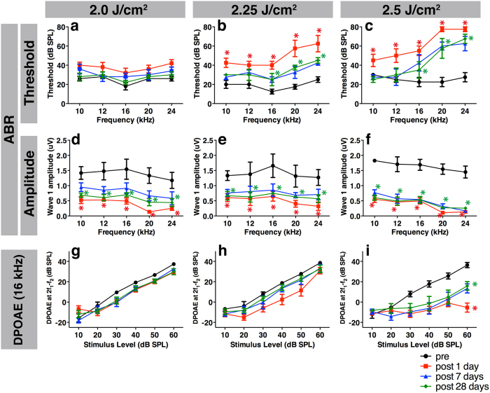
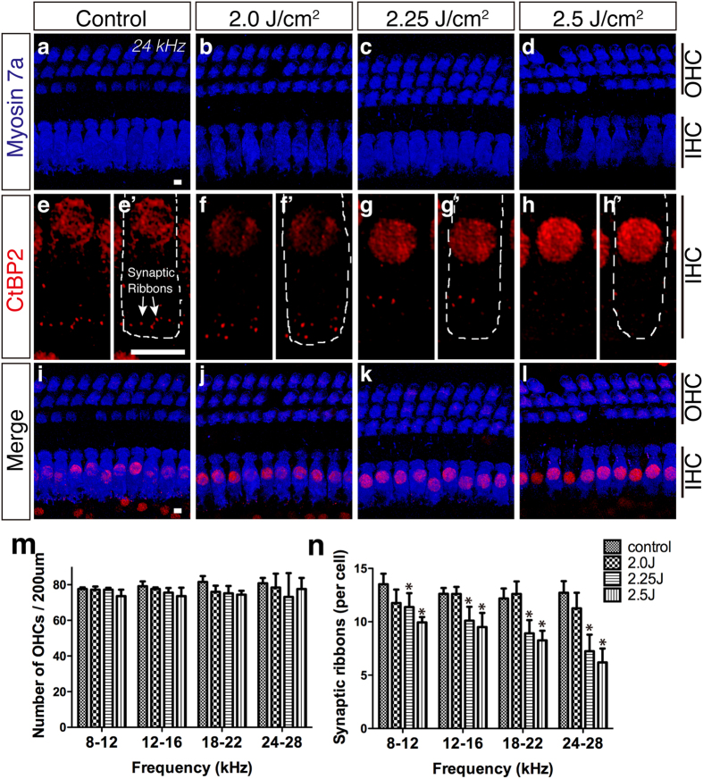
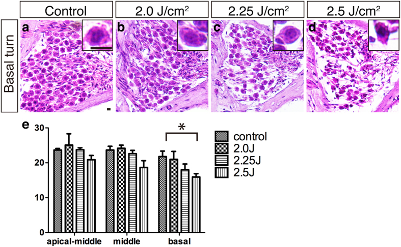
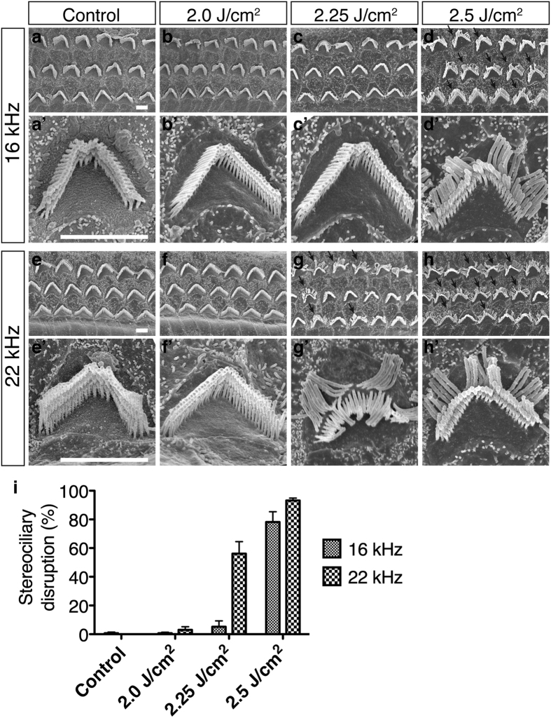
The ear is the organ that is most sensitive to blast overpressure, and ear damage is most frequently seen after blast exposure. Blast overpressure to the ear results in sensorineural hearing loss, which is untreatable and is often associated with a decline in the quality of life. In this study, we used a rat model to demonstrate the pathophysiological and structural changes in the inner ear that replicate pure sensorineural hearing loss associated with blast injury using laser-induced shock wave (LISW) without any conductive hearing loss. Our results indicate that threshold elevation of the auditory brainstem response (ABR) after blast exposure was primarily caused by outer hair cell dysfunction induced by stereociliary bundle disruption. The bundle disruption pattern was unique; disturbed stereocilia were mostly observed in the outermost row, whereas those in the inner and middle rows stereocilia remained intact. In addition, the ABR examination showed a reduction in wave I amplitude without elevation of the threshold in the lower energy exposure group. This phenomenon was caused by loss of the synaptic ribbon. This type of hearing dysfunction has recently been described as hidden hearing loss caused by cochlear neuropathy, which is associated with tinnitus or hyperacusis.


Figures

Figure 1
Figure 1. Generation and characteristics of LISW.
(a) Experimental setup for the LISW. Shock waves were generated by irradiating a laser target (black rubber) with a Q-switched Nd:YAG laser. Plasma formation occurred at the interface between a transparent material and the black rubber. (b) Image showing how to apply an LISW to induce inner ear injuries. (c) Typical temporal waveforms of LISWs generated at different laser fluences on the laser target. (d) Dependence of peak pressure and LISW impulse on laser fluence. LISW, laser-induced shock wave.
Figure 2
Figure 2. Measurement of hearing function using ABR and DPOAE before and after LISW exposure.
(ac) Significant ABR threshold shifts one day after LISW (filled squares) exposure were observed up to 1 month after exposure (filled diamonds) in the 2.25 (b) and 2.5 J/cm2 (c) groups compared to the pre-exposure thresholds (filled circles). (df) Despite the absence of ABR threshold elevation, the wave I amplitude was decreased in the 2.0 J/cm2 (d) group, which was also seen in the 2.25 (e) and 2.5 J/cm2 (f) groups. (gi) The DPOAE level in the 2.5 J/cm2 (i) group was significantly decreased, whereas no decrease was observed in the 2.0 (g) or 2.25 J/cm2 (h) group. Asterisks indicate significant differences (p < 0.05) compared to the pre-exposure values. Error bars indicate SEs of the means (n = 5 in each group). ABR, auditory brainstem response; DPOAE, distortion product otoacoustic emission; LISW, laser-induced shock wave.
Figure 3
Figure 3. Changes in the organ of Corti after LISW exposure.
(ai) Confocal images of immunohistochemistry for OHCs (ad, blue, anti-myosin 7a), synaptic ribbons (eh, red, anti-CtBP2; the white dotted line in e’–h’ shows the contour of the OHCs; anti-CtBP2 also stains the IHC nuclei), and merged images (i–l) at 24 kHz. (mn) The number of OHCs (m) and synaptic ribbons (n) 1 month after LISW exposure (n = 5 in each group). Despite ABR threshold shifts observed in the 2.25 and 2.5 J/cm2 groups (c,d), the number of OHCs remained steady 1 month after LISW exposure (m) compared to the control group (a). The numbers of synaptic ribbons were decreased by LISW in an energy-dependent manner (eh,n). Asterisks indicate significant differences (p < 0.05). Error bars indicate SEs of the means. Scale bar is 5 μm. ABR, auditory brainstem response; IHC, inner hair cell; LISW, laser-induced shock wave; OHC, outer hair cell.
Figure 4
Figure 4. Changes in the spiral ganglion after LISW exposure.
(ad) Images of the SGCs using HE-stained frozen sections from the cochlear basal turns. Insets show high-power views of a single SGC. (e) The number of SGCs 1 month after LISW exposure (n = 5 in each group). The concentration of SGCs at the basal turn in the 2.5 J/cm2 group was significantly decreased (d,e), compared to the control (a). The basal turn of the SGC in the 2.25 J/cm2 (d, inset) group shows shrinking compared to the control (a, inset). The asterisk indicates a significant difference (p < 0.05). Error bars indicate SEs of the means. Scale bar is 5 μm. LISW, laser-induced shock wave; SGC, spiral ganglion cell; SE, standard error.
Figure 5
Figure 5. Surface structures in the OHCs.
(ad) Scanning electron microscopy images of three rows of OHC stereocilia at the 16 kHz organ of Corti area. (a’–d’) high-power views of single OHC stereocilia. (eh) and (e’–h’) Images from the 22 kHz organ of Corti area. (i) The stereociliary disruption ratio on OHCs one month after LISW exposure (n = 3 in each group). Stereociliary bundle disruption, i.e., the outermost layer of bundles being broken from the root (arrows), was seen in the 2.25 J/cm2 (22 kHz area, g and g’) and 2.5 J/cm2 groups (16 and 22 kHz area, d, h, and d’, h’). Scale bar is 2 μm. Error bars indicate SEs of the means. OHC, outer hair cell; SE, standard error.

References

    1. Wolf S. J., Bebarta V. S., Bonnett C. J., Pons P. T. & Cantrill S. V. Blast injuries. Lancet 374, 405–415, doi: 10.1016/S0140-6736(09)60257-9 (2009). - DOI - PubMed
    1. Owens B. D. et al.. Combat wounds in operation Iraqi Freedom and operation Enduring Freedom. J Trauma 64, 295–299, doi: 10.1097/TA.0b013e318163b875 (2008). - DOI - PubMed
    1. DePalma R. G., Burris D. G., Champion H. R. & Hodgson M. J. Blast injuries. N Engl J Med 352, 1335–1342, doi: 10.1056/NEJMra042083 (2005). - DOI - PubMed
    1. Shah A., Ayala M., Capra G., Fox D. & Hoffer M. Otologic assessment of blast and nonblast injury in returning Middle East-deployed service members. Laryngoscope 124, 272–277, doi: 10.1002/lary.24169 (2014). - DOI - PubMed
    1. Gallun F. J. et al.. Implications of blast exposure for central auditory function: a review. J Rehabil Res Dev 49, 1059–1074 (2012). - PubMed

Publication types