. 2016 Aug 17:6:31646.
doi: 10.1038/srep31646.

The β-amyloid peptide compromises Reelin signaling in Alzheimer's disease

Affiliations

The β-amyloid peptide compromises Reelin signaling in Alzheimer's disease

Inmaculada Cuchillo-Ibañez et al. Sci Rep. .

Abstract

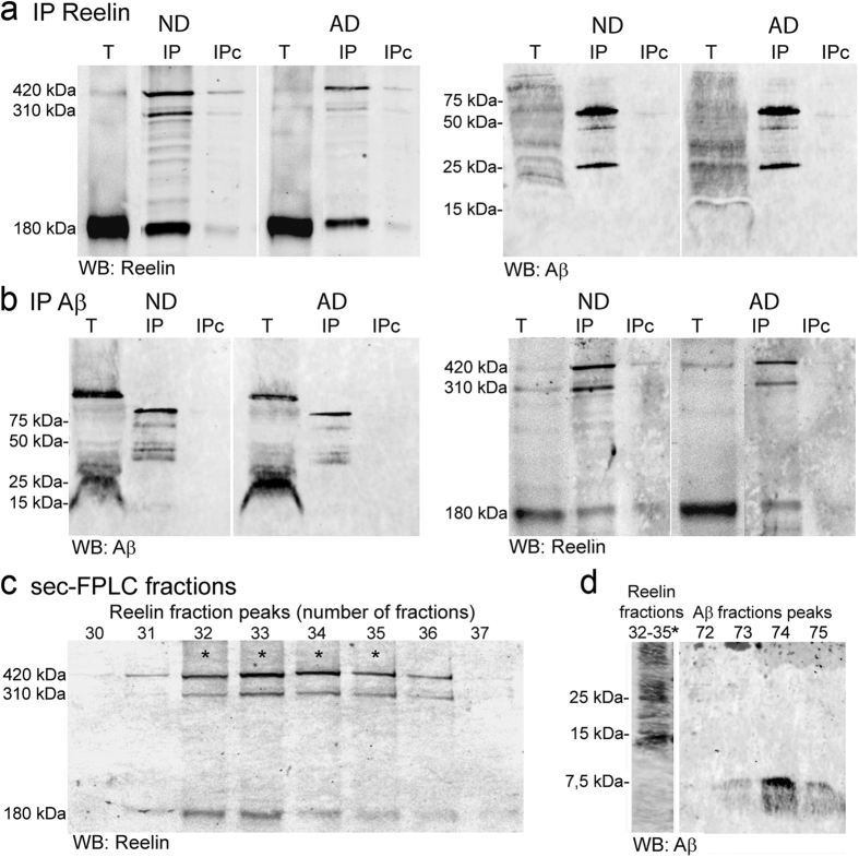
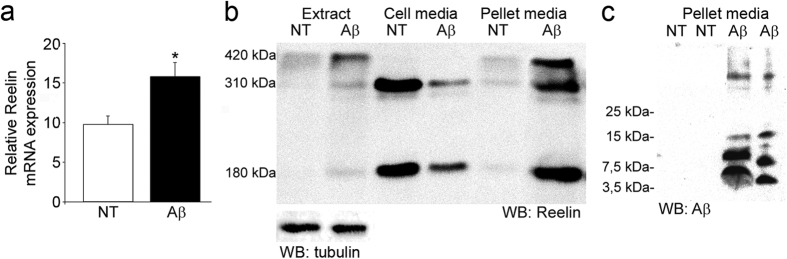
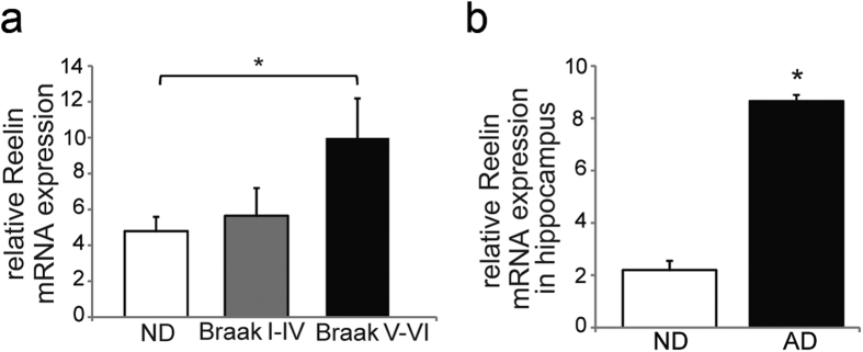
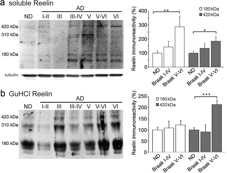
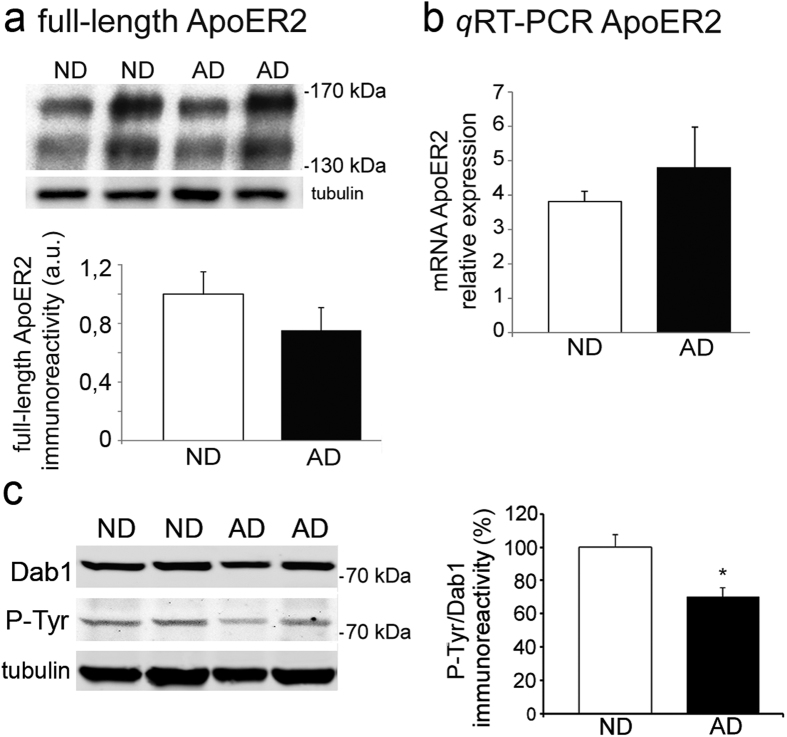
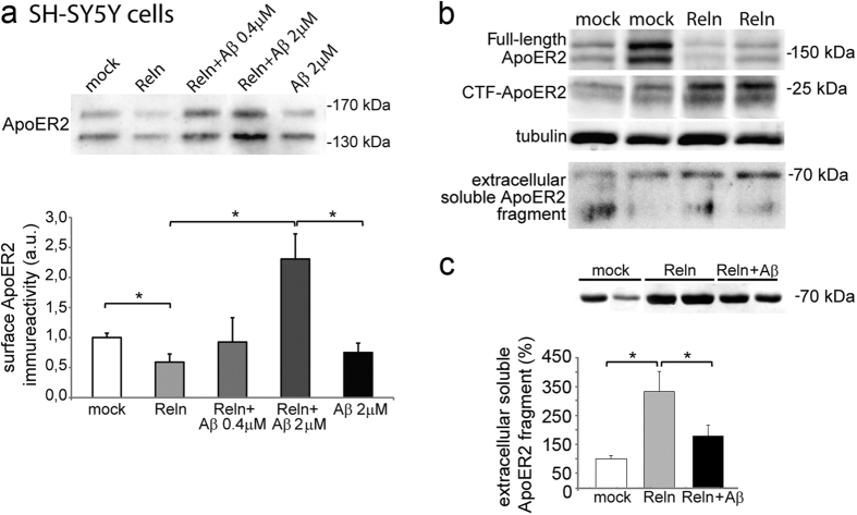
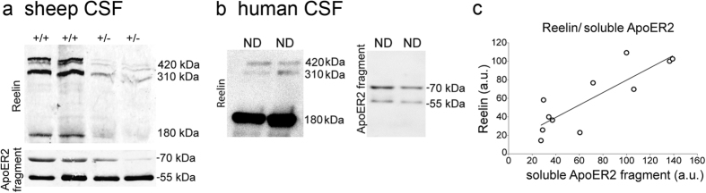
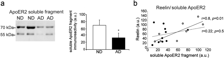
Reelin is a signaling protein that plays a crucial role in synaptic function, which expression is influenced by β-amyloid (Aβ). We show that Reelin and Aβ oligomers co-immunoprecipitated in human brain extracts and were present in the same size-exclusion chromatography fractions. Aβ treatment of cells led to increase expression of Reelin, but secreted Reelin results trapped together with Aβ aggregates. In frontal cortex extracts an increase in Reelin mRNA, and in soluble and insoluble (guanidine-extractable) Reelin protein, was associated with late Braak stages of Alzheimer's disease (AD), while expression of its receptor, ApoER2, did not change. However, Reelin-dependent induction of Dab1 phosphorylation appeared reduced in AD. In cells, Aβ reduced the capacity of Reelin to induce internalization of biotinylated ApoER2 and ApoER2 processing. Soluble proteolytic fragments of ApoER2 generated after Reelin binding can be detected in cerebrospinal fluid (CSF). Quantification of these soluble fragments in CSF could be a tool to evaluate the efficiency of Reelin signaling in the brain. These CSF-ApoER2 fragments correlated with Reelin levels only in control subjects, not in AD, where these fragments diminished. We conclude that while Reelin expression is enhanced in the Alzheimer's brain, the interaction of Reelin with Aβ hinders its biological activity.


Figures

Figure 1
Figure 1. Reelin and Aβ interact in human brain tissue.
Immunoprecipitation of human frontal cortex extracts, non-disease control (ND) and AD with (a) anti-Reelin (G10+CR50), or (b) anti-Aβ (4G8) antibodies (n = 3; T, total lysate). The immunoprecipitated proteins (IP) were probed with the 142 antibody against Reelin and the 6E10 antibody against Aβ. The Reelin antibody detects the 420 kDa full-length and the 310 and 180-kDa fragments. Extracts incubated with beads in the absence of antibody were negative controls (IPc). (c) Reelin blot from cortex extracts fractioned by size-exclusion. (d) Reelin-rich fractions were also pooled together and probed with the 4G8 antibody against Aβ, and analyzed for comparison with fractions containing small Aβ oligomers. Note that Reelin co-immunoprecipitates and interacts mostly with oligomeric Aβ species. One AD case is shown but similar elution profiles were obtained for ND cases.
Figure 2
Figure 2. Aβ42 increases Reelin expression in cell culture and is trapped into Aβ fibers.
(a) Differentiated SH-SY5Y cells were treated with 10 μM of Aβ42 and mRNA Reelin was determined and compared with non-treated (NT) cells. Data represents relative Reelin mRNA levels normalized to GAPDH and are expressed as mean values ± SEM of 10 independent determinations from at least 2 different experiments (*p < 0.001). (b) Western blots of SH-SY5Y cell extracts, the media and the pellet from the media of cells maintained in the presence or absence of Aβ42, and probed for Reelin (representative blot, n = 3) and α-tubulin from cell extracts as a loading control. (c) Proteins recovered in the pellet from the media were also probed with the 4G8 antibody against Aβ.
Figure 3
Figure 3. Increased Reelin expression in the brain at advanced Braak stages of AD.
AD subjects were categorized as early (I-II to IV) or advanced Braak stages (V to VI). (a) Relative Reelin mRNA expression analyzed by qRT-PCR in frontal cortex samples from ND (n = 11) and AD subjects (stages I to IV, n = 7; stages V to VI, n = 10). (b) Reelin mRNA was also analyzed in the hippocampus of ND controls (ND; n = 5) and AD samples corresponding to Braak stages IV to VI (n = 9). The values were calculated from relative standard curves and normalized to GAPDH from the same cDNA preparation, confirming the specificity of the PCR products from dissociation curves. The data represent the means ± SEM. *p < 0.05 using Student’s t-test.
Figure 4
Figure 4. An increase in Reelin protein in the frontal cortex at advanced Braak stages of AD.
Frontal cortex samples from ND (n = 11) and AD subjects (n=17) were homogenized in detergents diluted in Tris-saline buffer (soluble Reelin, (a) and the pellets recovered were re-extracted in Guanidine-HCl (GuHCl Reelin, (b). Western blots were probed with the 142 antibody (α-tubulin served as a loading control). AD subjects were categorized as early (I–II to IV; n = 7) or advanced Braak stages (V to VI; n = 10). The data represent the means ± SEM and were normalized with respect to the ND values. The data represent the means ± SEM. *p < 0.05, **p < 0.01, ***p < 0.001, using Student’s t test.
Figure 5
Figure 5. ApoER2 levels remain unaltered in the AD frontal cortex but Dab1 phosphorylation decreases.
(a) Quantification of ApoER2 (~130 kDa + ~170 kDa) in the ND (n = 10) and AD cortex (n = 12) in Western blots probed with a C-terminal ApoER2 antibody and α-tubulin as loading control. (b) Relative ApoER2 mRNA expression analyzed by qRT-PCR in ND (n = 8) and AD (n = 11) frontal cortex, calculated from standard curves and normalized to GAPDH. Data represent the mean ± SEM. None of the comparisons resulted in significant differences, considering the entire AD group or the advanced Braak stages. (c) Western blots of frontal cortex extracts from ND (n = 12) and AD (n = 15) revealed by fluorescence simultaneously for an anti-Dab1 antibody and an anti-phosphotyrosine (P-Tyr), and α-tubulin as loading control. Data represent the means ± SEM and were normalized with respect to the ND values. *p < 0.005 using a Student’s t test.
Figure 6
Figure 6. Aβ impairs the binding of Reelin to ApoER2.
(a) Aβ increases the presence of ApoER2 at the cytoplasmic membrane induced by Reelin. Quantification of biotinylated ApoER2 (~130 kDa + ~170 kDa) from untreated SH-SY5Y cells and those treated with Reelin alone (Reln; ~10 nM), with Aβ42 or with Reelin previously incubated with Aβ42 at the concentration indicated. Note that Aβ42 (2 μM) alone did not have noticeable effect. Data from 6 independent experiments are normalized with respect to the mock values and represented as the means ± SEM: *significantly different (p < 0.05) using a Student’s t test. (b) Reelin binding induces cleavage of the ApoER2 receptor in SH-SY5Y cells, generating a soluble ApoER2 fragment (~70 kDa) in the culture medium of treated cells that can be monitored with the 186 antibody against ApoER2. The processing of the full-length ApoER2 receptor was also assessed by the appearance of an intracellular C-terminal fragment (CTF). (c) The presence of Aβ (2 μM) weakens generation of the soluble 70 kDa ApoER2 fragments. Data represent the means ± SEM normalized with respect to the mock values (8 determinations from 2 separate experiments). *p < 0.005 using Mann-Whitney test.
Figure 7
Figure 7. Characterization of the soluble ApoER2 fragment present in CSF.
(a) Western blots of Reelin and soluble ApoER2 fragments of CSF from homozygous (+/+) and heterozygous (+/−) sheep carrying a mutation that reduces Reelin expression (representative blot, n = 2). (b) Western blot showing the presence of Reelin and soluble ApoER2 fragments in human CSF, and (c) the correlation between full-length Reelin and the 70-kDa soluble ApoER2 fragment in human non-disease (ND; n = 11 from different ages) CSF samples.
Figure 8
Figure 8. Decrease of the soluble ApoER2 fragment in AD CSF.
(a) Samples from age-matched ND controls (n = 8) and AD patients (n = 10) were assayed for Reelin by immnoblotting and ELISA and selected on the basis of a similar Reelin content (comparison is shown). (b) Representative blots and densitometric quantification of the 70 kDa soluble ApoER2 fragment assayed with the 186 antibody in samples. (c) Correlation between the soluble full-length Reelin and ApoER2 soluble fragment in ND (open circles) and AD (closed circles) CSF samples. The dashed line represents the lack of correlation in AD samples.

References

    1. Förster E. et al. Emerging topics in Reelin function. Eur J Neurosci, 31, 1511–1518 (2010). - PMC - PubMed
    1. D’Arcangelo G. et al. Reelin is a ligand for lipoprotein receptors. Neuron. 24, 471–479 (1999). - PubMed
    1. Hiesberger T. et al. Direct binding of Reelin to VLDL receptor and ApoE receptor 2 induces tyrosine phosphorylation of disabled-1 and modulates tau phosphorylation. Neuro. 24, 481–489 (1999). - PubMed
    1. Schmechel D. E. et al. Increased amyloid beta-peptide deposition in cerebral cortex as a consequence of apolipoprotein E genotype in late-onset Alzheimer disease. Proc Natl Acad Sci USA. 90, 9649–9653 (1993). - PMC - PubMed
    1. May P., Bock H. H., Nimpf J. & Herz J. Differential glycosylation regulates processing of lipoprotein receptors by gamma-secretase. J Biol Chem. 278, 37386–37392 (2003). - PubMed

Publication types

MeSH terms