. 2016 Oct 7;291(41):21584-21595.
doi: 10.1074/jbc.M116.730978. Epub 2016 Aug 16.

Protein Kinase A-independent Ras Protein Activation Cooperates with Rap1 Protein to Mediate Activation of the Extracellular Signal-regulated Kinases (ERK) by cAMP

Affiliations

Protein Kinase A-independent Ras Protein Activation Cooperates with Rap1 Protein to Mediate Activation of the Extracellular Signal-regulated Kinases (ERK) by cAMP

Yanping Li et al. J Biol Chem. .

Abstract

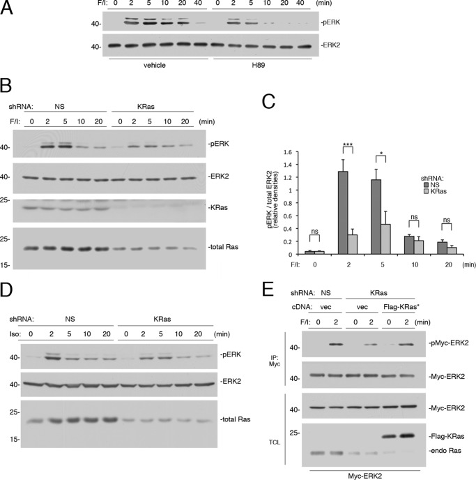
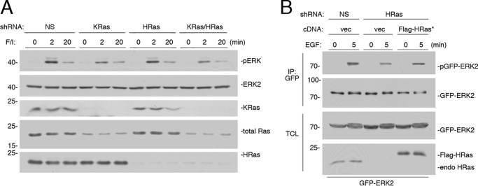
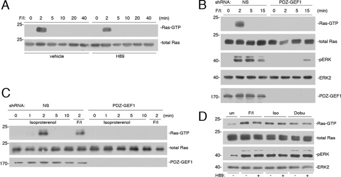
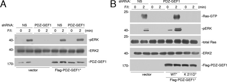
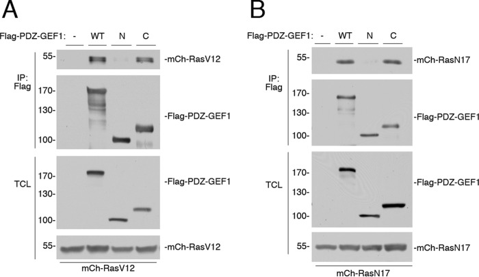
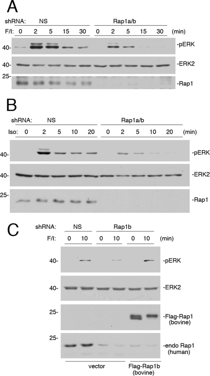
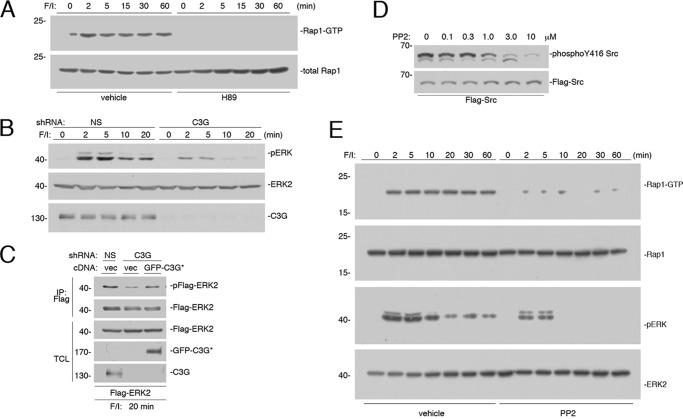
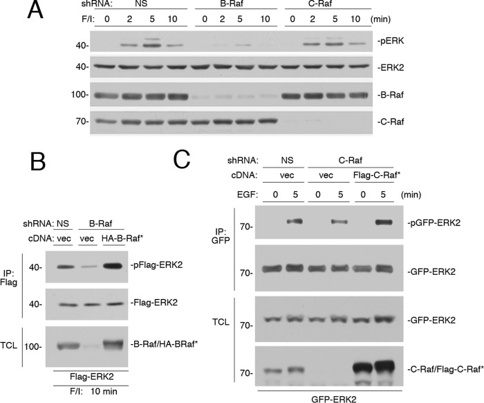
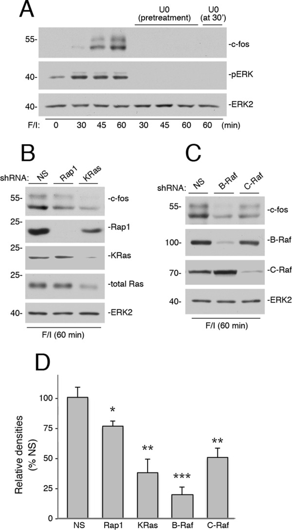
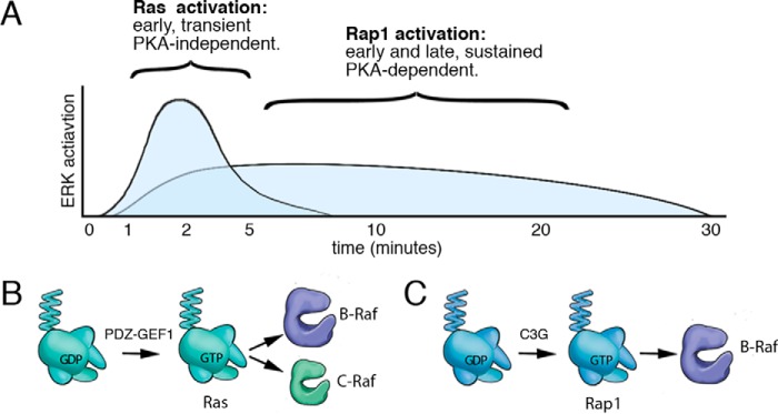
Cyclic adenosine monophosphate (cAMP) is an important mediator of hormonal stimulation of cell growth and differentiation through its activation of the extracellular signal-regulated kinase (ERK) cascade. Two small G proteins, Ras and Rap1, have been proposed to mediate this activation, with either Ras or Rap1 acting in distinct cell types. Using Hek293 cells, we show that both Ras and Rap1 are required for cAMP signaling to ERKs. The roles of Ras and Rap1 were distinguished by their mechanism of activation, dependence on the cAMP-dependent protein kinase (PKA), and the magnitude and kinetics of their effects on ERKs. Ras was required for the early portion of ERK activation by cAMP and was activated independently of PKA. Ras activation required the Ras/Rap guanine nucleotide exchange factor (GEF) PDZ-GEF1. Importantly, this action of PDZ-GEF1 was disrupted by mutation within its putative cyclic nucleotide-binding domain within PDZ-GEF1. Compared with Ras, Rap1 activation of ERKs was of longer duration. Rap1 activation was dependent on PKA and required Src family kinases and the Rap1 exchanger C3G. This is the first report of a mechanism for the cooperative actions of Ras and Rap1 in cAMP activation of ERKs. One physiological role for the sustained activation of ERKs is the transcription and stabilization of a range of transcription factors, including c-FOS. We show that the induction of c-FOS by cAMP required both the early and sustained phases of ERK activation, requiring Ras and Rap1, as well as for each of the Raf isoforms, B-Raf and C-Raf.

Keywords: Ras protein; Ras-related protein 1 (Rap1); cyclic AMP (cAMP); extracellular-signal-regulated kinase (ERK); guanine nucleotide exchange factor (GEF).


Figures

FIGURE 1.
FIGURE 1.
Rapid activation of ERKs by cAMP is PKA-independent and Ras-dependent. A, Hek293 cells were treated with F/I and pretreated with H89 or vehicle, as indicated. Activation of ERK was measured as described under “Experimental Procedures,” using phospho-ERK antibodies. The levels of ERK activation (pERK) and total ERK2 are shown. B, cells transfected with nonspecific (NS) shRNA or shRNA against human KRas were treated with F/I for the times indicated. The levels of pERK and total ERK2 are shown in the 1st two panels. KRas levels are shown in the 3rd panel. Total Ras levels are shown in the 4th panel, using a pan-Ras antibody. C, shRNA data from four independent experiments are shown as bar graphs, with ERK activation (pERK) normalized to total ERK2 levels and the pERK/ERK ratio presented as relative densities. Error bars show standard error. Significance was assessed by unpaired Student's t tests (ns, not significant, * = <0.05, and *** = <0.001). D, cells transfected with NS shRNA or shRNA against human KRas were treated with isoproterenol (Iso) for the times indicated. The levels of pERK, total ERK2, and total Ras (using a pan-Ras Ab) are shown. E, cells were transfected with Myc-ERK2 and either NS shRNA or shRNA against KRas, along with either vector or FLAG-tagged shRNA-resistant wild type KRas (FLAG-KRas*). Cells were treated with F/I for 2 min or left untreated and subjected to immunoprecipitation using agarose-coupled Myc Ab. The levels of Myc-ERK2 activation (pMyc-ERK2) and total Myc-ERK2 within the IP are shown. The levels of Myc-ERK2 and Ras proteins (total Ras) within the total cell lysate (TCL) are shown in the 3rd and 4th panels, respectively. Note that both FLAG-KRas and endogenous Ras are easily separated on this gel, with FLAG-KRas migrating more slowly.
FIGURE 2.
FIGURE 2.
KRas is the major Ras isoform downstream of cAMP. A, cells transfected with NS shRNA, shRNA against human KRas, shRNA against human HRas, or shRNAs against both KRas and HRas (KRas/HRas) were treated with F/I for the times indicated. The levels of ERK activation (pERK) and total ERK2 are shown in the 1st two panels. The levels of KRas, total Ras, and HRas are shown in the 3rd, 4th, and 5th panels, respectively. B, cells were transfected with GFP-ERK2 and NS shRNA or shRNA against HRas and either vector or FLAG-tagged shRNA-resistant HRas (FLAG-HRas*). Cells were treated with EGF for 5 min or left untreated, lysed, and subjected to GFP IP. The levels of activation of GFP-ERK2 (pGFP-ERK2) and the levels of total GFP-ERK2 are shown in the 1st and 2nd panels, respectively. The levels of GFP-ERK2 and HRas within the TCL are shown in the bottom two panels.
FIGURE 3.
FIGURE 3.
Ras activation by cAMP is PKA-independent and mediated by PDZ-GEF1. A, Hek293 cells were treated with F/I for the times indicated and pretreated with H89 or vehicle, as indicated. Ras activity (Ras-GTP) was measured as described under “Experimental Procedures” and shown in the 1st panel. Total Ras is shown in the 2nd panel. B, cells transfected with NS shRNA or shRNA against human PDZ-GEF1 were treated with F/I for the times indicated. Ras-GTP was measured as described, shown in the 1st panel. Total Ras is shown in the 2nd panel. The levels of pERK, total ERK2, and PDZ-GEF1 are shown in the bottom three panels, respectively. C, cells were transfected with NS shRNA or PDZ-GEF1 shRNA and treated with F/I or isoproterenol for the times indicated. The levels of Ras-GTP are shown in the 1st panel. Total Ras levels are shown in the 2nd panel, and the efficacy of the knockdown of PDZ-GEF1 is shown in the 3rd panel. D, newborn mouse cardiomyocytes were harvested and treated with F/I, isoproterenol (Iso), or dobutamine (Dobu) for 2 min, or left untreated (un), and either pretreated with H89 (+) or not pretreated (−), as indicated. Ras-GTP was measured in the 1st panel. Total Ras levels are shown in the 2nd panel. pERK and total ERK2 levels are shown in the 3rd and 4th panels, respectively.
FIGURE 4.
FIGURE 4.
cAMP-binding site within PDZ-GEF1 is required for cAMP activation of Ras. A, Hek293 cells were transfected with NS shRNA or shRNA against PDZ-GEF1 and either vector or FLAG-tagged shRNA-resistant wild type PDZ-GEF1 (WT*). Cells were treated with F/I for the times indicated. The levels of pERK and total ERK2 are shown in the 1st and 2nd panels, respectively. The levels of PDZ-GEF1 are shown in the 3rd panel. B, cells transfected with NS shRNA or shRNA against PDZ-GEF1 and either vector, FLAG-tagged shRNA-resistant wild type PDZ-GEF1 (WT*), or FLAG-tagged shRNA-resistant PDZ-GEF1 K211D mutant (K211D*). Cells were treated with F/I for the times indicated. The levels of Ras-GTP are shown in the 1st panel. The levels of pERK, total Ras, total ERK2, and FLAG-PDZ-GEF1 are shown in the lower four panels, respectively.
FIGURE 5.
FIGURE 5.
Ras binds to PDZ-GEF1. A, Hek293 cells were transfected with mCherry-HRasV12 (mCh-RasV12) and either vector (−), FLAG-PDZ-GEF1 (WT), a FLAG-tagged N-terminal fragment of PDZ-GEF1 spanning amino acids 1–716 (N), or a FLAG-tagged C-terminal fragment spanning amino acids 693–1499 (C), and cells were subjected to FLAG IP. The levels of mCh-RasV12 within the IP are shown in the 1st panel. The levels of FLAG-PDZ-GEF1 and fragments within the IP are shown in the 2nd panel. The levels of FLAG-PDZ-GEF1 and fragments within the TCL are shown in the 3rd panel. The levels of mCh-RasV12 within the TCL are shown in the 4th panel. B, cells were transfected with mCherry-HRasN17 (mCh-RasN17) and either vector (−), FLAG-PDZ-GEF1 (WT), the FLAG-tagged N-terminal fragment of PDZ-GEF1 (N), or the FLAG-tagged C-terminal fragment (C), and subjected to FLAG IP. The levels of mCh-RasN17 within the IP are shown in the 1st panel. The levels of FLAG-PDZ-GEF1 and fragments within the IP are shown in the 2nd panel. The levels of FLAG-PDZ-GEF1 and fragments within the TCL are shown in the 3rd panel. The levels of mCh-RasN17 within the TCL are shown in the 4th panel.
FIGURE 6.
FIGURE 6.
Small G protein Rap1 mediates the sustained portion of ERK activation by cAMP. A, Hek293 cells transfected with either NS or shRNA against Rap1a/b were treated with F/I for the times indicated. The levels of pERK and total ERK2 are shown in the 1st and 2nd panels, respectively. The efficacy of the Rap1 knockdown is shown in the 3rd panel. B, cells transfected with either NS shRNA or shRNA against Rap1a/b were treated with isoproterenol (Iso) for the times indicated. The levels of pERK and total ERK2 are shown in the 1st and 2nd panels, respectively. The efficacy of the Rap1 knockdown is shown in the 3rd panel. C, cells were transfected with NS shRNA or shRNA against human Rap1b and either vector or FLAG-tagged shRNA-resistant Rap1b (bovine FLAG-Rap1b). Cells were treated with F/I for 10 min or left untreated, lysed, and examined by Western blotting. The levels of activation of ERK (pERK) and levels of total ERK2 are shown in the 1st and 2nd panels, respectively. The levels of FLAG-Rap1b (bovine) and endogenous (endo) human Rap1 are shown in the bottom two panels, respectively.
FIGURE 7.
FIGURE 7.
PKA-dependent activation of Rap1 requires C3G. A, Hek293 cells were treated with F/I for the times indicated and pretreated with H89 or vehicle, as indicated. Rap1 activity (Rap1-GTP) was measured and shown in the 1st panel. Levels of total Rap1 are shown in the 2nd panel. B, cells transfected with either NS shRNA or shRNA against C3G were treated with F/I for the times indicated. The levels of pERK and total ERK2 are shown in the 1st and 2nd panels, respectively. The efficacy of the C3G knockdown is shown in the 3rd panel. C, cells were transfected with FLAG-ERK2 and either NS shRNA or shRNA against C3G and either vector or GFP-tagged shRNA-resistant C3G (GFP-C3G*), as indicated. Cells were treated with F/I for 20 min, lysed, and subjected to FLAG IP. The levels of FLAG-ERK2 activation (pFLAG-ERK2) and total FLAG-ERK2 within the IP are shown in the top two panels. The levels of total ERK2, GFP-C3G*, and C3G within the TCL are shown in the lower three panels, respectively. D, cells were transfected with FLAG-Src and treated with increasing doses of PP2 as indicated. The levels of phosphorylation of Y416 Src were determined using the phospho-Y416 antibody (top panel). The levels of FLAG-Src were determined using the FLAG antibody (bottom panel). E, cells were treated with F/I for the times indicated and pretreated with 2.5 μm PP2 or vehicle, as indicated. Rap1-GTP was measured in the 1st panel. Total levels of Rap1 are shown in the 2nd panel. The levels of pERK and total ERK2 are shown in the 3rd and 4th panels, respectively.
FIGURE 8.
FIGURE 8.
B-Raf is required for the sustained activation of ERKs by cAMP. A, Hek293 cells were transfected with NS shRNA, or shRNA against B-Raf or C-Raf, and treated with F/I for 0, 2, 5, and 10 min, as indicated. The endogenous levels of pERK and total ERK2, and the efficacies of the shRNAs are shown. B, cells were transfected with NS shRNA or shRNA against B-Raf. Cells were also transfected with FLAG-ERK2 and either vector or HA-tagged shRNA-resistant B-Raf (HA-B-Raf*). Cells were treated with F/I for 10 min, lysed, and subjected to FLAG IP. The levels of ERK activation (pFLAG-ERK2), total FLAG-ERK2 within the IP are shown in the 1st and 2nd panels. The levels of B-Raf within the TCL are shown. Note that both endogenous B-Raf and HA-B-Raf* can be seen as a doublet in the 3rd lane. C, cells were transfected with GFP-ERK2 and NS shRNA or shRNA against C-Raf and either vector or FLAG-tagged shRNA-resistant C-Raf (FLAG-C-Raf*). Cells were treated with EGF for 5 min or left untreated, lysed, and subjected to GFP IP. The levels of activation of GFP-ERK2 (pGFP-ERK2) and total GFP-ERK2 within the IP are shown in the 1st and 2nd panels, respectively. The levels of GFP-ERK2 within the TCL are shown in the 3rd panel. The levels of C-Raf and FLAG-C-Raf* within the TCL are shown in the 4th panel.
FIGURE 9.
FIGURE 9.
Ras, Rap1, C-Raf, and B-Raf are all required for cAMP-dependent induction of c-FOS protein. A, Hek293 cells were treated with F/I for the times indicated in the presence and absence of U0126. In one condition, U0126 was applied at 30 min after F/I, and cells were harvested at 60 min. The endogenous levels of c-FOS were detected by Western blotting (1st panel). The levels of pERK and total ERK2 are shown (2nd and 3rd panels). B, cells were transfected with NS shRNA or shRNA against human Ras and Rap1, as indicated, and treated with F/I for 60 min. The levels of c-FOS are shown in the upper panel. The levels of Rap1, KRas, and total Ras are shown in the middle three panels, respectively, Total ERK2 levels are shown as a loading control (bottom panel). C, cells were transfected with NS shRNA, or shRNA against human B-Raf, and C-Raf, as indicated, and treated with F/I for 60 min. The levels of c-FOS are shown in the 1st panel. The levels of B-Raf and C-Raf are shown in the 2nd and 3rd panels, respectively, Total ERK2 levels are shown as a loading control (4th panel). D, shRNA data from three independent experiments representing those shown in B and C are presented as bar graphs, with c-FOS levels calculated by ImageJ densitometry and presented as relative densities normalized to the average densities seen in NS-treated cells. Error bars show standard error. Significance of the differences between densities in each condition and those seen in NS controls was assessed by unpaired Student's t tests (NS = not significant, * = <0.05, and ** = <0.01, and *** = <0.001).
FIGURE 10.
FIGURE 10.
Model of cAMP activation of Ras and Rap1. A, time course of ERK activation by cAMP is shown. cAMP activation of ERKs has both a Ras and a Rap1 component. The Ras component is rapid and transient, contributing to the early phase (2–5 min) of ERK activation. The Rap1 component is more modest in magnitude but is both early (2–5 min) and sustained (10–30 min). Although Rap1 and Ras both contribute to the early rapid component of ERK activation, Rap1 is solely responsible for the later sustained component. The Ras component does not require PKA, whereas the Rap1 component does. B, PKA-independent activation of Ras via cAMP occurs via cAMP-activated GEFs (PDZ-GEF1 in Hek293 cells). This results in the direct binding of Ras-GTP to the Ras-binding domains of B-Raf and C-Raf. Because B-Raf is more abundant than C-Raf in Hek293 cells, the action of B-Raf predominates at these early time points of ERK activation. C, PKA-dependent activation of Rap1 via cAMP occurs via cAMP/PKA-activated GEFs (chiefly C3G in Hek293 cells). This results in the binding of Rap1-GTP to B-Raf. Because Rap1 can only activate B-Raf, only B-Raf participates in the sustained phase of ERK activation.

Similar articles

Cited by

References

    1. Gomez E., Pritchard C., and Herbert T. P. (2002) cAMP-dependent protein kinase and Ca2+ influx through L-type voltage-gated calcium channels mediate Raf-independent activation of extracellular regulated kinase in response to glucagon-like peptide-1 in pancreatic beta-cells. J. Biol. Chem. 277, 48146–48151 - PubMed
    1. Ehses J. A., Pelech S. L., Pederson R. A., and McIntosh C. H. (2002) Glucose-dependent insulinotropic polypeptide activates the Raf-Mek1/2-ERK1/2 module via a cyclic AMP/cAMP-dependent protein kinase/Rap1-mediated pathway. J. Biol. Chem. 277, 37088–37097 - PubMed
    1. Fujita T., Meguro T., Fukuyama R., Nakamuta H., and Koida M. (2002) New signaling pathway for parathyroid hormone and cyclic AMP action on extracellular-regulated kinase and cell proliferation in bone cells. Checkpoint of modulation by cyclic AMP. J. Biol. Chem. 277, 22191–22200 - PubMed
    1. Van Kolen K., Dautzenberg F. M., Verstraeten K., Royaux I., De Hoogt R., Gutknecht E., and Peeters P. J. (2010) Corticotropin releasing factor-induced ERK phosphorylation in AtT20 cells occurs via a cAMP-dependent mechanism requiring EPAC2. Neuropharmacology 58, 135–144 - PubMed
    1. Le Péchon-Vallée C., Magalon K., Rasolonjanahary R., Enjalbert A., and Gérard C. (2000) Vasoactive intestinal polypeptide and pituitary adenylate cyclase-activating polypeptides stimulate mitogen-activated protein kinase in the pituitary cell line GH4C1 by a 3′,5′-cyclic adenosine monophosphate pathway. Neuroendocrinology 72, 46–56 - PubMed

MeSH terms

LinkOut - more resources