

Is size an essential criterion to define tumefactive plaque? MR features and clinical correlation in multiple sclerosis
- PMID: 27531859
- PMCID: PMC5033102
- DOI: 10.1177/1971400916665385
Is size an essential criterion to define tumefactive plaque? MR features and clinical correlation in multiple sclerosis
Abstract
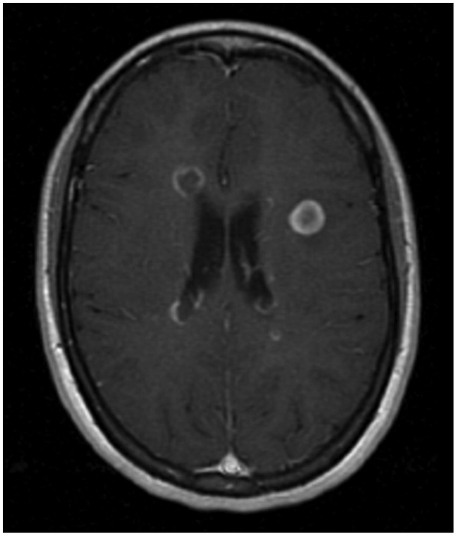
Tumefactive multiple sclerosis is an inflammatory demyelinating disease of the central nervous system. It has recently been described as a rare subtype of multiple sclerosis (MS) characterised by the appearance of solitary or multiple space-occupying lesions associated with imaging characteristics mimicking neoplasm. Atypical features include plaque size >2 cm with mass effect, oedema, and/or ring enhancement on magnetic resonance (MR) images.This study is a retrospective review designed to evaluate the prevalence of tumefactive plaques in a selected population of 440 MS patients referred to our MS centre in Southern Italy between 2005 and 2014. We analysed the radiographic features of lesions ranging in size from 0.5 to 2 cm to establish whether smaller plaques with MR characteristics similar to tumefactive plaques present different symptoms, disease evolution and prognosis. We also aimed to ascertain if MR features suggestive of biological aggressiveness could be useful prognostic criteria for a correct diagnosis of the disease and subsequent treatment. Our data suggest that lesions 0.5-2 cm and >2 cm have similar MR features and clinical evolution.
Keywords: Multiple sclerosis; magnetic resonance imaging; tumefactive demyelinating lesions.
© The Author(s) 2016.
Figures


Similar articles
-
Clinical and radiographic spectrum of pathologically confirmed tumefactive multiple sclerosis.Brain. 2008 Jul;131(Pt 7):1759-75. doi: 10.1093/brain/awn098. Epub 2008 Jun 5. Brain. 2008. PMID: 18535080 Free PMC article.
-
Clinical and radiological characteristics of tumefactive demyelinating lesions: follow-up study.Mult Scler. 2012 Oct;18(10):1448-53. doi: 10.1177/1352458512438237. Epub 2012 Mar 14. Mult Scler. 2012. PMID: 22419670
-
Conventional and advanced magnetic resonance imaging in tumefactive demyelination.Acta Radiol. 2011 Dec 1;52(10):1159-68. doi: 10.1258/ar.2011.110007. Epub 2011 Oct 24. Acta Radiol. 2011. PMID: 22025739
-
Tumefactive Demyelinating Lesions in Multiple Sclerosis and Associated Disorders.Curr Neurol Neurosci Rep. 2016 Mar;16(3):26. doi: 10.1007/s11910-016-0626-9. Curr Neurol Neurosci Rep. 2016. PMID: 26847090 Review.
-
Tumefactive demyelinating lesions: A comprehensive review.Mult Scler Relat Disord. 2017 May;14:72-79. doi: 10.1016/j.msard.2017.04.003. Epub 2017 Apr 9. Mult Scler Relat Disord. 2017. PMID: 28619436 Review.
Cited by
-
A rare case of tumefactive demyelination of brain: A case report and literature review.Clin Case Rep. 2023 Dec 21;11(12):e8369. doi: 10.1002/ccr3.8369. eCollection 2023 Dec. Clin Case Rep. 2023. PMID: 38130854 Free PMC article.
-
Imaging in Pediatric Multiple Sclerosis : An Iconographic Review.Clin Neuroradiol. 2021 Mar;31(1):61-71. doi: 10.1007/s00062-020-00929-8. Epub 2020 Jul 16. Clin Neuroradiol. 2021. PMID: 32676699 Review.
-
Demyelinating lesions behaving like aggressive tumours on advanced MRI techniques.Neuroradiol J. 2019 Apr;32(2):103-107. doi: 10.1177/1971400919826394. Epub 2019 Jan 22. Neuroradiol J. 2019. PMID: 30667319 Free PMC article.
-
Clinical spectrum and prognosis of pathologically confirmed atypical tumefactive demyelinating lesions.Sci Rep. 2023 May 13;13(1):7773. doi: 10.1038/s41598-023-34420-4. Sci Rep. 2023. PMID: 37179394 Free PMC article.
-
Tumor-like Lesions in Primary Angiitis of the Central Nervous System: The Role of Magnetic Resonance Imaging in Differential Diagnosis.Diagnostics (Basel). 2024 Mar 14;14(6):618. doi: 10.3390/diagnostics14060618. Diagnostics (Basel). 2024. PMID: 38535038 Free PMC article. Review.
References
MeSH terms
LinkOut - more resources
Full Text Sources
Other Literature Sources
Medical
