. 2017 Jan;101(1):321-328.
doi: 10.1189/jlb.3A0416-166R. Epub 2016 Aug 16.

SiglecF+Gr1hi eosinophils are a distinct subpopulation within the lungs of allergen-challenged mice

Affiliations

SiglecF+Gr1hi eosinophils are a distinct subpopulation within the lungs of allergen-challenged mice

Caroline M Percopo et al. J Leukoc Biol. 2017 Jan.

Abstract

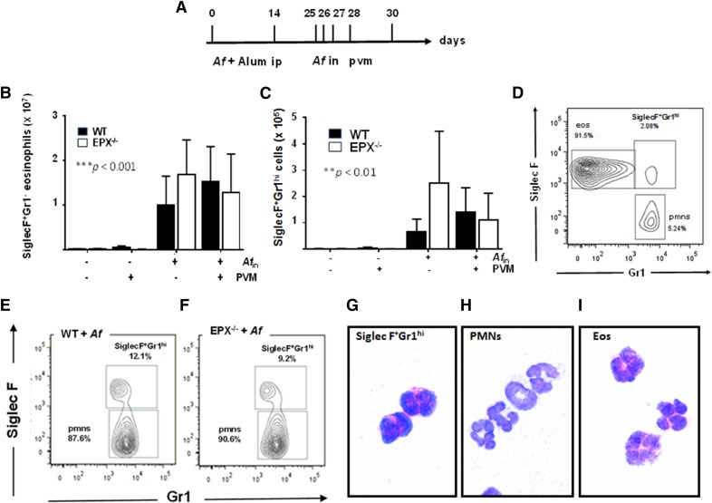
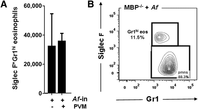
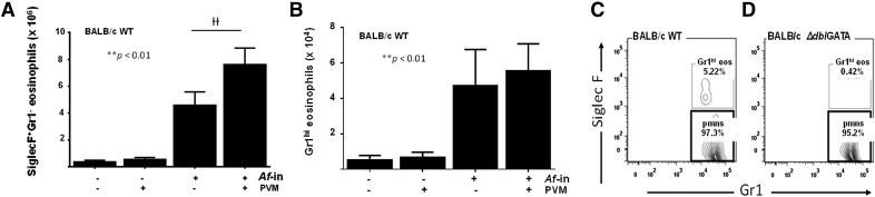
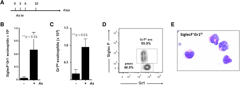
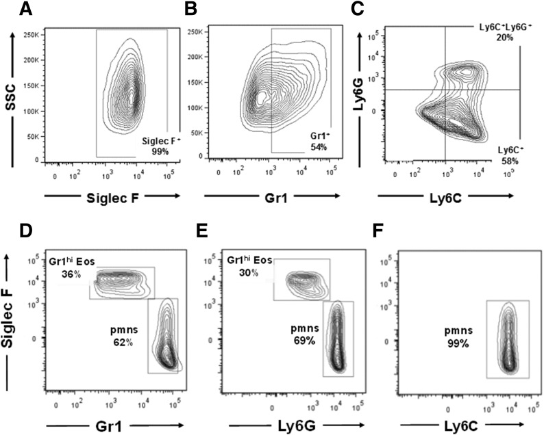
Although eosinophils as a group are readily identified by their unique morphology and staining properties, flow cytometry provides an important means for identification of subgroups based on differential expression of distinct surface Ags. Here, we characterize an eosinophil subpopulation defined by high levels of expression of the neutrophil Ag Gr1 (CD45+CD11c-SiglecF+Gr1hi). SiglecF+Gr1hi eosinophils, distinct from the canonical SiglecF+Gr1- eosinophil population, were detected in allergen-challenged wild-type and granule protein-deficient (EPX-/- and MBP-1-/-) mice, but not in the eosinophil-deficient ΔdblGATA strain. In contrast to Gr1+ neutrophils, which express both cross-reacting Ags Ly6C and Ly6G, SiglecF+Gr1hi eosinophils from allergen-challenged lung tissue are uniquely Ly6G+ Although indistinguishable from the more-numerous SiglecF+Gr1- eosinophils under light microscopy, FACS-isolated populations revealed prominent differences in cytokine contents. The lymphocyte-targeting cytokines CXCL13 and IL-27 were identified only in the SiglecF+Gr1hi eosinophil population (at 3.9 and 4.8 pg/106 cells, respectively), as was the prominent proinflammatory mediator IL-13 (72 pg/106 cells). Interestingly, bone marrow-derived (SiglecF+), cultured eosinophils include a more substantial Gr1+ subpopulation (∼50%); Gr1+ bmEos includes primarily a single Ly6C+ and a smaller, double-positive (Ly6C+Ly6G+) population. Taken together, our findings characterize a distinct SiglecF+Gr1hi eosinophil subset in lungs of allergen-challenged, wild-type and granule protein-deficient mice. SiglecF+Gr1hi eosinophils from wild-type mice maintain a distinct subset of cytokines, including those active on B and T lymphocytes. These cytokines may facilitate eosinophil-mediated immunomodulatory responses in the allergen-challenged lung as well as in other distinct microenvironments.

Keywords: allergy; cytokines; flow cytometry; inflammation.


Figures

Figure 1.
Figure 1.. SiglecF+Gr1hi eosinophils are an independent subpopulation detected in lungs of wild-type and eosinophil–peroxidase gene-deleted (EPX−/−) mice.
(A) Protocol I: Af sensitization on d 0 and d 14, followed by Af or PBS challenge (d 25, 26, 27). Mice were evaluated on d 30 as indicated. SiglecF+Gr1 eosinophils (CD45+CD11cSiglecF+Gr1) (B) and SiglecF+Gr1hi cells (CD45+CD11cSiglecF+Gr1hi) (C) in single-cell lung suspensions from Af-sensitized and Af- or PBS-challenged mice as indicated in (A). ***P < 0.001, **P < 0.01 Af-sensitization and Af challenge vs. Af-sensitization and PBS challenge, 2-way ANOVA, n = 5–13 mice/group. (D) SiglecF+Gr1hi cells, as distinct from SiglecF+Gr1 eosinophils, represent ∼2% of the total CD45+CD11c leukocyte population isolated from the lungs of allergen-sensitized and -challenged mice. (E and F) SiglecF+Gr1hi cells represent ∼10% of the CD45+CD11cGr1hi cells isolated from lungs of Af-sensitized and -challenged, wild-type mice and EPX−/− mice, respectively. CD45+CD11cSiglecF+Gr1hi cells (G), CD45+CD11cSiglecFGr1hi (polymorphonuclear neutrophils; neutrophils) (H), and CD45+CD11cSiglecF+Gr1 (Eos; eosinophils) (I) isolated from lung single-cell suspensions from Af-sensitized and -challenged mice by flow cytometry and cell sorting (FACS); original magnification ×100.
Figure 2.
Figure 2.. SiglecF+Gr1hi eosinophils are detected in lungs of eosinophil MBP-1−/− mice.
(A) SiglecF+Gr1hi eosinophils in single-cell lung suspensions from Af-sensitized and -challenged MBP-1−/− mice both with and without subsequent virus infection; n = 5–6 mice/group. (B) SiglecF+Gr1hi eosinophils represent >10% of the CD45+CD11cGr1hi cells isolated from lungs of Af-sensitized and -challenged, wild-type mice and MBP-1−/− mice.
Figure 3.
Figure 3.. SiglecF+Gr1hi eosinophils are detected in response to Af-sensitization and challenge in wild-type (WT) BALB/c, but not eosinophil-deficient, ΔdblGATA mice.
SiglecF+Gr1 eosinophils (A) and SiglecF+Gr1hi eosinophils (B) in single-cell lung suspensions from Af-sensitized and -challenged mice, as described in Fig. 1; **P < 0.01 Af-sensitization and Af-challenge vs. Af-sensitization and PBS challenge, ††P < 0.01 PVM infected vs. diluent control, 2-way ANOVA, n = 3–5 mice/group. C. SiglecF+Gr1hi eosinophils are an independent population and represent ∼5% of the CD45+CD11cGr1+ cells isolated from lungs of Af-sensitized and -challenged BALB/c mice. (D) SiglecF+Gr1hi cells cannot be detected in Af-sensitized and -challenged, eosinophil-deficient ΔdblGATA mice.
Figure 4.
Figure 4.. SiglecF+Gr1hi eosinophils are detected in response to intranasal challenge with Alternaria alternata in wild-type BALB/c mice.
(A) Protocol II: Aa intranasal challenge on d 0, 3, and 6; mice were evaluated on d 10 as indicated. SiglecF+Gr1 eosinophils (B) and SiglecF+Gr1hi eosinophils (C) in single-cell lung suspensions from mice challenged with Aa, **P < 0.01 Mann-Whitney U test, n = 4–6 mice/group. (D) SiglecF+Gr1hi eosinophils represent 50% of the CD45+CD11cGr1+ cells isolated from lungs of the Aa-challenged BALB/c mice. (E) CD45+CD11cSiglecF+Gr1hi eosinophils isolated from lung single-cell suspensions from Aa-challenged mice by flow cytometry; original magnification ×100.
Figure 5.
Figure 5.. Ly6C/Ly6G Ag profiles of bmEos and Gr1+ eosinophils from allergen-challenged lung.
Eosinophils generated in culture from unselected bmEos from C57BL/6 mice are SiglecF+ (99%) (A), Gr1+ (54%) (B), and Ly6C+ or Ly6C+Ly6G+ (C) SiglecF+Gr1hi (D) eosinophils identified in lung suspensions from mice challenged with Aa (see Fig. 4A) are Ly6G+ (E) and Ly6C (F).

References

    1. Lee J. J., Rosenberg H. F.. 2013. Eosinophils in Health and Disease. Elsevier, Amsterdam.
    1. Rosenberg H. F., Dyer K. D., Foster P. S. (2013) Eosinophils: changing perspectives in health and disease. Nat. Rev. Immunol. 13, 9–22. - PMC - PubMed
    1. Jacobsen E. A., Lee N. A., Lee J. J. (2014) Re-defining the unique roles for eosinophils in allergic respiratory inflammation. Clin. Exp. Allergy 44, 1119–1136. - PMC - PubMed
    1. Lee J. J., Jacobsen E. A., McGarry M. P., Schleimer R. P., Lee N. A. (2010) Eosinophils in health and disease: the LIAR hypothesis. Clin. Exp. Allergy 40, 563–575. - PMC - PubMed
    1. Chu V. T., Beller A., Rausch S., Strandmark J., Zänker M., Arbach O., Kruglov A., Berek C. (2014) Eosinophils promote generation and maintenance of immunoglobulin-A-expressing plasma cells and contribute to gut immune homeostasis. Immunity 40, 582–593. - PubMed

MeSH terms