. 2016 Dec;65(12):3667-3679.
doi: 10.2337/db16-0081. Epub 2016 Aug 16.

CIN85 Deficiency Prevents Nephrin Endocytosis and Proteinuria in Diabetes

Affiliations

CIN85 Deficiency Prevents Nephrin Endocytosis and Proteinuria in Diabetes

Beina Teng et al. Diabetes. 2016 Dec.

Abstract

Diabetic nephropathy (DN) is the major cause of end-stage renal disease worldwide. Podocytes are important for glomerular filtration barrier function and maintenance of size selectivity in protein filtration in the kidney. Podocyte damage is the basis of many glomerular diseases characterized by loss of interdigitating foot processes and decreased expression of components of the slit diaphragm. Nephrin, a podocyte-specific protein, is the main component of the slit diaphragm. Loss of nephrin is observed in human and rodent models of diabetic kidney disease. The long isoform of CIN85 (RukL) is a binding partner of nephrin that mediates nephrin endocytosis via ubiquitination in podocytes. Here we demonstrate that the loss of nephrin expression and the onset of proteinuria in diabetic mice correlate with an increased accumulation of ubiquitinated proteins and expression of CIN85/RukL in podocytes. CIN85/RukL deficiency preserved nephrin surface expression on the slit diaphragm and reduced proteinuria in diabetic mice, whereas overexpression of CIN85 in zebrafish induced severe edema and disruption of the filtration barrier. Thus, CIN85/RukL is involved in endocytosis of nephrin in podocytes under diabetic conditions, causing podocyte depletion and promoting proteinuria. CIN85/RukL expression therefore shows potential to be a novel target for antiproteinuric therapy in diabetes.


Figures

Figure 1
Figure 1
Time course of CIN85, CD2AP, and nephrin expression after glucose or mannitol stimulation in murine and human podocytes. Human and murine podocytes were exposed to 30 mmol/L high glucose or mannitol as osmotic control at the times indicated (0–48 h). Western blotting analysis depicts the expression of CIN85, CD2AP, and nephrin in murine podocytes (A and B) and human podocytes (C and D) (results are representative for three independent experiments). Expression level of CIN85, CD2AP, and nephrin were quantified by densitometry (right panels). High glucose augmented CIN85 expression in a time-dependent manner and reached the highest level at 48 h in murine (A) and human (C) podocytes, whereas a downregulated expression of CD2AP and nephrin was detected as early as 24 h after stimulation. Values are means ± SEM of three independent experiments expressed as percentage of control (0 h), where the ratio in control was defined as 100%. *P ≤ 0.05, **P ≤ 0.01 vs. control (0 h) by unpaired t test.
Figure 2
Figure 2
Increased glomerular CIN85 expression in mice and a patient with DN. A: CIN85 and SUMO-1 were visualized by dual immunofluorescence staining on cryosections of C57BL/6J and STZ-induced type 1 diabetic mice (n = 5 per group). In wild-type mice, colocalization of SUMO-1 and CIN85 indicate posttranscriptional modification of CIN85 by SUMOylation. In the renal cortex section of diabetic animals, SUMO-1 is predominantly localized in the nucleus, and colocalization with CIN85 is not detected. Original magnification ×63. B: Dual immunofluorescence staining with CIN85 and nephrin antibodies was performed on frozen kidney cortex sections of C57BL/6J and diabetic mice. In diabetic kidney sections, expression of CIN85 (red) was significantly increased, whereas the nephrin expression (green) was reduced. Colocalization of nephrin and CIN85 is depicted in yellow. CIN85 is partially colocalized with nephrin, indicating expression of CIN85 in projection of the slit diaphragm (white arrows). The pictures are representative for most of the glomeruli in the C57BL/6J and diabetic mice (n = 5 per group). Original magnification ×63. C: Representative immunoperoxidase staining for CIN85 in renal biopsy sections from a normal kidney (upper panels) and from a patient with DN (lower panels). The white arrows depict the distribution of CIN85 in podocytes. CIN85-positive podocytes could only be detected in the glomeruli of patients with DN (n = 5) but not in the glomeruli from normal control kidneys. Original magnification ×100. D: Downregulated SUMOylation of CIN85 in glomeruli of diabetic mice. Antibodies against SUMO-1 were used for immunoprecipitation (IP) experiments to detect interaction with glomerular lysates from CD2AP-knockout mice, nondiabetic C57BL/6J mice (−), and type 1 diabetic mice (+ indicates blood glucose level between 12 and 18 mmol/dL, ++ indicates blood glucose level above 18 mmol/dL). E: Nephrin and CIN85 are binding partners in vivo. Endogenous nephrin and CIN85 were precipitated using antibodies against nephrin and CIN85 on glomerular lysates isolated from C56BL/6J (−) and type 1 diabetic mice (+) (n = 5 per group). IgG antibody was used as a negative control, and whole glomerular lysate was used as input. F: Increased interaction between nephrin and CIN85 upon high glucose stimulation in human podocytes in vitro. The immunoprecipitation was performed with cell lysates of human podocytes with indicated treatment. Western blot analysis indicated a significantly enhanced association between nephrin and CIN85 48 h after high glucose treatment.
Figure 3
Figure 3
CIN85 exon2 deletion ameliorates proteinuria and glomerular matrix accumulation in diabetic mice. A: Immunofluorescence staining of mouse glomeruli using antibodies against nephrin (green) and ubiquitin (red). Ubiquitin partially colocalized with nephrin (yellow) in diabetic C57BL/6J mice. Ubiquitin-positive podocytes were increased in 16-week-old diabetic wild-type mice, whereas nephrin expression was downregulated. Nephrin expression was preserved in CIN85Δex2 mice, and substantially fewer ubiquitin-positive podocytes were detected under diabetic conditions compared with C57BL/6J diabetic mice. Original magnification ×60. B: Albuminuria in C57BL/6J mice was determined by analysis of spot urine samples of mice 16 weeks after low-dose STZ injection (n = 5 in first three conditions, n = 12 in CIN85Δex2 diabetic mice). The error bars show the mean ± SEM. ***P ≤ 0.001 by unpaired t test. C: Ubiquitination assay using immunoprecipitation (IP) with antibody against nephrin and ubiquitin showed the ubiquitinated nephrin content increased 1 h after high glucose stimulation. D: Representative images of glomeruli stained by immunofluorescence using anti-collagen IV. Collagen IV staining indicated increased matrix accumulation caused by diabetes. Original magnification ×60. E: Glomerular collagen IV expression in glomeruli from the indicated mice was semiquantitatively scored in a blinded fashion: score 1, very mild; score 2, mild; score 3, moderate; score 4, intense. The error bars show the mean ± SEM (n = 5 per condition). *P ≤ 0.05; n.s., not statistically significant by unpaired t test.
Figure 4
Figure 4
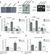
Impaired nephrin endocytosis in the absence of CIN85 after high glucose exposure. To perform the cell-based nephrin endocytosis assay, we generated conditional immortalized podocytes with CIN85 exon 2 deletion. A: Light microscopic morphology of cultured podocytes. CIN85Δex2 podocytes grown under nonpermissive conditions (37°C without γ-interferon) display similar morphology as wild-type and CD2AP−/− murine podocytes. Original magnification ×200. B: Western blot analysis showed a complete absence of full-length CIN85 in CIN85Δex2 podocytes. Full-length CIN85 was not detectable in wild-type podocytes but was seen in CD2AP-knockout podocytes. C: PCR analysis using primers against full-length CIN85 indicated the deletion of CIN85 exon 2 in murine podocytes. D and E: Nephrin molecules expressed on the surface of differentiated wild-type, CD2AP−/−, and CIN85Δex2 murine podocytes were labeled with nephrin antibody, followed by incubation with high glucose and mannitol for 2 h and 24 h to induced endocytosis. F and G: An adenoviral system was used to transfect murine and human podocytes with CIN85-flag. Nephrin expressed on the cell surface was labeled with antibody against nephrin. The labeled podocytes were treated with high glucose for 2 h to induce internalization of nephrin. The error bars show the mean ± SEM (n = 5 per condition). *P ≤ 0.05, **P ≤ 0.01, ***P ≤ 0.001; n.s., not statistically significant by unpaired t test.
Figure 5
Figure 5
CIN85 expression impaired the integrity of the filtration barrier in zebrafish. Fertilized zebrafish eggs were injected at the 1- to 4-cell stage with murine CIN85 or CD2AP-capped mRNA in the indicated concentrations. A: Phenotype of zebrafish after CD2AP and CIN85 mRNA injection at 120 h after fertilization. The injected zebrafish larvae showed a very severe generalized edema and pericardial effusion. B: Categorization of zebrafish phenotype. At 120 h after fertilization, the phenotype of larvae was categorized into four groups: P1, no edema; P2, mild edema; P3, severe edema; and P4, very severe edema (n > 50 per condition). Compared with CD2AP mRNA–injected fish with 90% P1 phenotype, 7.5 ng CIN85 mRNA induced edema in ∼40% of embryos, and 10 ng CIN85 mRNA led to edema in >60% of embryos (n > 50 per condition). C: Fluorescent images of the retinal vessel plexus. Bar graph presenting fluorescence intensity of eGFP-DBP in the fish eye under indicated conditions was analyzed by ImageJ software. The CIN85 overexpression induced proteinuria showed in a dose-dependent manner. At least 50 animals were measured for each condition. The error bars show the mean ± SEM. *P ≤ 0.05, ***P ≤ 0.001 by unpaired t test. D: Expression of CD2AP and CIN85 capped-mRNA was determined using Western blot analysis. E: Ultrastructural analysis of the zebrafish glomerular structures using transmission electron microscopy. Under normal conditions, the secondary foot process of podocytes can be visualized (*). The black arrows depict slit diaphragms. GEC, glomerular endothelial cells. Scale bars: 500 nm. Control fish and fish injected with CD2AP mRNA displayed normal foot processes, whereas in zebrafish injected with CIN85 mRNA, podocyte foot processes and slit diaphragms are lost (effacement).
Figure 6
Figure 6
Schema of CIN85-induced nephrin endocytosis under diabetic conditions. In the presence of CD2AP, the complexes on the slit diaphragm are stabilized. Diabetes/high glucose downregulates CD2AP expression, which leads to increased full-length CIN85 expression. CIN85 is required for cbl-mediated ubiquitination and trafficking of nephrin. Nephrin endocytosis leads to proteinuria. Deletion of CIN85 preserves the nephrin expression and ameliorates the proteinuria and glomerular matrix accumulation.

Comment in

Similar articles

Cited by

References

    1. Fioretto P, Mauer M. Histopathology of diabetic nephropathy. Semin Nephrol 2007;27:195–207 - PMC - PubMed
    1. Jefferson JA, Shankland SJ, Pichler RH. Proteinuria in diabetic kidney disease: a mechanistic viewpoint. Kidney Int 2008;74:22–36 - PubMed
    1. Susztak K, Raff AC, Schiffer M, Böttinger EP. Glucose-induced reactive oxygen species cause apoptosis of podocytes and podocyte depletion at the onset of diabetic nephropathy. Diabetes 2006;55:225–233 - PubMed
    1. Stieger N, Worthmann K, Teng B, et al. . Impact of high glucose and transforming growth factor-β on bioenergetic profiles in podocytes. Metabolism 2012;61:1073–1086 - PubMed
    1. Teng B, Duong M, Tossidou I, Yu X, Schiffer M. Role of protein kinase C in podocytes and development of glomerular damage in diabetic nephropathy. Front Endocrinol (Lausanne) 2014;5:179. - PMC - PubMed

MeSH terms