Review
. 2016 Aug 2:10:81.
doi: 10.3389/fncom.2016.00081. eCollection 2016.

Burst Firing in the Electrosensory System of Gymnotiform Weakly Electric Fish: Mechanisms and Functional Roles

Affiliations
Review

Burst Firing in the Electrosensory System of Gymnotiform Weakly Electric Fish: Mechanisms and Functional Roles

Michael G Metzen et al. Front Comput Neurosci. .

Abstract

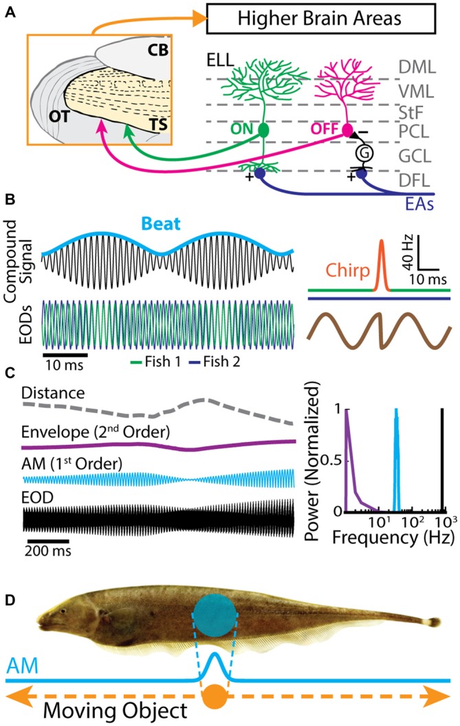
Neurons across sensory systems and organisms often display complex patterns of action potentials in response to sensory input. One example of such a pattern is the tendency of neurons to fire packets of action potentials (i.e., a burst) followed by quiescence. While it is well known that multiple mechanisms can generate bursts of action potentials at both the single-neuron and the network level, the functional role of burst firing in sensory processing is not so well understood to date. Here we provide a comprehensive review of the known mechanisms and functions of burst firing in processing of electrosensory stimuli in gymnotiform weakly electric fish. We also present new evidence from existing data showing that bursts and isolated spikes provide distinct information about stimulus variance. It is likely that these functional roles will be generally applicable to other systems and species.

Keywords: burst firing; directional selectivity; envelope; feature detection; neural coding; weakly electric fish.


Figures

Figure 1
Figure 1
Electrosensory circuitry and natural stimuli. (A) Peripheral electrosensory afferents (EAs) enter the hindbrain at the deep fiber layer (DFL) of the electrosensory lateral line lobe (ELL) and project onto two types of pyramidal neurons (ON: green; OFF: magenta) within the pyramidal cell layer (PCL). ON type cells have a basilar dendrite that connects directly to the EAs, while OFF type cells lack such a basilar dendrite and instead receive disynaptic input via local interneurons (G) within the granule cell layer (GCL). The apical dendrites of both types extend through the stratum tractus fibrosum (StF) to the molecular layers of the ELL (VML, ventral molecular layer; DML, dorsal molecular layer). Both types of neurons send projections to higher brain areas, such as the midbrain torus semicircularis (TS). (B) Left: the electric organ discharges (EODs) of two fish (green and blue) interfere and thus create a sinusoidal beat (cyan) whose frequency is equal to the EOD frequency difference between the two fish. Right: during an electro-communication call (i.e., a chirp), the emitter fish’s EOD frequency (top green trace) transiently increases for a brief period of time (top orange trace), while the receiver fish’s EOD frequency (top blue trace) remains constant. The chirp results in a phase reset of the beat (bottom brown trace). (C) Left: EOD waveform from Apteronotus leptorhynchus (black) with amplitude modulation (AM, cyan) and envelope (purple) waveforms. We note that the envelope corresponds to the depth of modulation of the EOD AM that is due to relative movement (dashed gray line) between individuals. Right: shown are the frequency contents of the full signal (black), the AM (cyan), and the envelope (purple). (D) EOD AM (cyan) originating from an object (orange) that is moving along the fish’s body (dashed orange arrows) and the corresponding electric image projected onto the skin (cyan).
Figure 2
Figure 2
Electrosensory afferents (EAs) are composed of two sub-populations: bursting and non-bursting. (A) Primary afferents from peripheral electroreceptors project onto pyramidal neurons within the hindbrain. (B) Example recording of a non-bursting EA. (C) Example recording from an EA that displays burst firing (red). (D) Return map of the same neuron shown in (B). Inset: interspike interval (ISI) distribution. (E) Return map of the same neuron shown in (C). Inset: ISI distribution. (F) Segregating a population (n = 94) based on burst fraction (i.e., fraction of ISIs below a threshold corresponding to the inverse of the EOD frequency indicated by the arrow ~2 ms) reveals two subpopulations of EAs (Two-sample Kolmogorov-Smirnov test, p ≪ 10–3). (G) Plotting firing probability as a function of burst fraction yields a positive correlation (r = 0.74). Also shown is the firing probability as a function of burst fraction for an equivalent Poisson process (blue curve). The data plotted in (B–G) are from Metzen and Chacron (2015).
Figure 3
Figure 3
Bursting in neurons in the hindbrain ELL. (A) ELL pyramidal cells receive input from the electrosensory primary afferents and project to the midbrain. (B) Schematic showing the distribution of sodium (Na+, magenta), and two subtypes of small-conductance potassium (blue: SK1; green: SK2) channels. Na+ channels are located in the soma as well as the proximal dendrite, SK1 channels are located in the proximal and distal dendrite, whereas SK2 channels are only expressed in the soma. Neuromodulators, such as serotonin (5-HT) and acetylcholine (ACh), influence spiking. (C) A somatic and dendritic burst of spikes recorded separately in two cells (somatic spikes are truncated). The slowdown in dendritic spike repolarization is due to inactivation of a dendritic K+ conductance and results in a potentiation of the somatic depolarizing afterpotential (DAP; arrows). When the DAP reaches threshold for a high-frequency spike doublet, the second spike fails to backpropagate. This allows the afterhyperpolarization (AHP) to terminate the burst. (D) Example in vivo recording of an ELL pyramidal cell under control conditions. (E) The same neuron as in (E) displays bursting after treatment with the Ca2+ chelator BAPTA. The arrows indicate the ramp depolarizations. Inset: a DAP is seen after electrically stimulating serotonergic pathways (blue line). (F) ISI distribution under control condition (black) and after BAPTA treatment (red) showing a decrease in the cell’s absolute refractory period and the emergence of a second peak (red arrows). The burst threshold used to segregate bursts and isolated spikes for ELL pyramidal cells was 10 ms. (G) ISI return map under control condition (black) and after BAPTA treatment (red) showing a transition to a bursting regime. The data plotted in (D–G) are from Toporikova and Chacron (2009).
Figure 4
Figure 4
Serotonin increases electrosensory pyramidal neuron excitability. (A) Schematic showing the setup used to apply serotonin focally. Shown are the recording electrode that is positioned near a pyramidal neuron and the pipette containing serotonin that is positioned close to this neuron’s dendritic tree. (B) Schematic showing how stimulation of the raphe nuclei was achieved. Shown is a dorsal view of the animal’s brain with the recording pipette and the stimulation electrode. CCb, corpus cerebelli; EGP, eminentia granularis posterior; Tel, telencephalon; OT, optic tectum. (C) Top: spiking activity from an example ELL pyramidal neuron recorded in vitro under control conditions (left) and after serotonin application (right). Note that the application of serotonin induces burst firing (arrows). Bottom: spiking activity from an example ELL pyramidal neuron recorded in vivo under baseline (left) and raphe nuclei stimulation (right). Note the increased burst firing (blue) after raphe stimulation. (D) Left: population-averaged burst fraction (i.e., the fraction of ISIs < 10 ms) before stimulation (black), and after raphe stimulation (blue, n = 13). Right: release of serotonin through raphe stimulation led to a significant reduction in the medium component of the AHP (mAHP) in pyramidal neurons. Asterisks indicate statistical significance at the p = 0.05 level using a paired t-test. (E) Stimulus waveform showing a chirp at the center (top), raster plot (middle) showing spike times (gray), and the first spike occurring immediately after the small chirp (red) as well as the corresponding peristimulus time histogram (PSTH; bottom) before (left) and after (right) raphe stimulation. (F) Bar graphs showing the population-averaged normalized first spike latency (left, n = 13) and the normalized SD of the first spike latency (right, n = 13) before (black) and after raphe stimulation (blue). (G) Top: stimulus waveform, an example control recording of a pyramidal cell (burst spikes in black) and a recording from the same cell after raphe stimulation (burst spikes in blue). Bottom: Population-averaged vector strength values as a function of stimulus frequency before (black, n = 6) and after (blue, n = 6) raphe stimulation. (H) EOD frequency in response to a jamming stimulus as a function of time before (control, black) and after serotonin (serotonin, red) injection. Note the higher increase in EOD frequency after serotonin injection compared to the control condition. Data plotted are from Deemyad et al. (2013).
Figure 5
Figure 5
Bursting in neurons in the midbrain TS. (A) Neurons within the TS receive input from ELL pyramidal cells. (B) Summary of inputs to and outputs from TS layers. Note that TS layer 6 receives only input from the frequency modulation (FM) pathway that is not considered here. (C) Example recording of a non-bursting TS neuron (upper trace) and its return map (lower plot). The ISI distribution of this neuron shows a single peak (arrow) at around 100 ms (inset). (D) Example recording of a TS cell in bursting mode (upper trace). Arrows indicate the bursts of action potentials riding on top of a calcium spike. The return map displays clusters of dots close to the origin, the abscissa and the ordinate, indicating burst firing. Inset: the ISI distribution of this neuron shows two prominent peaks, as indicated by the arrows. Data plotted in (C,D) are from Chacron et al. (2009), Chacron and Fortune (2010), Khosravi-Hashemi et al. (2011), Khosravi-Hashemi and Chacron (2012).
Figure 6
Figure 6
Neurons in the midbrain TS respond to a moving object. (A) Schematic showing the stimulation protocol. The gray sphere represents the moving object that was moved sinusoidally back and forth along the fish at a distance of about 1 cm lateral to the fish. The orange arrow indicates the tail-to-head direction, whereas the red arrow indicates the head-to-tail direction. The resulting local EOD AM and the spread of the electric image projected onto the skin are shown in blue. (B) Example in vivo recordings from a bursting TS (left) and a non-bursting TS neuron (right) to a moving object. Action potentials (green ticks) with ISIs that were shorter than the burst threshold were identified as belonging to bursts (magenta ticks), whereas those that were not were identified as isolated spikes (blue ticks). Burst events are indicated as yellow stars. (C) Raster plot from an example directionally selective bursting TS neuron. The spikes that belong to bursts are shown in magenta, whereas isolated spikes are shown in blue. Bottom: normalized PSTH for this same neuron computed from all spikes (both bursts and isolated spikes, green line), bursts (magenta line), and isolated spikes (blue line). Inset: population-averaged directional biases obtained for bursts (magenta), all spikes (green), and isolated spikes (blue). Asterisks indicate statistical significance at the p = 0.05 level using a signed-rank test. (D) Normalized PSTH for an example neuron where bursts and isolated spikes code for opposite directions of movement (arrows) computed from all spikes (green line), bursts (magenta line), and isolated spikes (blue line). The curves have been normalized by their maximum values. Directional bias values were 0.6, 0.5, and −0.63 for burst, all spikes, and isolated spikes, respectively. Data plotted in (B–D) are from Chacron et al. (2009), Chacron and Fortune (2010), Khosravi-Hashemi et al. (2011), Khosravi-Hashemi and Chacron (2012).
Figure 7
Figure 7
Burst firing can improve the gain of EAs in response to envelopes. (A) Example time dependent firing rates obtained for all spikes (green), bursts (magenta), and isolated spikes (blue) of EAs with a low firing probability (left), intermediate firing probability (middle) and high firing probability (right) to a sinusoidal envelope (top, purple). (B) Population-averaged gain (top) and phase (bottom) curves as a function of envelope frequency for EAs with low (left), intermediate (middle) and high (right) firing probabilities. Gain and phase curves for all spikes (green), bursts (magenta) and isolated spikes (blue) are shown. Data plotted in (A,B) are from Metzen and Chacron (2015).

Similar articles

Cited by

References

    1. Adrian E. D. (1941). Afferent discharges to the cerebral cortex from peripheral sense organs. J. Physiol. 100, 159–191. 10.1113/jphysiol.1941.sp003932 - DOI - PMC - PubMed
    1. Aumentado-Armstrong T., Metzen M. G., Sproule M. K. J., Chacron M. J. (2015). Electrosensory midbrain neurons display feature invariant responses to natural communication stimuli. PLoS Comput. Biol. 11:e1004430. 10.1371/journal.pcbi.1004430 - DOI - PMC - PubMed
    1. Ávila-Akerberg O., Chacron M. J. (2011a). In vivo conditions influence the coding of stimulus features by bursts of action potentials. J. Comput. Neurosci. 31, 369–383. 10.1007/s10827-011-0313-4 - DOI - PMC - PubMed
    1. Ávila-Akerberg O., Chacron M. J. (2011b). Nonrenewal spike train statistics: causes and functional consequences on neural coding. Exp. Brain Res. 210, 353–371. 10.1007/s00221-011-2553-y - DOI - PMC - PubMed
    1. Ávila-Akerberg O., Krahe R., Chacron M. J. (2010). Neural heterogeneities and stimulus properties affect burst coding in vivo. Neuroscience 168, 300–313. 10.1016/j.neuroscience.2010.03.012 - DOI - PMC - PubMed

Grants and funding

LinkOut - more resources