. 2016 Aug 16:4:45.
doi: 10.1186/s40425-016-0149-6. eCollection 2016.

A hypofractionated radiation regimen avoids the lymphopenia associated with neoadjuvant chemoradiation therapy of borderline resectable and locally advanced pancreatic adenocarcinoma

Affiliations

A hypofractionated radiation regimen avoids the lymphopenia associated with neoadjuvant chemoradiation therapy of borderline resectable and locally advanced pancreatic adenocarcinoma

Todd Crocenzi et al. J Immunother Cancer. .

Abstract

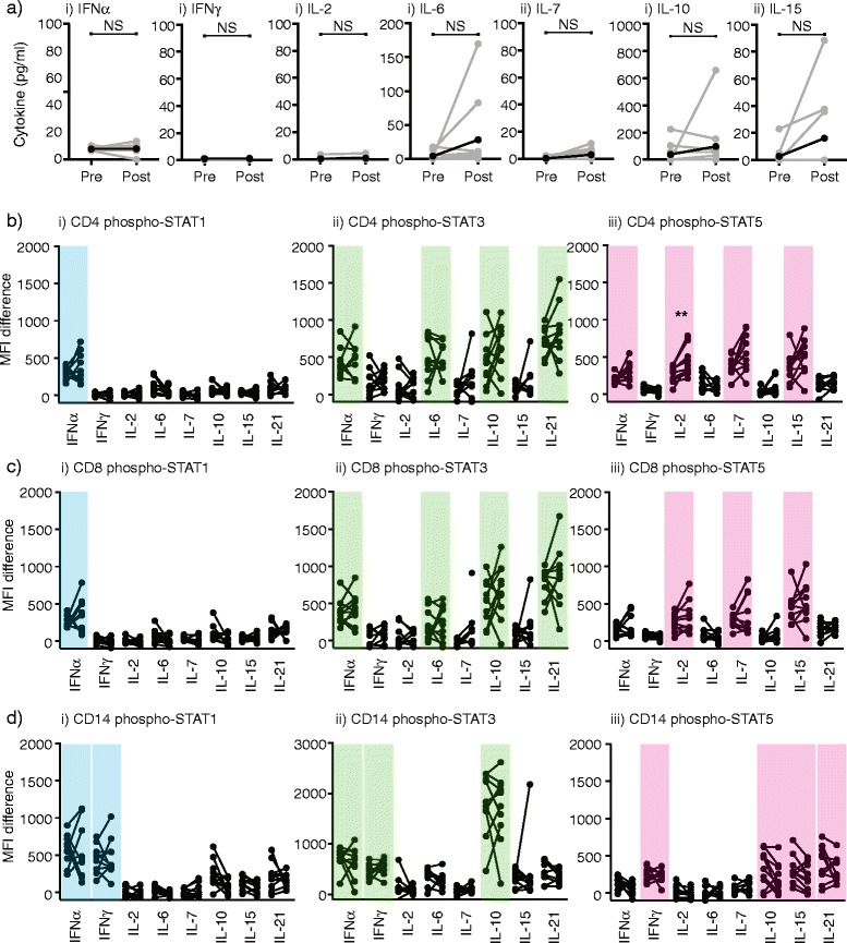
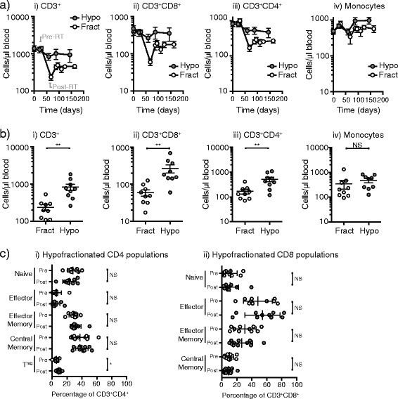
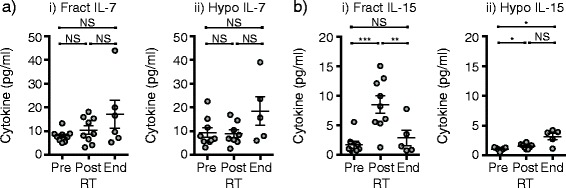
Background: Preclinical studies have shown synergy between radiation therapy and immunotherapy. However, in almost all preclinical models, radiation is delivered in single doses or short courses of high doses (hypofractionated radiation). By contrast in most clinical settings, radiation is delivered as standard small daily fractions of 1.8-2 Gy to achieve total doses of 50-54 Gy (fractionated radiation). We do not yet know the optimal dose and scheduling of radiation for combination with chemotherapy and immunotherapy.

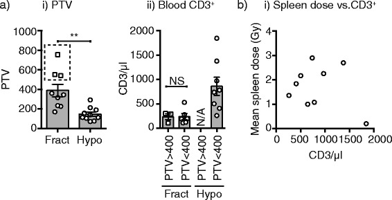
Methods: To address this, we analyzed the effect of neoadjuvant standard fractionated and hypofractionated chemoradiation on immune cells in patients with locally advanced and borderline resectable pancreatic adenocarcinoma.

Results: We found that standard fractionated chemoradiation resulted in a significant and extended loss of lymphocytes that was not explained by a lack of homeostatic cytokines or response to cytokines. By contrast, treatment with hypofractionated radiation therapy avoided the loss of lymphocytes associated with conventional fractionation.

Conclusion: Hypofractionated neoadjuvant chemoradiation is associated with reduced systemic loss of T cells.

Trial registration: ClinicalTrials.gov NCT01342224, April 21, 2011; NCT01903083, July 2, 2013.

Keywords: Chemotherapy; Fractionation; Gemcitabine; Homeostatic repopulation; IL-15; IL-7; Immunotherapy; Lymphocytes; Lymphodepletion; Radiation.


Figures

Fig. 1
Fig. 1
Effect of conventional neoadjuvant chemoradiation on immune cells in patient blood. a Absolute numbers of i) CD3+, ii) CD8+, iii) CD4+ and iv) CD4+CD25+ T cells as well as v) SSCintCD14+ monocytes and vi) SSChiCD15+ granulocytes by flow cytometry of fresh peripheral blood over the course of the study. Individual patients are gray, the mean is black. Dotted lines show sampling times and shows periods of neoadjuvant and adjuvant chemotherapy (red rectangles), neoadjuvant chemoradiation (blue bar) according to the trial schema provided in Additional file 1: Figure S1. b Analysis of the proportions of i) CD4 and ii) CD8 subpopulations in PBMC collected pretreatment (open circles) and post-treatment (closed circles). Key: NS – not significant. * p < 0.05
Fig. 2
Fig. 2
Role of homeostatic repopulation through cytokines and cytokine responses. a Patient serum collected prior to treatment (pre) and at the end of treatment (post) were tested for cytokine levels by multiplex assay. Individual patients are gray, the mean is black. b-d PBMC collected prior to treatment and at the end of treatment were treated with a range of cytokines for and analyzed for i) pSTAT1, ii) pSTAT3 or iii) pSTAT5 expression by intracellular flow cytometry. Surface staining of these mixed populations identified pSTAT activation in (b) CD4+ T cells, (c) CD8+ T cells or (d) CD14+ monocytes. Individual patient’s pre and post treatment values are connected. Graphs show change in pSTAT MFI over control (vehicle alone) stimulated cells. Colors highlight STATs that respond to a particular stimulation in each cell type. Key: NS – not significant. * p < 0.05, ** p < 0.01
Fig. 3
Fig. 3
Effect of hypofraction on immune cells in patient blood. a Absolute numbers of i) CD3+, ii) CD8+, iii) CD4+ and iv) SSCintCD14+ monocytes by flow cytometry of fresh peripheral blood in patients on a conventionally fractionated regimen (Fract, open symbols) versus a hypofractionated regimen (Hypo, closed symbols). The samples immediately pre-RT and immediately post-RT are highlighted on the first graph. b comparison of absolute numbers of i) CD3+, ii) CD8+, iii) CD4+ and iv) SSCintCD14+ monocytes by flow cytometry of fresh peripheral blood immediately following completion of radiation therapy in patients on a conventionally fractionated regimen (Fract, open symbols) versus a hypofractionated regimen (Hypo, closed symbols). c Analysis of the proportions of i) CD4 and ii) CD8 subpopulations in PBMC collected pretreatment (open circles) and post-treatment with a hypofractionated regimen (closed circles)
Fig. 4
Fig. 4
Effect of treatment regimen on early homeostatic cytokines. Patient serum collected prior to treatment (pre), immediately following completion of radiation therapy (post) and at the end of treatment (end) from patients on i) a conventionally fractionated regimen (Fract) versus ii) a hypofractionated regimen (Hypo) were tested for (a) IL-7 and (b) IL-15 cytokine levels by ultrasensitive multiplex assay. Key: NS – not significant. * p < 0.05, ** p < 0.01, *** p < 0.001
Fig. 5
Fig. 5
Link between planning target volume (PTV), spleen dose and T cell count. a) i) Graph showing the PTV for patients receiving a conventionally fractionated regimen (Fract) versus a hypofractionated regimen (Hypo). A subset of patients receiving a conventionally fractionated regimen exhibited a PTV greater than 400 (square symbols). ii) Post-chemoRT blood T cell counts are shown for patients with PTV greater than or less than 400 receiving a fractionated regimen (Fract) versus a hypofractionated regimen (Hypo). b) Relationship between mean spleen dose (Gy) and post-chemoRT blood T cell counts for patients receiving a hypofractionated regimen. Each symbol represents one patient. Key: NS – not significant. ** p < 0.01

References

    1. Siegel RL, Miller KD, Jemal A. Cancer statistics, 2015. CA Cancer J Clin. 2015;65(1):5–29. doi: 10.3322/caac.21254. - DOI - PubMed
    1. Sutton JM, Abbott DE. Neoadjuvant therapy for pancreas cancer: past lessons and future therapies. World J Gastroenterol. 2014;20(42):15564–79. doi: 10.3748/wjg.v20.i42.15564. - DOI - PMC - PubMed
    1. Callery M, Chang K, Fishman E, Talamonti M, Traverso W, Linehan D. Pretreatment assessment of resectable and borderline resectable pancreatic cancer: expert consensus statement. Ann Surg Oncol. 2009;16(7):1727–33. doi: 10.1245/s10434-009-0408-6. - DOI - PubMed
    1. Conroy T, Desseigne F, Ychou M, Bouche O, Guimbaud R, Becouarn Y, et al. FOLFIRINOX versus gemcitabine for metastatic pancreatic cancer. N Engl J Med. 2011;364(19):1817–25. doi: 10.1056/NEJMoa1011923. - DOI - PubMed
    1. Von Hoff DD, Ervin T, Arena FP, Chiorean EG, Infante J, Moore M, et al. Increased survival in pancreatic cancer with nab-paclitaxel plus gemcitabine. N Engl J Med. 2013;369(18):1691–703. doi: 10.1056/NEJMoa1304369. - DOI - PMC - PubMed

Associated data