Demonstration of Non-Gaussian Restricted Diffusion in Tumor Cells Using Diffusion Time-Dependent Diffusion-Weighted Magnetic Resonance Imaging Contrast
- PMID: 27532028
- PMCID: PMC4970563
- DOI: 10.3389/fonc.2016.00179
Demonstration of Non-Gaussian Restricted Diffusion in Tumor Cells Using Diffusion Time-Dependent Diffusion-Weighted Magnetic Resonance Imaging Contrast
Abstract
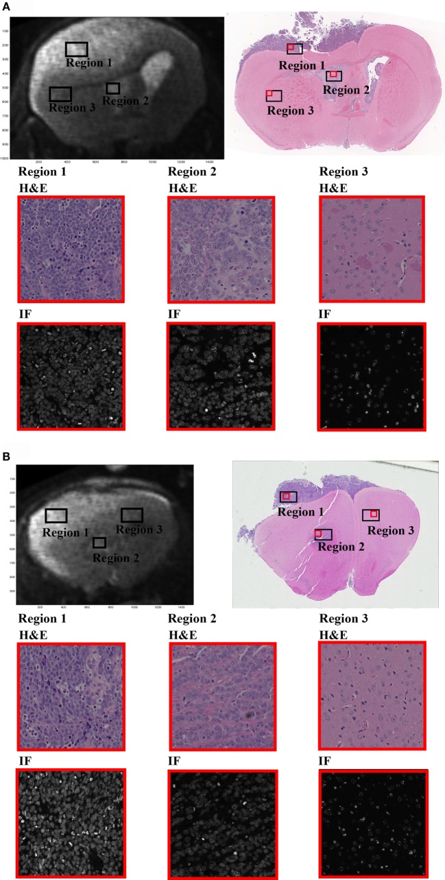
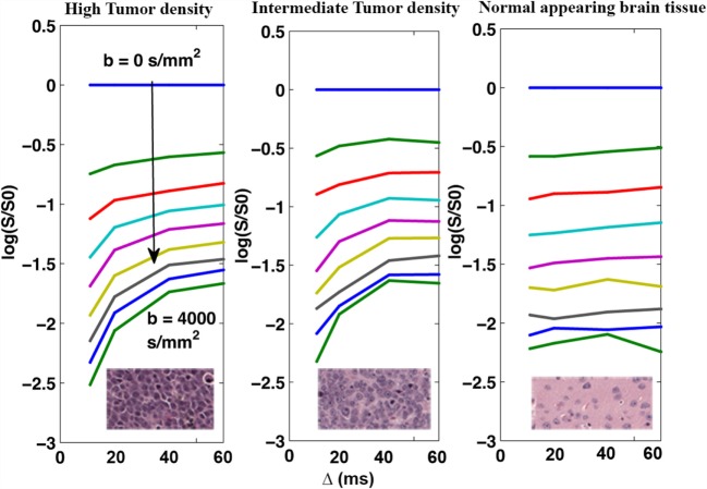
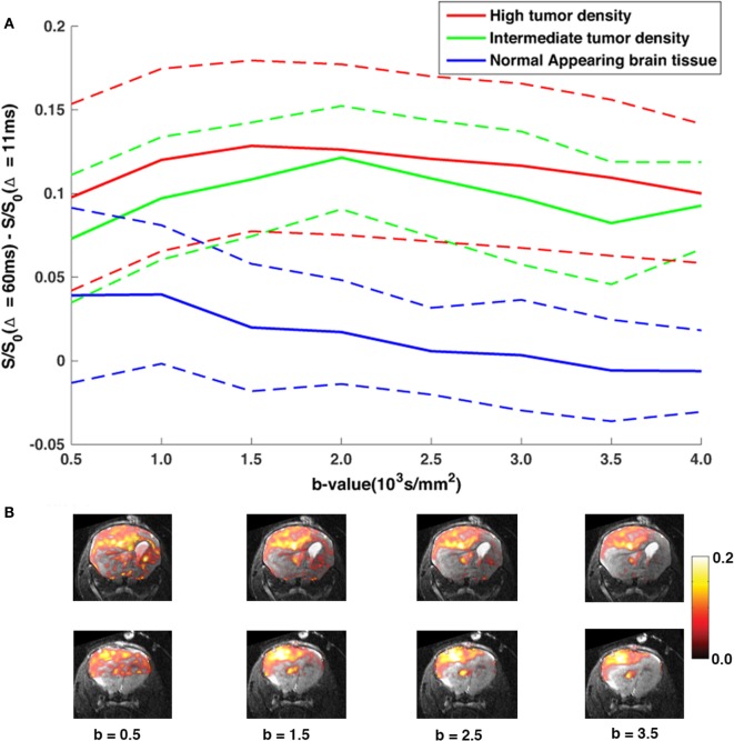
The diffusion-weighted magnetic resonance imaging (DWI) technique enables quantification of water mobility for probing microstructural properties of biological tissue and has become an effective tool for collecting information about the underlying pathology of cancerous tissue. Measurements using multiple b-values have indicated biexponential signal attenuation, ascribed to "fast" (high ADC) and "slow" (low ADC) diffusion components. In this empirical study, we investigate the properties of the diffusion time (Δ)-dependent components of the diffusion-weighted (DW) signal in a constant b-value experiment. A xenograft gliobastoma mouse was imaged using Δ = 11 ms, 20 ms, 40 ms, 60 ms, and b = 500-4000 s/mm(2) in intervals of 500 s/mm(2). Data were corrected for EPI distortions, and the Δ-dependence on the DW-signal was measured within three regions of interest [intermediate- and high-density tumor regions and normal-appearing brain (NAB) tissue regions]. In this study, we verify the assumption that the slow decaying component of the DW-signal is non-Gaussian and dependent on Δ, consistent with restricted diffusion of the intracellular space. As the DW-signal is a function of Δ and is specific to restricted diffusion, manipulating Δ at constant b-value (cb) provides a complementary and direct approach for separating the restricted from the hindered diffusion component. We found that Δ-dependence is specific to the tumor tissue signal. Based on an extended biexponential model, we verified the interpretation of the diffusion time-dependent contrast and successfully estimated the intracellular restricted ADC, signal volume fraction, and cell size within each ROI.
Keywords: DWI; MRI; cancer; diffusion time-dependence; glioblastoma multiforme; restricted diffusion; xenograft tumor model.
Figures


Similar articles
-
Diffusion restriction in the human spinal cord characterized in vivo with high b-value STEAM diffusion imaging.Neuroimage. 2013 Nov 15;82:416-25. doi: 10.1016/j.neuroimage.2013.05.122. Epub 2013 Jun 5. Neuroimage. 2013. PMID: 23747289
-
Mathematical models for the diffusion magnetic resonance signal abnormality in patients with prion diseases.Neuroimage Clin. 2014 Nov 29;7:142-54. doi: 10.1016/j.nicl.2014.11.017. eCollection 2015. Neuroimage Clin. 2014. PMID: 25610776 Free PMC article.
-
Comparisons of multi b-value DWI signal analysis with pathological specimen of breast cancer.Magn Reson Med. 2012 Sep;68(3):890-7. doi: 10.1002/mrm.23277. Epub 2011 Dec 8. Magn Reson Med. 2012. PMID: 22161802
-
Diffusion-weighted MR of the brain: methodology and clinical application.Radiol Med. 2005 Mar;109(3):155-97. Radiol Med. 2005. PMID: 15775887 Review. English, Italian.
-
High b-value q-space analyzed diffusion-weighted MRS and MRI in neuronal tissues - a technical review.NMR Biomed. 2002 Nov-Dec;15(7-8):516-42. doi: 10.1002/nbm.778. NMR Biomed. 2002. PMID: 12489099 Review.
Cited by
-
Restriction spectrum imaging with elastic image registration for automated evaluation of response to neoadjuvant therapy in breast cancer.Front Oncol. 2023 Sep 15;13:1237720. doi: 10.3389/fonc.2023.1237720. eCollection 2023. Front Oncol. 2023. PMID: 37781199 Free PMC article.
-
Assessing tumor microstructure with time-dependent diffusion imaging: Considerations and feasibility on clinical MRI and MRI-Linac.Med Phys. 2025 Jan;52(1):346-361. doi: 10.1002/mp.17453. Epub 2024 Oct 10. Med Phys. 2025. PMID: 39387639 Free PMC article.
-
Discrimination Between Benign and Malignant Lesions With Restriction Spectrum Imaging MRI in an Enriched Breast Cancer Screening Cohort.J Magn Reson Imaging. 2025 Apr;61(4):1876-1887. doi: 10.1002/jmri.29599. Epub 2024 Sep 18. J Magn Reson Imaging. 2025. PMID: 39291552 Free PMC article.
-
Effects of diffusion time on non-Gaussian diffusion and intravoxel incoherent motion (IVIM) MRI parameters in breast cancer and hepatocellular carcinoma xenograft models.Acta Radiol Open. 2018 Jan 11;7(1):2058460117751565. doi: 10.1177/2058460117751565. eCollection 2018 Jan. Acta Radiol Open. 2018. PMID: 29372076 Free PMC article.
-
Restriction Spectrum Imaging Improves Risk Stratification in Patients with Glioblastoma.AJNR Am J Neuroradiol. 2017 May;38(5):882-889. doi: 10.3174/ajnr.A5099. Epub 2017 Mar 9. AJNR Am J Neuroradiol. 2017. PMID: 28279985 Free PMC article.
References
-
- Stejskal E, Tanner J. Spin diffusion measurements: spin echoes in the presence of a time-dependent field gradient. J Chem Phys (1965) 42(1):288.10.1063/1.1695690 - DOI
Grants and funding
LinkOut - more resources
Full Text Sources
Other Literature Sources
Research Materials