. 2016 Aug 17;11(8):e0161159.
doi: 10.1371/journal.pone.0161159. eCollection 2016.

Interactive Tooth Separation from Dental Model Using Segmentation Field

Affiliations

Interactive Tooth Separation from Dental Model Using Segmentation Field

Zhongyi Li et al. PLoS One. .

Abstract

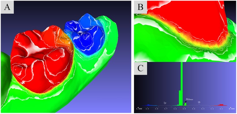
Tooth segmentation on dental model is an essential step of computer-aided-design systems for orthodontic virtual treatment planning. However, fast and accurate identifying cutting boundary to separate teeth from dental model still remains a challenge, due to various geometrical shapes of teeth, complex tooth arrangements, different dental model qualities, and varying degrees of crowding problems. Most segmentation approaches presented before are not able to achieve a balance between fine segmentation results and simple operating procedures with less time consumption. In this article, we present a novel, effective and efficient framework that achieves tooth segmentation based on a segmentation field, which is solved by a linear system defined by a discrete Laplace-Beltrami operator with Dirichlet boundary conditions. A set of contour lines are sampled from the smooth scalar field, and candidate cutting boundaries can be detected from concave regions with large variations of field data. The sensitivity to concave seams of the segmentation field facilitates effective tooth partition, as well as avoids obtaining appropriate curvature threshold value, which is unreliable in some case. Our tooth segmentation algorithm is robust to dental models with low quality, as well as is effective to dental models with different levels of crowding problems. The experiments, including segmentation tests of varying dental models with different complexity, experiments on dental meshes with different modeling resolutions and surface noises and comparison between our method and the morphologic skeleton segmentation method are conducted, thus demonstrating the effectiveness of our method.


Conflict of interest statement

Competing Interests: The authors have declared that no competing interests exist.

Figures

Fig 1
Fig 1. The proposed pipeline for dental mesh segmentation.
Fig 2
Fig 2. The single segmentation field with two constraint sites.
(A): Visualization of the molar-target segmentation with two constraint sites. (B): Visualization of the variation of field values on the concave seam between molar and gingiva. (C): The segmentation result under a single segmentation field.
Fig 3
Fig 3. Multiple segmentation field with four constraint sites.
(A): Visualization of the molar-target segmentation with four pairs of constraint sites. (B): Visualization of the variation of field values on the concave seam between molar and gingiva. (C): The segmentation result under multiple segmentation field.
Fig 4
Fig 4. Cutting boundary selection.
(A): All sampled counter lines. (B): The counter lines on concave region. (C): The histogram of vertex amount distribution on segmentation field values.
Fig 5
Fig 5. The easy-to-use interactive tool.
Fig 6
Fig 6. The segmentation results of employing our approach on ten dental models with crowding problems varying from “mild crowding” to “severe crowding”.
(A), (B) and (C) show the consecutive segmentation operations.
Fig 7
Fig 7. The segmentation results of employing our approach on three dental models with different surface resolutions.
From up to down ((A) to (C)), each dental model is with 301964, 50000 and 10000 faces, respectively.
Fig 8
Fig 8. The segmentation results of employing our approach on three dental models with different noises.
From up to down ((A) to (C)), the dental model is coupled with 0.05, 0.2 and 0.5 mean edge-length Gaussian noise, respectively.
Fig 9
Fig 9. The comparison between our method and the segmentation algorithm presented by [28].
(A) and (C) are the segmentation result of [28] with model case B and case D in Fig 6, and (B) and (D) are the correspond segmentation results of our method. The average time consumption of single tooth separation in A and C were 42629ms and 159208ms, and the correspond time consumption in B and D were 1319ms and 3279ms, respectively.
Fig 10
Fig 10. The concavity-sensitive weighting scheme produces more denser contour lines at tooth boundary areas compared with other weighting schemes, hence leads to better segmentation result.
(Left)((A), (D) and (G)) are segmentation field, (Middle)((B), (E) and (H)) are detailed contour lines in tooth boundary ares, (Right)((C), (F) and (I)) are relevant segmentation results under different weighting schemes.
Fig 11
Fig 11. Average time consumption of single tooth segmentation in experiments illustrated in Fig 6.
Blue, red, green and purple lines indicate the time consumed by segmentation field computation, cutting line selection, tooth separation and coloring and total time, respectively (in ms).

References

    1. Barnfather K, Brunton P. Restoration of the upper dental arch using Lava all-ceramic crown and bridgework. British dental journal. 2007;202(12):731–735. 10.1038/BDJ.2007.534 - DOI - PubMed
    1. Bell RB. Computer planning and intraoperative navigation in cranio-maxillofacial surgery. Oral and maxillofacial surgery clinics of North America. 2010;22(1):135–156. 10.1016/j.coms.2009.10.010 - DOI - PubMed
    1. Grauer D, Wiechmann D, Heymann GC, SWIFT J, EDWARD J. Computer-Aided Design/Computer-Aided Manufacturing Technology in Customized Orthodontic Appliances. Journal of Esthetic and Restorative Dentistry. 2012;24(1):3–9. 10.1111/j.1708-8240.2011.00500.x - DOI - PubMed
    1. Kihara T, Tanimoto K, Michida M, Yoshimi Y, Nagasaki T, Murayama T, et al. Construction of orthodontic setup models on a computer. American Journal of Orthodontics and Dentofacial Orthopedics. 2012;141(6):806–813. 10.1016/j.ajodo.2011.10.027 - DOI - PubMed
    1. Hoffman DD, Richards WA. Parts of recognition. Cognition. 1984;18(1):65–96. 10.1016/0010-0277(84)90022-2 - DOI - PubMed