

Cystatin C Shifts APP Processing from Amyloid-β Production towards Non-Amyloidgenic Pathway in Brain Endothelial Cells
- PMID: 27532339
- PMCID: PMC4988779
- DOI: 10.1371/journal.pone.0161093
Cystatin C Shifts APP Processing from Amyloid-β Production towards Non-Amyloidgenic Pathway in Brain Endothelial Cells
Abstract
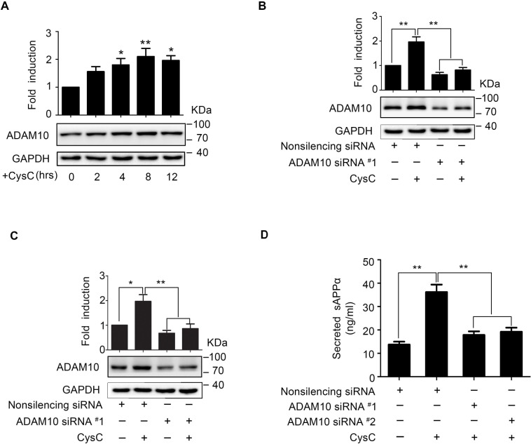
Amyloid-β (Aβ), the major component of neuritic plaques in Alzheimer's disease (AD), is derived from sequential proteolytic cleavage of amyloid protein precursor (APP) by secretases. In this study, we found that cystatin C (CysC), a natural cysteine protease inhibitor, is able to reduce Aβ40 secretion in human brain microvascular endothelial cells (HBMEC). The CysC-induced Aβ40 reduction was caused by degradation of β-secretase BACE1 through the ubiquitin/proteasome pathway. In contrast, we found that CysC promoted secretion of soluble APPα indicating the activated non-amyloidogenic processing of APP in HBMEC. Further results revealed that α-secretase ADAM10, which was transcriptionally upregulated in response to CysC, was required for the CysC-induced sAPPα secretion. Knockdown of SIRT1 abolished CysC-triggered ADAM10 upregulation and sAPPα production. Taken together, our results demonstrated that exogenously applied CysC can direct amyloidogenic APP processing to non-amyloidgenic pathway in brain endothelial cells, mediated by proteasomal degradation of BACE1 and SIRT1-mediated ADAM10 upregulation. Our study unveils previously unrecognized protective role of CysC in APP processing.
Conflict of interest statement
Figures


Similar articles
-
Effects of Folic Acid on Secretases Involved in Aβ Deposition in APP/PS1 Mice.Nutrients. 2016 Sep 9;8(9):556. doi: 10.3390/nu8090556. Nutrients. 2016. PMID: 27618097 Free PMC article.
-
C6 Glioma-Secreted NGF and FGF2 Regulate Neuronal APP Processing Through Up-Regulation of ADAM10 and Down-Regulation of BACE1, Respectively.J Mol Neurosci. 2016 Jul;59(3):334-42. doi: 10.1007/s12031-015-0690-7. Epub 2015 Nov 27. J Mol Neurosci. 2016. PMID: 26614345
-
Effects of senescence and angiotensin II on expression and processing of amyloid precursor protein in human cerebral microvascular endothelial cells.Aging (Albany NY). 2018 Jan 15;10(1):100-114. doi: 10.18632/aging.101362. Aging (Albany NY). 2018. PMID: 29348391 Free PMC article.
-
Alzheimer's disease.Subcell Biochem. 2012;65:329-52. doi: 10.1007/978-94-007-5416-4_14. Subcell Biochem. 2012. PMID: 23225010 Review.
-
BACE1: the beta-secretase enzyme in Alzheimer's disease.J Mol Neurosci. 2004;23(1-2):105-14. doi: 10.1385/JMN:23:1-2:105. J Mol Neurosci. 2004. PMID: 15126696 Review.
Cited by
-
Comprehensive Profiling of Secretome Formulations from Fetal- and Perinatal Human Amniotic Fluid Stem Cells.Int J Mol Sci. 2021 Apr 2;22(7):3713. doi: 10.3390/ijms22073713. Int J Mol Sci. 2021. PMID: 33918297 Free PMC article.
-
Soluble Expression of Recombinant Human Cystatin C and Comparison of the Ni Column and Magnetic Bead Purification.Protein J. 2020 Feb;39(1):85-95. doi: 10.1007/s10930-019-09873-0. Protein J. 2020. PMID: 31625059
-
Human cystatin C induces the disaggregation process of selected amyloid beta peptides: a structural and kinetic view.Sci Rep. 2023 Nov 27;13(1):20833. doi: 10.1038/s41598-023-47514-w. Sci Rep. 2023. PMID: 38012338 Free PMC article.
-
Expression and function of β-site amyloid precursor protein-cleaving enzyme 2 in vascular endothelium.Am J Physiol Heart Circ Physiol. 2025 Aug 1;329(2):H291-H302. doi: 10.1152/ajpheart.00126.2025. Epub 2025 Jun 23. Am J Physiol Heart Circ Physiol. 2025. PMID: 40549561 Free PMC article.
-
Procyanidins and Alzheimer's Disease.Mol Neurobiol. 2019 Aug;56(8):5556-5567. doi: 10.1007/s12035-019-1469-6. Epub 2019 Jan 16. Mol Neurobiol. 2019. PMID: 30649713 Review.
References
-
- Scheltens P, Blennow K, Breteler MM, de Strooper B, Frisoni GB, Salloway S, et al. (2016) Alzheimer's disease. Lancet. - PubMed
-
- Greenberg SM, Gurol ME, Rosand J, Smith EE (2004) Amyloid angiopathy-related vascular cognitive impairment. Stroke 35: 2616–2619. - PubMed
-
- Deane R, Wu Z, Sagare A, Davis J, Du Yan S, Hamm k, et al. (2004) LRP/amyloid beta-peptide interaction mediates differential brain efflux of Abeta isoforms. Neuron 43: 333–344. - PubMed
MeSH terms
Substances
LinkOut - more resources
Full Text Sources
Other Literature Sources
