. 2016 Aug 17;11(8):e0160057.
doi: 10.1371/journal.pone.0160057. eCollection 2016.

Prolonged Exposure of Primary Human Muscle Cells to Plasma Fatty Acids Associated with Obese Phenotype Induces Persistent Suppression of Muscle Mitochondrial ATP Synthase β Subunit

Affiliations

Prolonged Exposure of Primary Human Muscle Cells to Plasma Fatty Acids Associated with Obese Phenotype Induces Persistent Suppression of Muscle Mitochondrial ATP Synthase β Subunit

Lee Tran et al. PLoS One. .

Abstract

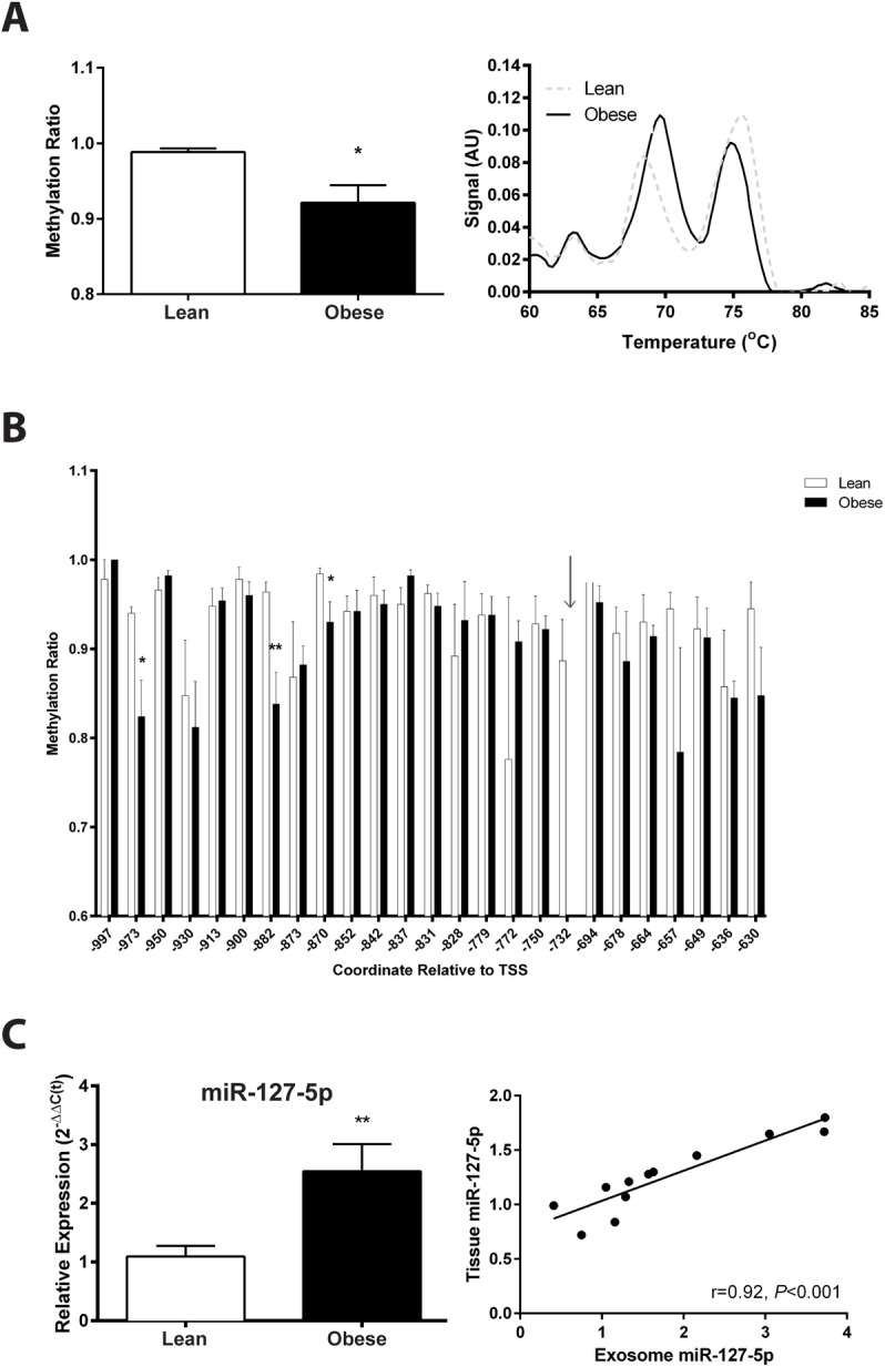
Our previous studies show reduced abundance of the β-subunit of mitochondrial H+-ATP synthase (β-F1-ATPase) in skeletal muscle of obese individuals. The β-F1-ATPase forms the catalytic core of the ATP synthase, and it is critical for ATP production in muscle. The mechanism(s) impairing β-F1-ATPase metabolism in obesity, however, are not completely understood. First, we studied total muscle protein synthesis and the translation efficiency of β-F1-ATPase in obese (BMI, 36±1 kg/m2) and lean (BMI, 22±1 kg/m2) subjects. Both total protein synthesis (0.044±0.006 vs 0.066±0.006%·h-1) and translation efficiency of β-F1-ATPase (0.0031±0.0007 vs 0.0073±0.0004) were lower in muscle from the obese subjects when compared to the lean controls (P<0.05). We then evaluated these same responses in a primary cell culture model, and tested the specific hypothesis that circulating non-esterified fatty acids (NEFA) in obesity play a role in the responses observed in humans. The findings on total protein synthesis and translation efficiency of β-F1-ATPase in primary myotubes cultured from a lean subject, and after exposure to NEFA extracted from serum of an obese subject, were similar to those obtained in humans. Among candidate microRNAs (i.e., non-coding RNAs regulating gene expression), we identified miR-127-5p in preventing the production of β-F1-ATPase. Muscle expression of miR-127-5p negatively correlated with β-F1-ATPase protein translation efficiency in humans (r = - 0.6744; P<0.01), and could be modeled in vitro by prolonged exposure of primary myotubes derived from the lean subject to NEFA extracted from the obese subject. On the other hand, locked nucleic acid inhibitor synthesized to target miR-127-5p significantly increased β-F1-ATPase translation efficiency in myotubes (0.6±0.1 vs 1.3±0.3, in control vs exposure to 50 nM inhibitor; P<0.05). Our experiments implicate circulating NEFA in obesity in suppressing muscle protein metabolism, and establish impaired β-F1-ATPase translation as an important consequence of obesity.


Conflict of interest statement

Competing Interests: The authors have declared that no competing interests exist.

Figures

Fig 1
Fig 1. Decreased total protein synthesis in muscle and increased amino acid concentrations in plasma in obese subjects.
(A) Fractional synthesis rate (FSR) for total muscle protein in the lean (BMI<25) and obese (BMI>30) subjects. (B) Fasting plasma amino acid concentrations. Data shown are means ±SE, and significance was *P<0.05, **P<0.01, and ***P<0.001 by Student’s unpaired t-test.
Fig 2
Fig 2. Concurrent assessment of glucose uptake and nascent protein synthesis in primary human myotubes reveals independent regulatory mechanisms.
(A) Average responses for glucose uptake and protein synthesis. (B) Corresponding recovery experiments for myotubes exposed to Obese NEFA (Pre) and then incubated for an additional five days in normal growth media (Post). Data shown are means ±SE, n = 6 replicates/group, 10−3 cells/experiment, *P<0.05, **P<0.01, and ***P<0.001 by Student’s unpaired t-test or one-way ANOVA with Tukey’s posthoc analysis.
Fig 3
Fig 3. Assessment of growth and differentiation parameters suggest different responses between NEFA exposure groups.
(A&C) MyoD1 mRNA was quantified as a marker of myoblast proliferation, and (B&D) myogenin mRNA was used as a marker of myotube differentiation in muscle biopsy or primary culture samples, respectively. (E) A modified fusion index was determined for each cell based on DNA content and categorized based on frequency clusters. (F) Percentages of cells identified in each fusion category were compared. Individual cell populations were analyzed for glucose uptake (G) and nascent protein synthesis (H). Data shown are means ±SE, n = 6 replicates/group, *P<0.05, **P<0.01, and ***P<0.001 by one-way ANOVA with Tukey’s posthoc or two-way ANOVA with Bonferroni’s posthoc analysis.
Fig 4
Fig 4. Quantification of ATP5B mRNA and β-F1-ATPase protein expression shows decreased protein translation in obesity.
(A) The mRNA expression for ATP5B and ATP5E are shown, respectively. (B) Summary of β-F1-ATPase protein expression and representative blots are depicted. Positive bands for β-F1-ATPase are seen at ~56 kDa and for GAPDH at ~37 kDa. (C) Translation efficiency was compared between lean (n = 6) and obese (n = 7) subjects. Prolonged NEFA exposure on myotube β-F1-ATPase translation was determined in skeletal muscle explants (n = 6/group). (D) ATP5B mRNA was quantified for each NEFA-exposed group, (E) differences in β-F1-ATPase protein expression in response to NEFA exposure were observed, and (F) translation efficiency was calculated. (G) An additional myotube group was exposed to Obese NEFA (Pre) and then incubated for an additional five days in normal growth media (Post), and the cells were analyzed for GLUT-4 and β-F1-ATPase protein expressions. Data shown are means ±SE, and significance was *P<0.05, **P<0.01, and ***P<0.001 by Student’s unpaired t-test, or by one-way ANOVA with Tukey’s posthoc analysis.
Fig 5
Fig 5. Obesity increases specific skeletal muscle miRNA expression.
(A) Expression of miR-127-5p (left) and miR-101-3p (right) were quantified in lean (n = 6) and obese (n = 6) subjects. (B) Changes in myotube miR-127-5p in response to repeated exposure to NEFA extracts were determined (n = 6 replicates/group). Alexa Fluor-488 conjugated locked nucleic acid (LNA) oligos were used to inhibit miR-127-5p in primary myotubes from an obese subject. (C) Response to graded concentrations of inhibitors (10nM-100nM) are shown for miR-127-5p expression and ATP5B mRNA expression. (D) Observed responses for β-F1-ATPase protein expression and (E) translation efficiency. Data shown are means ±SE, and significance was *P<0.05 and **P<0.01 by one-way ANOVA with Tukey’s posthoc analysis.
Fig 6
Fig 6. Epigenetic regulation and distribution of miR-127-5p.
The 1kB region adjacent to the TSS of miR-127-5p contains four CpG islands, which may be responsible for sustaining expression of the miRNA. (A) High Resolution Melt (HRM) was used to analyze overall DNA methylation status of the 1 kB region upstream of the miR-127-5p TSS in muscle biopsies in the lean (n = 6) and obese (n = 7) subjects. (B) Results from bisulfite sequencing of the first two CpG islands of the 1 kB region identified 26 methylation sites in lean (n = 5) and obese (n = 5) subjects. No results could be obtained for obese subjects at the site indicated by the arrow. (C) Total RNA was extracted from serum exosomes of lean (n = 6) and obese (n = 6) subjects, analyzed for miR-127-5p expression (left), and correlation with skeletal muscle expression was determined (right). Data shown are means ±SE, *P<0.05 and **P<0.01 by Student’s unpaired t-test.

References

    1. Mokdad AH, Ford ES, Bowman BA, Dietz WH, Vinicor F, Bales VS, et al. Prevalence of obesity, diabetes, and obesity-related health risk factors, 2001. JAMA. 2003;289: 76–79. - PubMed
    1. Delarue J, Magnan C. Free fatty acids and insulin resistance. Curr Opin Clin Nutr Metab Care. 2007;10: 142–148. - PubMed
    1. Guillet C, Delcourt I, Rance M, Giraudet C, Walrand S, Bedu M, et al. Changes in basal and insulin and amino acid response of whole body and skeletal muscle proteins in obese men. J Clin Endocrinol Metab. 2009;94: 3044–3050. 10.1210/jc.2008-2216 - DOI - PubMed
    1. Katsanos CS, Mandarino LJ. Protein metabolism in human obesity: a shift in focus from whole-body to skeletal muscle. Obesity (Silver Spring). 2011;19: 469–475. - PubMed
    1. Guillet C, Masgrau A, Boirie Y. Is protein metabolism changed with obesity? Curr Opin Clin Nutr Metab Care. 2011;14: 89–92. 10.1097/MCO.0b013e328341389e - DOI - PubMed

MeSH terms