. 2016 Aug 17:38:2D.18.1-2D.18.27.
doi: 10.1002/cpsc.11.

Efficient Generation of Viral and Integration-Free Human Induced Pluripotent Stem Cell-Derived Oligodendrocytes

Affiliations

Efficient Generation of Viral and Integration-Free Human Induced Pluripotent Stem Cell-Derived Oligodendrocytes

Araceli Espinosa-Jeffrey et al. Curr Protoc Stem Cell Biol. .

Abstract

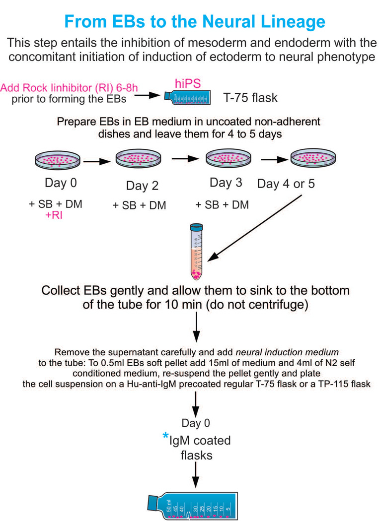
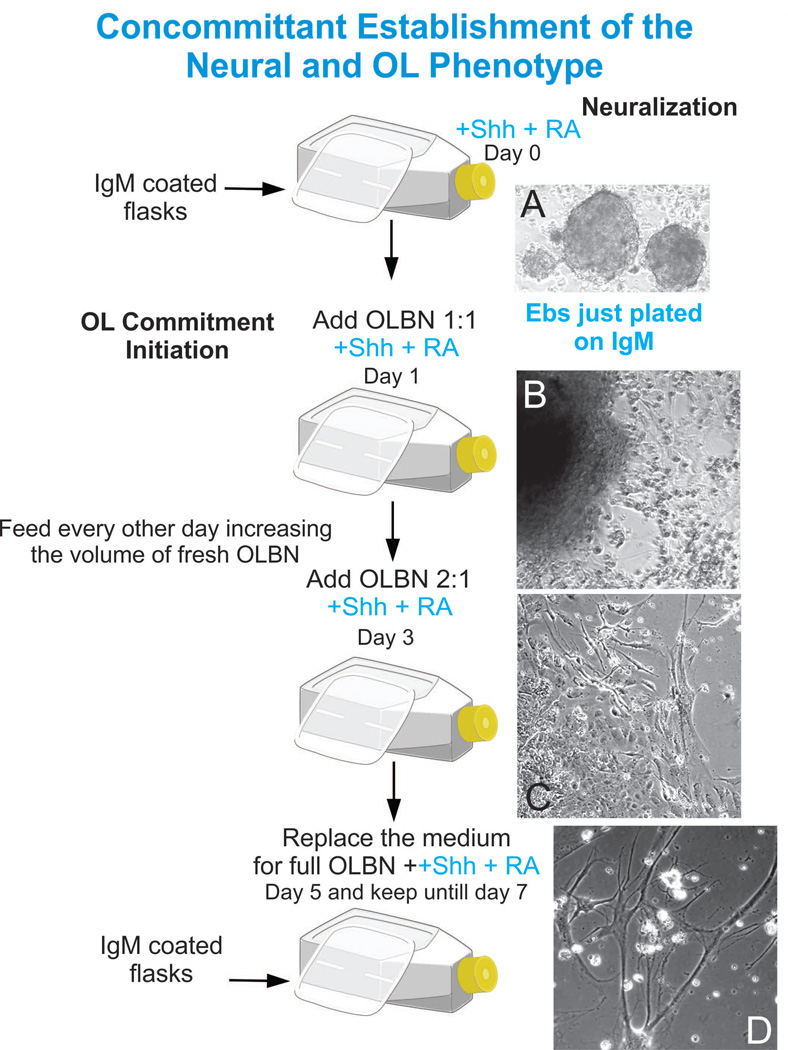
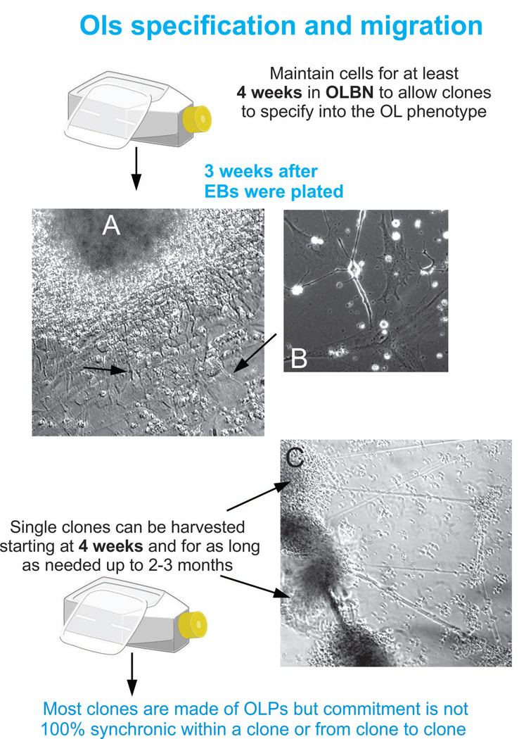
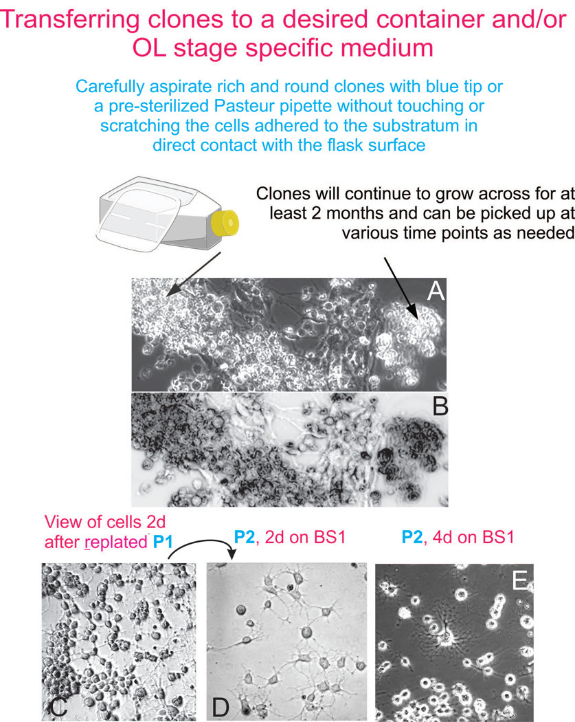
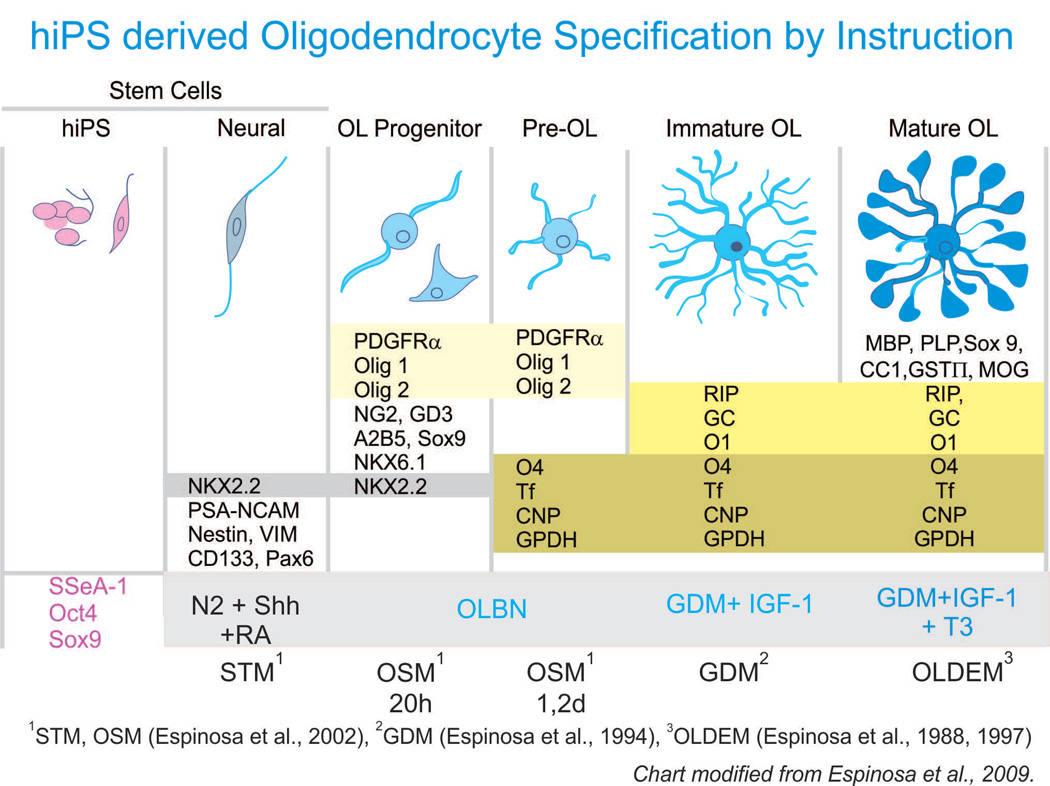
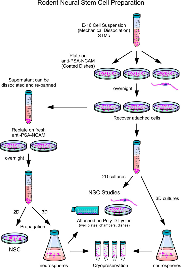
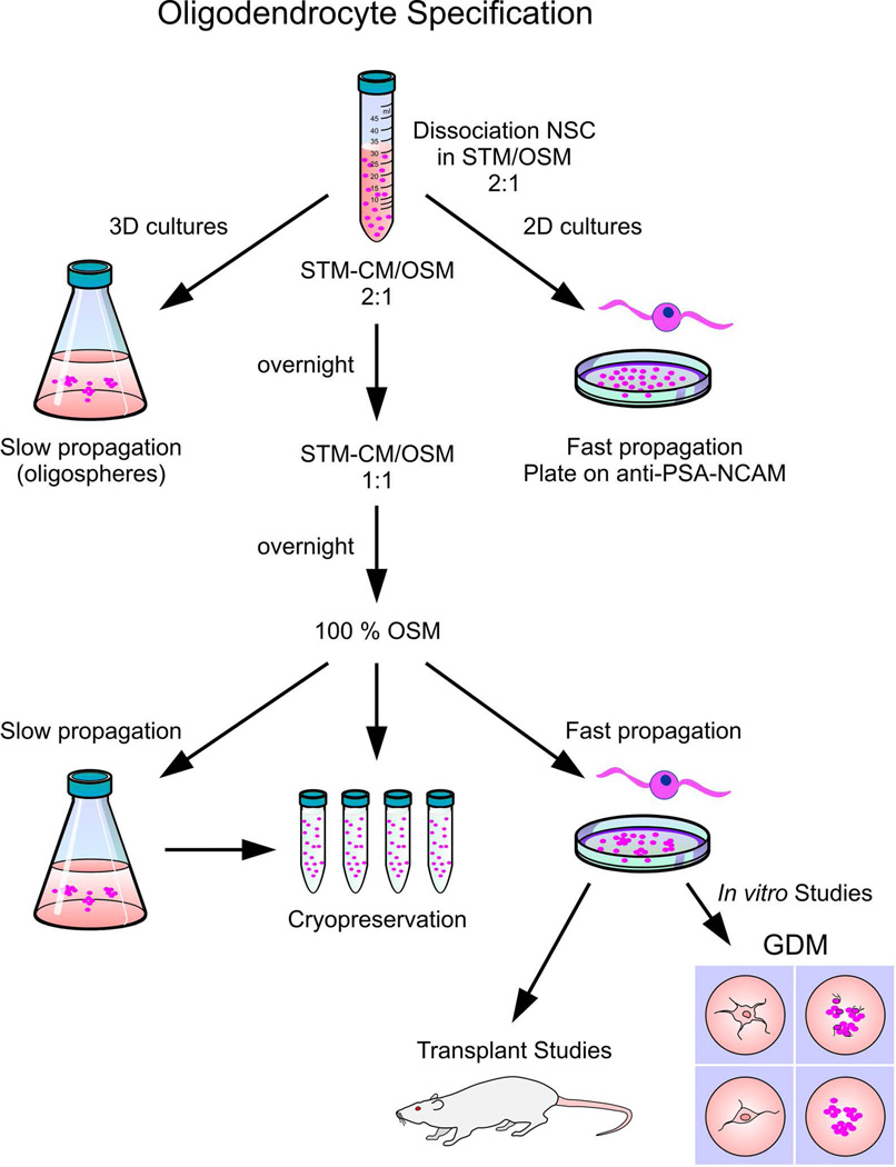
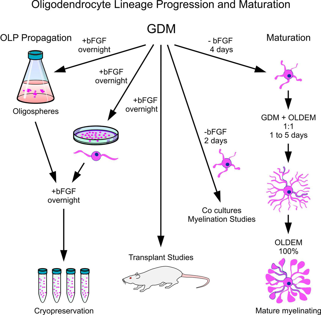
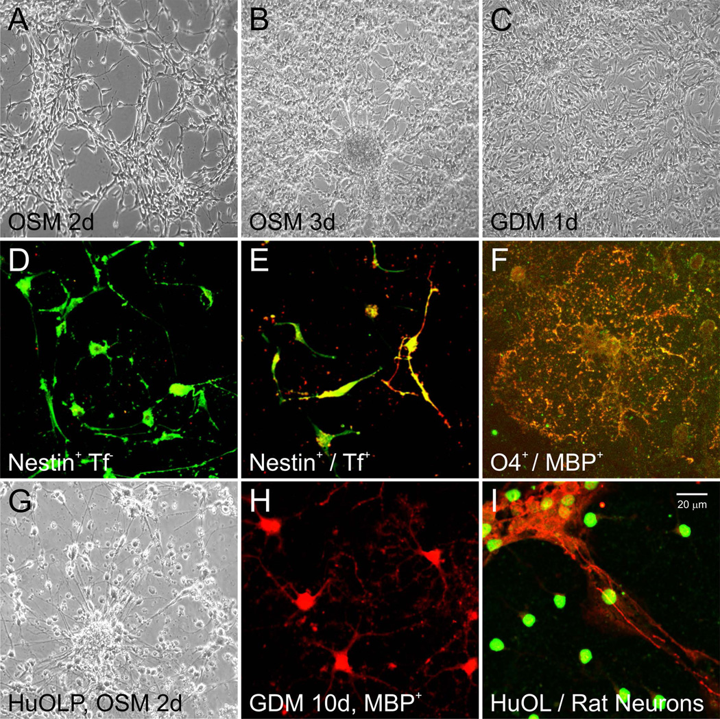
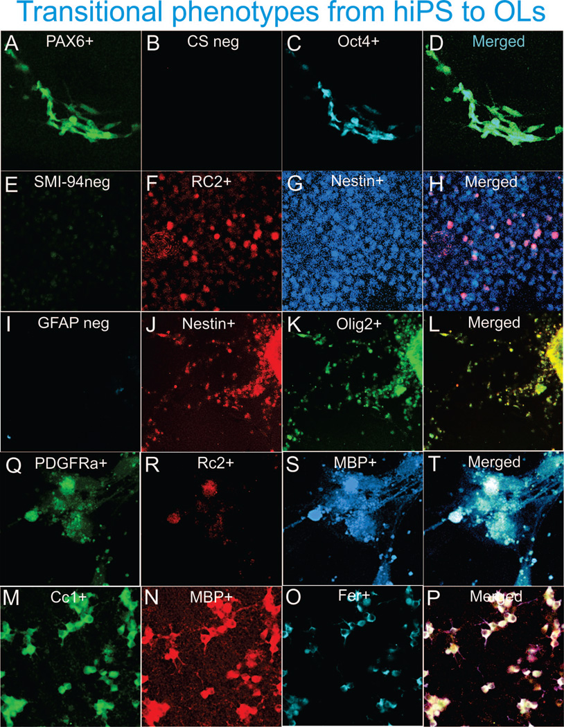
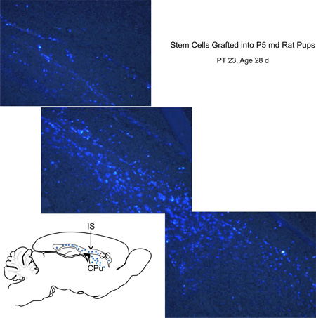
Here we document three highly reproducible protocols: (1) a culture system for the derivation of human oligodendrocytes (OLs) from human induced pluripotent stem cells (hiPS) and their further maturation-our protocol generates viral- and integration-free OLs that efficiently commit and move forward in the OL lineage, recapitulating all the steps known to occur during in vivo development; (2) a method for the isolation, propagation and maintenance of neural stem cells (NSCs); and (3) a protocol for the production, isolation, and maintenance of OLs from perinatal rodent and human brain-derived NSCs. Our unique culture systems rely on a series of chemically defined media, specifically designed and carefully characterized for each developmental stage of OL as they advance from OL progenitors to mature, myelinating cells. We are confident that these protocols bring our field a step closer to efficient autologous cell replacement therapies and disease modeling. © 2016 by John Wiley & Sons, Inc.

Keywords: NSC; chemically defined media; human induced pluripotent stem cells; lineage progression; neural stem cells; neurospheres; oligodendrocyte maturation; oligodendrocyte specification; oligospheres.


Conflict of interest statement

The authors acknowledge no conflict of interest

Figures

Figure 1
Figure 1
Ectoderm selection can be performed by exposing hiPS to SB431542 (SB) and dorsomorphin (DM), which instruct ectoderm formation preferentially, inhibiting endodermal and mesodermal commitment. All reagents should be freshly prepared as the stock solutions decay fast (between 1 [SB] and 3 [DM] months). Thus, it is suggested to create a stock of hiPS-derived OL while the reagents are fresh. For the procedures described here, the coating of choice was IgM. Nonetheless, if the EBs have been exposed to 4°C while in suspension they would clump together and the vast majority will not adhere to the IgM-coated surface. In this case it is imperative to use Matrigel or poly-D-lysine.
Figure 2
Figure 2
Once the EBs are transferred to the IgM-coated flask, the culture medium to favor neuralization is the N2 medium, which properties will be enhanced by Shh and RA. A sustained level of these two components leaves virtually no room for other cell types present in the cultures (i.e. flat, fibroblast-like cells, panel C) to prosper. Bi-refringent cells populate both the substratum and flat cells that are often used as anchorage zones (panel D).
Figure 3
Figure 3
With time in OLBN, flat cells tend to disappear and numerous cells migrate away from the mother clone (panels A and B). These bipolar cells extend their processes to migrate or to connect with other cells while serving as bridges for other cells to migrate on their extensions (panel C). As time in culture increases, cells tend to migrate using other cell processes rather than the substratum.
Figure 4
Figure 4
Transferring Clones successfully. The main aspect to consider is not to scratch the substratum with the yellow tip, aspirating a clone carefully will allow flat cells to remain in the mother flask while bringing the entire clone of round, bright cells to the new cell culture container (panel A). OL will proliferate and arrange as small clones, as well as single cells in a very homogeneous looking culture (panel C). With this cell culture medium-based selection method, there is no need to use FACS to enrich the cultures in OLP cells, and therefore, the cells are healthy and yield large numbers. Cells can be re-plated (transferred to new culture containers) using the same stage specific medium, or the next stage specific medium such as BS1 (panel D). With time, cells tend to become multipolar (panel E).
Figure 5
Figure 5. hiPS-derived OL specification by instruction through the specific culture medium
Human pluripotent stem cells can be obtained from skin fibroblasts, by introducing the 4 transcription factors described by Takahashi and Yamanaka (2006). Then, the components of the culture medium direct and determine the fate of hiPS. Our culture medium previously designed to favor the OL phenotype when starting from neural stem cells (Espinosa et al., 2002), needed to be enriched with the specific molecules present during the development of the CNS to successfully direct human induced pluripotent stem cells to the neural and OL phenotype (see pink highlighted area). Once this transition has been accomplished, OL undergo sequential morphological changes from OLP and acquire characteristics inherent to a functional OL. A partial list of OL markers below each developmental stage is not exhaustive but represents frequently used markers to identify OLs and their specific developmental stage. The main difference between the culture medium for lineage progression and maintenance of hiPS-derived OL is that they need N2 while in the progenitor stage, and IGF-1 + T3 when a mature phenotype is desired. When starting cultures from brain-derived neural stem cells, we have previously described the neural stem cell medium (STM; Espinosa-Jeffrey et al., 2002); OL specification medium (OSM; Espinosa-Jeffrey et al., 2002); glia defined medium (GDM; Espinosa de los Monteros and de Vellis, 1996); OLDEM (Espinosa de los Monteros et al,1988, 1997). Chart Modified from (Espinosa-Jeffrey et al., 2009).
Figure 6
Figure 6. Rodent neural stem cell preparation
Following dissection, the cell suspension is plated on anti-PSA-NCAM antibody–coated dishes and allowed to adhere. The process can be performed repeatedly to increase the numbers of neural stem cells, as two-dimensional cultures or three-dimensional “sphere” cultures (shown on the left side of the diagram). Every time cells are propagated, use anti-PSA-NCAM-coated dishes. Alternatively, cells can be propagated and immediately used for cell culture experiments (as shown on the right side of the diagram). While we prefer to use committed OL progenitors for cell transplants, other investigators also use uncommitted progenitors for grafting.
Figure 7
Figure 7
OL specification by instruction. The transition from NSC to committed OL lineage is brief, but sequential rather than abrupt. In order for cells to survive, they must acclimate to their new environment. OLP can be propagated to create frozen stocks as three-dimensional “oligosphere” cultures (shown on the left of the diagram), or frozen without propagation (as shown in the sequence in the center of the diagram). OLP can also be propagated in two-dimensional cultures for cryopreservation, for specific cell culture experiments, or for cell replacement therapies (as shown on the right side of the diagram).
Figure 8
Figure 8
OL lineage progression and maturation. After commitment of NSC to the OL lineage, cells are propagated at the OLP stage to create a frozen stock (steps indicated on the left portion of the flow chart) or processed further for transplantation studies (as shown by the middle arrow on the diagram). To allow OLP to further mature along the OL lineage and become myelinating cells, OLP cell cultures are transitioned into OLDEM for at least 48 h. Once OL have reached this stage of maturation, they are excellent for cell culture studies but are not recommended for cell grafting. Detachment from the substrate can damage the numerous delicate cell processes, therefore these cultures are no longer a quality source for cell transplants.
Figure 9
Figure 9
Phase contrast view of neural stem cells derived from embryonic day 16 rat brain at passage number 2 (P2). NSC were plated and maintained in OSM for 2 days (A), 3 days (B), or 3 days in OSM then switched to GDM for 1 day (C). Cells still proliferate while in OSM. When cells from (A or B) are plated and maintained in OSM on poly-D-lysine-coated coverslips for 1 day, they start to display a bipolar or multipolar morphology (D), and most express the immature precursor marker, nestin (green) but not Tf (red), an early marker for OL. After 2 days in OSM, bipolar nestin+ cells co-express transferrin (Tf) (E). After 4 days in OSM, cells were switched to GDM for 1 day; they developed numerous cell processes and co-expressed sulfatides (recognized by the anti-O4 antibody, green) and myelin basic protein (MBP; red) (F). Panels G to I are human cells. (G) Phase-contrast view of human NSCs (HFB-2050) acclimated and expanded in STM, then replated and maintained in OSM for 2 days. (H) OL derived from human NSCs (HFB-2050) were specified to the OL lineage with OSM and maintained in GDM for 10 days. OL matured and started to express MBP (red). (I) Rat cortical neurons (NFM-200-red) were cultured for 10 days, then human OLP derived from NSC (HFB-2050) were added in co-culture for 24 hr. These cells were labeled with human nuclei marker (HuNu, green).
Figure 10
Figure 10. hiPS derived OL express neural progenitor and OL cell lineage and stage specific markers recapitulating their development in vivo
Two days after EB’s were plated cells express and (panels A–D; developmental stage corresponding to Figure 2 panel B). By day 6 (panels F–H) expression of a marker for radial glia (F) at different intensity could be observed with a concomitant expression of (H) by virtually all cells (panel G), and not all nestin + cells bared the RC2 staining (stage corresponding to Figure 2 panel C). The co-expression of and can be observed within the next three weeks (panels J–L) most nestin expressing cells co-express Olig2 (panel L; developmental stage corresponding to Figure 2 panel C). When clones are harvested 4 weeks after starting their fate specification and re-plated on poly-D-Lysine in BS1 and allowed to become mature i.e. 6 days after seeding (panels M–P) cells expressing faintly (M) in their extended membrane and a few cells still bearing it in their cell body, they appear to be losing the RC2 staining (N) while intensely expressing in the cell body as well as, in their extended membrane sheath (O) the colocalization of markers is shown in panel (P). OLs in these cultures also express intensely in their cell body and immediate processes the mature markers and as well as (panels Q–R). Panel B shows control serum for Alexa-fluor 408,540 and 633. Panel I shows immunoreactivity for is absent from these cultures.
Figure 11
Figure 11
Human OL derived from (HFB-2050) human fetal NSC were labeled with fluorescent fast blue (FB; Sigma, cat. no. F-5756). A total of 60,000 cells were grafted into the corpus callosum (CC) of P(5) rat pups born to a myelin-deficient (md) carrier mother. At 23 days after grafting, samples were harvested and examined. Grafted NSC survived and migrated extensively within the host brain parenchyma extending along the corpus callosum (CC) and caudate putamen (CPu). In the sketch, dots represent the location where FB+ cells were found. The sketch represents a sagittal view of the transplanted rat brain at 28 days of age, IS indicates where cells were implanted.

Similar articles

Cited by

References

    1. Ahn SM, Byun K, Kim D, Lee K, Yoo JS, Kim SU, Jho EH, Simpson RJ, Lee B. Olig2-induced neural stem cell differentiation involves downregulation of Wnt signaling and induction of Dickkopf-1 expression. PLoS ONE. 2008;3(12):e3917. - PMC - PubMed
    1. Biancotti JC, Lavon N. Derivation, expansion, and characterization of human embryonic stem cell lines from aneuploid embryos. Methods in Molecular Biology (Clifton, N.J.) 2012;873:163–178. - PubMed
    1. Bjugstad KB, Redmond DE, Jr, Teng YD, Elsworth JD, Roth RH, Blanchard BC, Snyder EY, Sladek JR., Jr Neural stem cells implanted into MPTP-treated monkeys increase the size of endogenous tyrosine hydroxylase-positive cells found in the striatum: a return to control measures. Cell Transplant. 2005;14(4):183–192. - PubMed
    1. Bjugstad KB, Teng YD, Redmond DE, Jr, Elsworth JD, Roth RH, Cornelius SK, Snyder EY, Sladek JR., Jr Human neural stem cells migrate along the nigrostriatal pathway in a primate model of Parkinson's disease. Exp. Neurol. 2008;211(2):362–369. - PMC - PubMed
    1. Bottenstein JE, Sato GH. Growth of a rat neuroblastoma cell line in serum-free supplemented medium. Proc. Natl. Acad Sci. U.S.A. 1979;76:514–517. - PMC - PubMed

Publication types

MeSH terms

LinkOut - more resources