Review
. 2016 Aug 16;45(2):240-54.
doi: 10.1016/j.immuni.2016.08.003.

Complement-Mediated Regulation of Metabolism and Basic Cellular Processes

Affiliations
Review

Complement-Mediated Regulation of Metabolism and Basic Cellular Processes

Christoph Hess et al. Immunity. .

Abstract

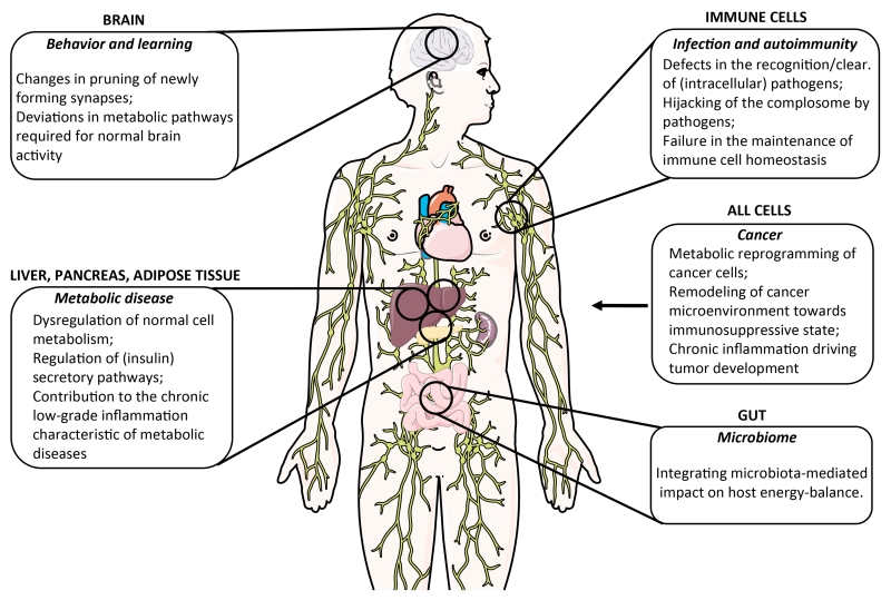
Complement is well appreciated as a critical arm of innate immunity. It is required for the removal of invading pathogens and works by directly destroying them through the activation of innate and adaptive immune cells. However, complement activation and function is not confined to the extracellular space but also occurs within cells. Recent work indicates that complement activation regulates key metabolic pathways and thus can impact fundamental cellular processes, such as survival, proliferation, and autophagy. Newly identified functions of complement include a key role in shaping metabolic reprogramming, which underlies T cell effector differentiation, and a role as a nexus for interactions with other effector systems, in particular the inflammasome and Notch transcription-factor networks. This review focuses on the contributions of complement to basic processes of the cell, in particular the integration of complement with cellular metabolism and the potential implications in infection and other disease settings.


Figures

Figure 1
Figure 1. Key metabolic pathways and their link with the complement system
Glycolysis, braking down glucose to pyruvate, is yielding low amounts of ATP, yet provides important intermediary metabolites for anabolic reactions and shapes T cell immune functioning. In mitochondria, full oxidation of pyruvate greatly increases the bioenergetic efficiency of cells. C3b–CD46-medited signaling enhances both glycolysis and mitochondrial respiration (1). Oxygen is toxic and inevitably linked with oxidative molecular damage brought by reactive oxygen species (ROS). However, evolution has incorporated ROS as indispensible signaling molecules, and triggering intracellular C5aR1 via C5a during T cell activation impacts on oxygen metabolism and mediates increased production of ROS (2). Mitochondria are furthermore key organelles in orchestrating programmed cell death, which is triggered by the release of cytochrome c and increased ROS production. In T cell homeostasis, complement drives pro-survival pathways. Specifically, C3a generated intracellularly binds it receptor, C3aR, expressed on lysosomes, thereby maintaining constant low-level activation of the mechanistic target of rapamycin (mTOR), which in turn controls glycolysis and mitochondrial function and biogenesis (3). Yet, context specifically, complement also plays critical roles in the regulation of apoptosis: While C3b–CD46-mediated signals counteract apoptosis in stimulated cells via Bcl-2 expression induction (4), local generation of C5a can activate the same anti-apoptotic Bcl-2 pathway but also promote apoptosis via autocrine C5aR1 signaling via the extracellular signal regulated kinase (ERK) pathway (5). Of note, the role(s) of the alternative C5aR2 in these pathways is currently least well understood. Gluc=glucose; G6P=glucose-6-phosphate; F1,6BP=fructose,1,6,-bisphosphate; G3P=glyceraldehyde-3-phosphate; Pyr=pyruvate; Lac=lactate; TCA=tricarboxylic acid cycle; ETC=electron transport chain.
Figure 2
Figure 2. Distinct locations of complement activation
A. Liver-derived, systemically circulating complement can be activated through the classical, lectin or alternative pathway. Via the formation of C3 convertases (C4bC2a for the classical and lectin pathways and C3bBb for the alternative pathway), these pathways lead to generation of C3b and C3a. Upon subsequent generation of C5 convertase (C4bC2aC3b for the classical and lectin pathways, C3bBbC3b for the alternative pathway), C5b and C5a are produced, with surface-bound C5b initiating the formation and insertion of the MAC on pathogens (or other target membranes). B. Local complement activation is triggered when activating signals (here, TCR stimulation or TLR activation on APCs (not shown)) initiate generation and secretion of C3, C5, Factor B, and Factor D, leading to C3 and C5 convertase formation in the extracellular space and on the cell surface – and ultimately the generation of the complement activation fragments C3a, C3b, C5b and C5a (C3 and C5 can also be cleaved by proteases in the extracellular space). These complement fragments bind to their respective receptors on the T cell surface and induce cellular responses. Note that such autocrine activation is also supported by preformed C3 and C5 activation fragments that are generated intracellularly (C), and rapidly transported to the cell surface to mediate autocrine signaling from that location. C. Intracellular complement activation in CD4+ T cells (and possibly other cells) occurs through the action of the C3-cleaving protease CTSL for C3, while the protease activating C5 is currently undefined. The resulting C3a and C5a fragments engage intracellular C3aR and C5aR1, respectively, and mediate effector function. Intracellular C3aR signaling occurs on lysosomes, while the intracellular compartment(s) expressing C5aR1 is, respectively are not yet defined. Self-tissue is protected from inappropriate complement deposition through fluid phase and cell-bound regulators (not shown in this schematic). C3b and C4b are inactivated by serine protease factor I, which requires one of several cofactor proteins (cell-bound CD46 and CR1 (CD35), or fluid phase factors H and C4bp). The C3 and C5 convertases are regulated through disassembly by regulators that have decay accelerating activity (DAA) (membrane-bound CD55 and CR1 (CD35), factor H and C4bp). The formation of the MAC is controlled by CD59 and protein S (vitronectin). CTSL, cathepsin L; MASP2, mannose-binding lectin serine protease 2; TCR, T cell receptor.
Figure 3
Figure 3. Model of the role of complement in Th1 cell homeostasis and effector function
In resting T cells, the ‘tonic’ generation of intracellular C3a via cathepsin L sustains C3aR stimulation expressed on lysosomes and the low level activation of mTOR required for T cell survival. Resting T cells also generate low levels of C5a, and although tonic C5a activity is also required for T cell survival, the mechanism underlying this function is unclear. T cell receptor activation and CD28 co-stimulation of resting T cells induces the local generation of the CD46 ligand C3b and increased expression of CD46 isoforms bearing CYT-1 (1). Autocrine CD46 CYT-1-driven signals then lead to up-regulation of the glucose transporter, GLUT1, and the amino acid (AA) channel, LAT1, allowing for increased influx of glucose and AAs into the cell (2). In parallel, CD46 CYT-1-mediated signals induce increased expression of LAMTOR5, and via this assembly of the lysosome-based machinery enabling amino acid sensing via mTORC1, which then leads to the induction of glycolysis and OXPHOS required for IFN-γ secretion (3). CD46-mediated signals also trigger increased intracellular C5a generation, which supports mitochondrial metabolic activity and ROS production critical to normal Th1 induction (4). Of note, complement-driven ROS production (via intracellular C5aR1 activation) and mTORC1 activity also activate the NLRP3 inflammasome in TCR-stimulated T cells, a process that supports Th1 expansion via IL-1β functioning in an autocrine fashion (not shown). During Th1 contraction and induction of IL-10 co-expression, CD46 isoform expression reverts to a CYT-2 predominant pattern (through a mechanism that is currently unknown) and this is accompanied by reduced expression of GLUT1 and LAT1, down-regulation of glycolysis and OXPHOS and re-instatement of C3a-driven low level mTOR activity. Furthermore, autocrine engagement of the surface-expressed C5aR2 via C5a-C5adesArg secreted upon T cell activation contributes to negative regulation of mitochondrial activity and reduction of ROS (through inhibition of intracellular C5aR1 activity and/or a yet undefined mechanism). (Adapted from Kolev et al., Immunity 2015).
Figure 4
Figure 4. ‘Feedback model’ of the complement–metabolism system in T cells
During organismal homeostasis, intracellularly generated complement fragments sustain T cell self-regulation through the fine-tuning of basal metabolic activity, and this quiescent state is supported by complement driven assembly of the IL-7R and restraining of Notch activity (1). Together with cognate T cell activation, signals indicative of disrupted homeostasis (2), initiate complement-driven metabolic reprogramming, IL-2R assembly, Notch and NLRP3 inflammasome activation and context-specific activation of the cell (3). This enables an efficient immune response, which in turn closes a feedback-loop to the sensing system by modifying the input on the sensor-module – namely removal of cognate pathogen and thus the inflammatory trigger (4). Metabolic reprogramming back towards steady-state metabolism thus occurs (5). The same basic principle of a complement-dependent ‘sensing–response–feedback system’ would also apply to other immune cells (for example, antigen-presenting cells, where TLR activation may induce complement-mediated metabolic programming) and in cell-autonomous models, e.g. when intracellular sensing of C3 fragments deposited onto cell-invading pathogens triggers a mitochondrial antiviral signaling protein-driven immune response, leading to degradation of the pathogens by the proteasome. Of note, we have predominantly integrated into this schematic the effector systems networking with complement that have a defined metabolic role in immune cell activation.
Figure 5
Figure 5. Potential novel contributions of complement dysregulation to human disease
Novel areas and aspects of human cell, tissue and organ function, where deviations from normal complement activity contribute to disease induction, pathogenesis, and/or failure of disease resolution. We have focused on disease settings where the new emerging roles of complement in cell metabolism and death are likely critical drivers.

References

    1. Alexander JJ, Jacob A, Bao L, Macdonald RL, Quigg RJ. Complement-dependent apoptosis and inflammatory gene changes in murine lupus cerebritis. J Immunol. 2005;175:8312–8319. - PubMed
    1. Amara U, Rittirsch D, Flierl M, Bruckner U, Klos A, Gebhard F, Lambris JD, Huber-Lang M. Interaction between the coagulation and complement system. Adv Exp Med Biol. 2008;632:71–79. - PMC - PubMed
    1. Amsen D, Helbig C, Backer RA. Notch in T Cell Differentiation: All Things Considered. Trends Immunol. 2015;36:802–814. - PubMed
    1. Arbore G, West EE, Spolski R, Robertson AA, Klos A, Rheinheimer C, Dutow P, Woodruff TM, Yu ZX, O’Neill LA, et al. T helper 1 immunity requires complement-driven NLRP3 inflammasome activity in CD4(+) T cells. Science. 2016;352:aad1210. - PMC - PubMed
    1. Asgari E, Le Friec G, Yamamoto H, Perucha E, Sacks SS, Kohl J, Cook HT, Kemper C. C3a modulates IL-1beta secretion in human monocytes by regulating ATP efflux and subsequent NLRP3 inflammasome activation. Blood. 2013;122:3473–3481. - PubMed