. 2016 Aug 25;536(7617):460-3.
doi: 10.1038/nature19074. Epub 2016 Aug 17.

The TRPM2 ion channel is required for sensitivity to warmth

The TRPM2 ion channel is required for sensitivity to warmth

Chun-Hsiang Tan et al. Nature. .

Abstract

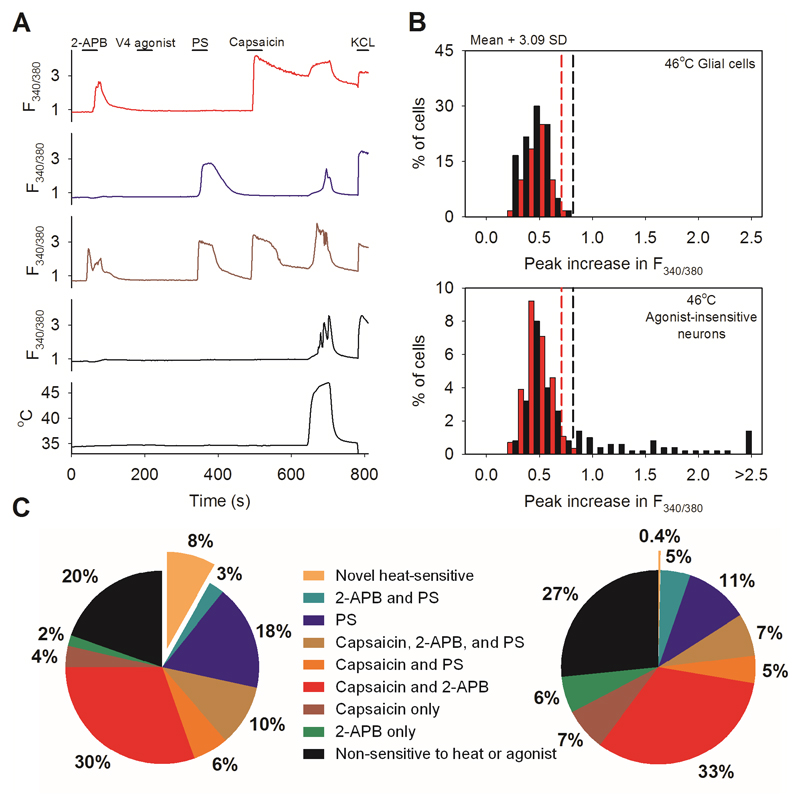
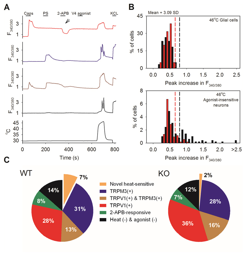
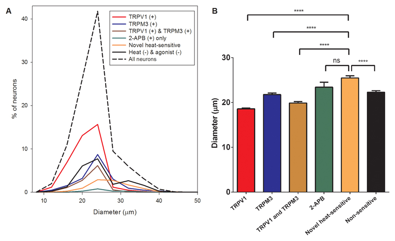
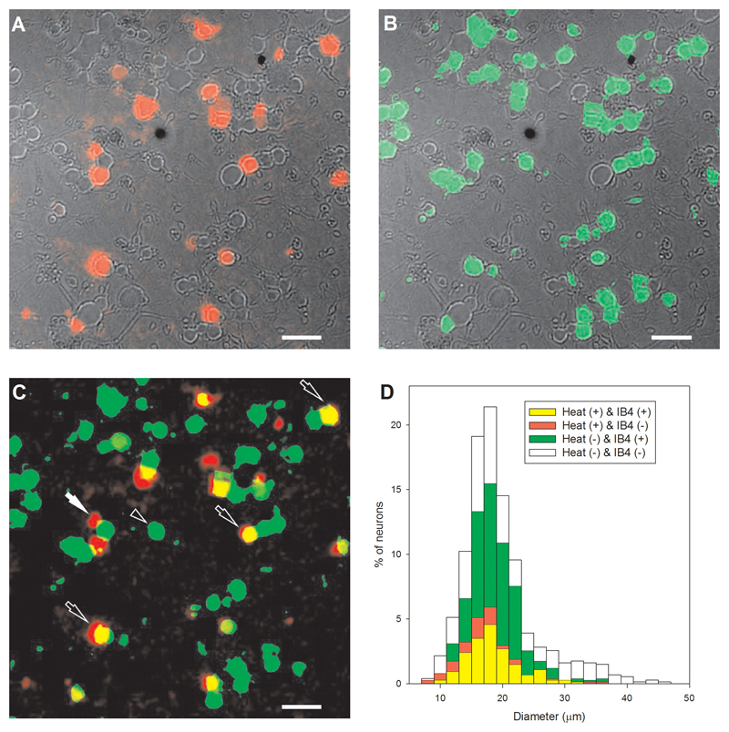
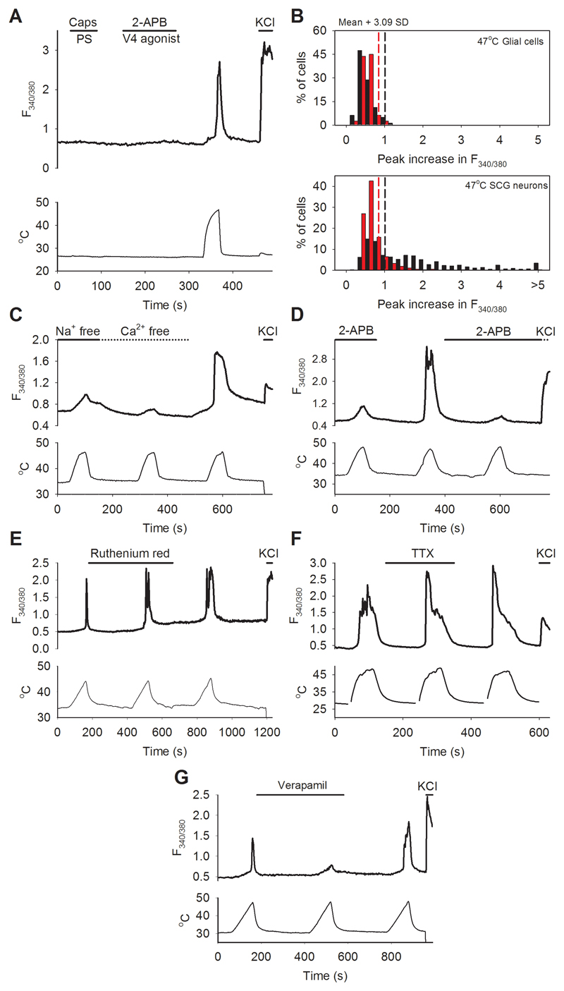
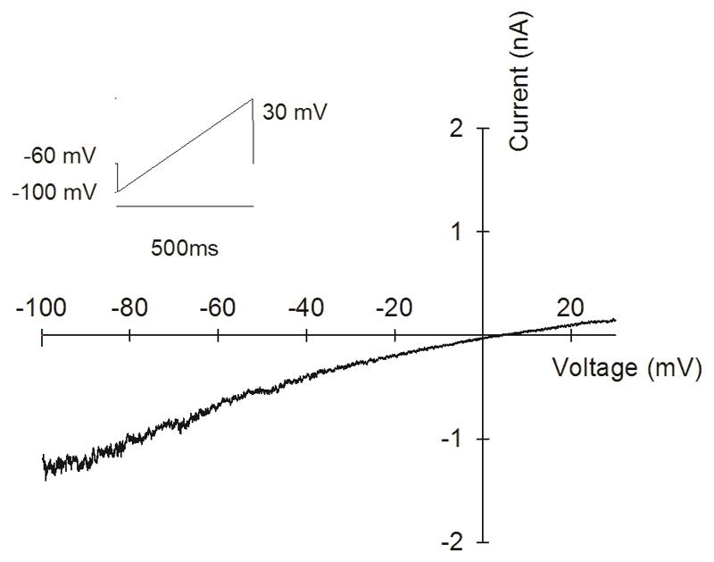
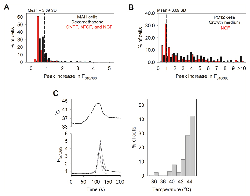
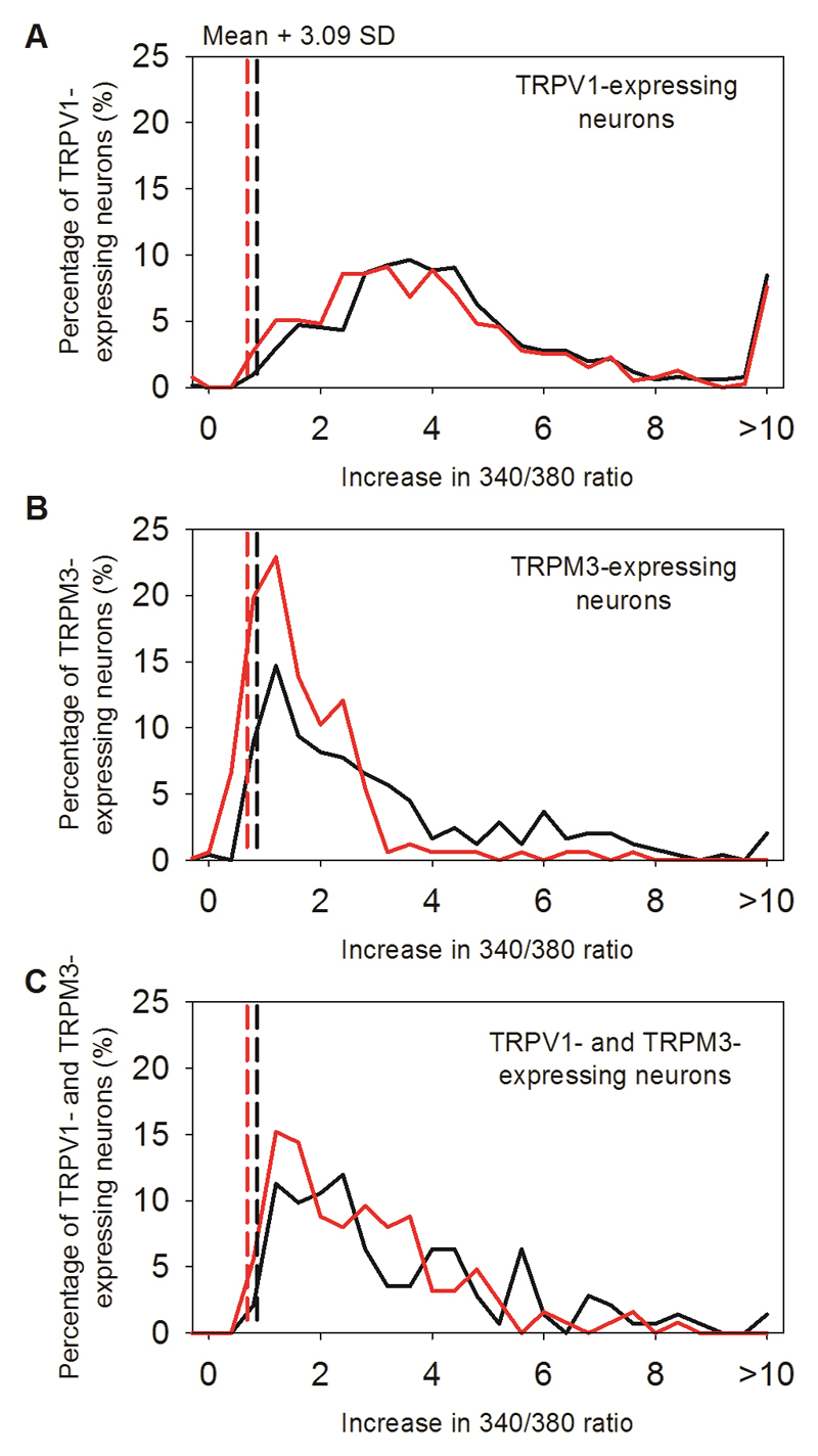
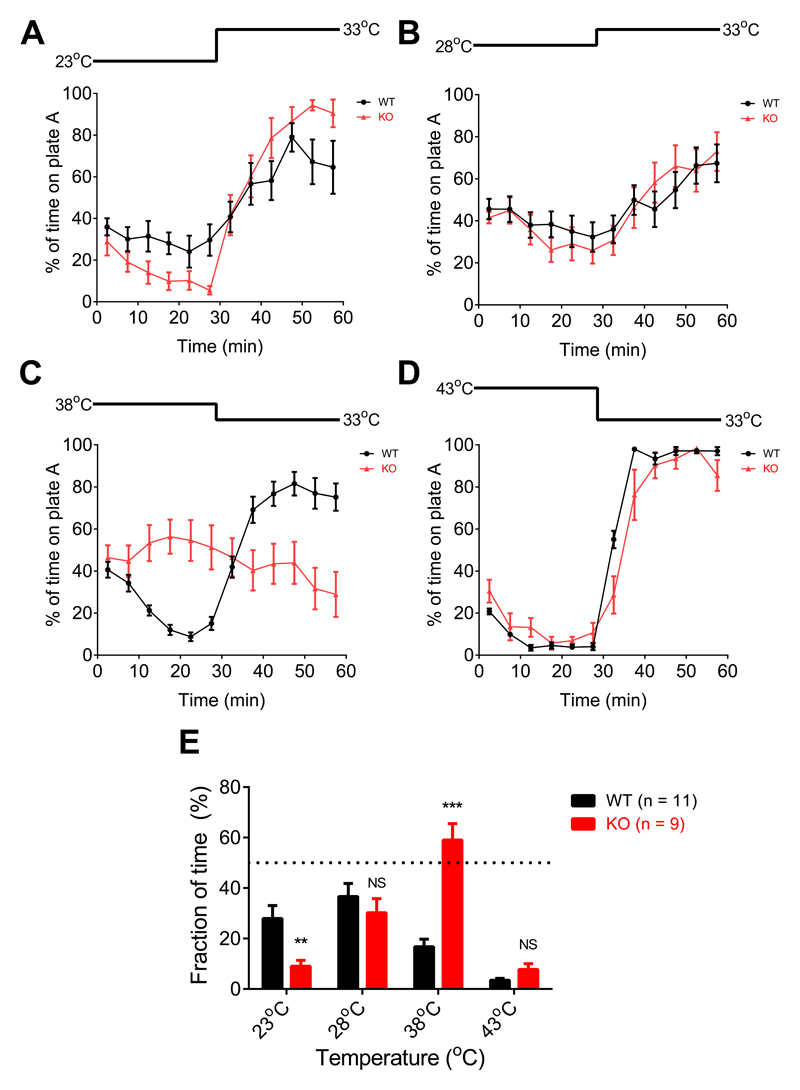
Thermally activated ion channels are known to detect the entire thermal range from extreme heat (TRPV2), painful heat (TRPV1, TRPM3 and ANO1), non-painful warmth (TRPV3 and TRPV4) and non-painful coolness (TRPM8) through to painful cold (TRPA1). Genetic deletion of each of these ion channels, however, has only modest effects on thermal behaviour in mice, with the exception of TRPM8, the deletion of which has marked effects on the perception of moderate coolness in the range 10-25 °C. The molecular mechanism responsible for detecting non-painful warmth, in particular, is unresolved. Here we used calcium imaging to identify a population of thermally sensitive somatosensory neurons which do not express any of the known thermally activated TRP channels. We then used a combination of calcium imaging, electrophysiology and RNA sequencing to show that the ion channel generating heat sensitivity in these neurons is TRPM2. Autonomic neurons, usually thought of as exclusively motor, also express TRPM2 and respond directly to heat. Mice in which TRPM2 had been genetically deleted showed a striking deficit in their sensation of non-noxious warm temperatures, consistent with the idea that TRPM2 initiates a 'warm' signal which drives cool-seeking behaviour.


Figures

Extended Data Fig. 1
Extended Data Fig. 1. Effect of altering the order of agonist application in DRG neurons
A. Method for detecting novel heat-sensitive somatosensory neurons. Representative traces showing increases of [Ca2+]i (F340/380 ratio, ordinate) in DRG neurons in response to the TRPV1-3 agonist 2-APB (250μM), to the specificTRPV4 agonist PF-4674114 (V4 agonist, 200 nM), to the specific TRPV1 agonist capsaicin (Caps, 1μM), to the TRPM3 agonist pregnenolone sulphate (PS, 100μM), to a heat ramp from 35°C to 46°C (temperature protocol shown at bottom), and to KCl (50 mM). Other details as in Fig. 1. From top: TRPV1-expressing neuron responding to 2-APB, capsaicin (1 μM) and heat (red, 30% of 500 neurons); TRPM3-expressing neuron responding to PS (blue, 100 μM) and heat (18%); TRPV1- and TRPM3- co-expressing neuron responding to 2-APB, PS, capsaicin, and heat (brown, 10%); neuron unresponsive to TRP channel agonists but showing a response to heat and therefore expressing a novel heat sensitive ion channel (black, 8% of total). No neuron responded to the specific TRPV4 agonist PF-4674114 (200nM). B. Heat has a small effect on the fura-2 fluorescence ratio, so we eliminated neurons in which an increase of fluorescence ratio was due simply to this physical effect by comparing the increase of fura-2 fluorescence ratio in neurons with that in glial cells in the same culture. Maximum increases in F340/380 ratio in response to a heat ramp from 35°C to 46°C in WT glial cells (black bars, top, n=60) and in WT DRG neurons not responding to known thermo-TRP agonists (black bars, bottom, from same images as the glial cells in top panel, n=139). Vertical black dashed line in top panel shows mean + 3.09 SD (cumulative probability value of 99.9%) of the increase in the F340/380 ratio in glial cells; this value is taken as the maximum increase in F340/380 ratio caused by effect of heat on fura-2 and is used as the cut-off value for defining novel heat-sensitive neurons present in the same culture dish (vertical black dashed line in lower panel). Similar results from separate culture of TRPM2-/- glia (n = 40) and neurons (n= 76) shown in red. The proportion of novel heat-sensitive neurons was significantly reduced from 8% (41/500) in WT to 0.4% (1/282) in TRPM2-/- (p≦0.0001; Fisher’s exact test). The increases in F340/380 ratio of novel heat-sensitive neurons above the cut-off value in response to heat (smallest increase = 0.019854) are all higher than that of the single heat-responding TRPM2-/- neuron (0.019084). C. Pie charts showing the percentage of novel heat-sensitive neurons responding to TRP ion channel agonists and to heat in WT DRG neurons (left) and DRG neurons from TRPM2-/- mice (right). Deletion of TRPM2 reduces the percentage of novel heat-sensitive neurons from 8% to 0.4%. Cell numbers: A,B,C: 500 DRG neurons from one WT mouse on 3 coverslips and 282 DRG neurons from one TRPM2-/- mouse on 2 coverslips were imaged. No further replicates performed.
Extended Data Fig. 2
Extended Data Fig. 2. Effect of starting heat ramp at a lower temperature in DRG neurons
Identical experiment to that shown in Fig. 1A-C except that the temperature ramp started from 30°C. A. Agonist and heat-responsive neurons as in Fig. 1A. From top: TRPV1-expressing neuron responding to capsaicin (1 μM) and heat (red, 28% of 491 neurons); TRPM3-expressing neuron responding to PS (blue, 100 μM) and heat (31%); TRPV1- and TRPM3- co-expressing neuron responding to capsaicin, PS and heat (brown, 13%); neuron unresponsive to TRP channel agonists but showing a response to heat and therefore expressing a novel heat sensitive ion channel (black, 7% of total). A small number of neurons (8%) responded to 2-APB (250μM) but not to other agonists, and 14% of DRG neurons did not respond to any of the agonists nor to heat (not shown). No neuron responded to the specific TRPV4 agonist PF-4674114 (200nM). B. Maximum increases in F340/380 ratio in response to a heat ramp from 30°C to 46°C in WT glial cells (black bars, top, n=60) and in heat-sensitive WT DRG neurons not responding to known thermo-TRP agonists (black bars, bottom, from same images as the glial cells in top panel, n=103). Vertical black dashed line in top panel shows mean + 3.09 SD (cumulative probability value of 99.9%) of the increase in the F340/380 ratio in glial cells; this value is taken as the maximum increase in F340/380 ratio caused by effect of heat on fura-2 and is used as the cut-off value for defining novel heat-sensitive neurons present in the same culture dish (vertical black dashed line in lower panel). Similar results from separate culture of TRPM2-/- glia (n = 60) and neurons (n= 73) shown in red. The proportion of novel heat-sensitive neurons was significantly reduced from 7% (103/491) in WT to 2% (73/522) in TRPM2-/- (p≦0.0001; Fisher’s exact test). The mean increase in F340/380 ratio of novel heat-sensitive neurons above the cut-off values in response to heat was also significantly reduced from 1.237 ± 0.09207 in WT (n=36) to 0.7959 ± 0.03767 in TRPM2-/- (n=8) (p=0.0313; two-tailed unpaired t test). C. Pie charts showing the percentage of novel heat-sensitive and TRPV1- or TRPM3- expressing neurons in WT DRG neurons (left) and DRG neurons from TRPM2-/- mice (right). Cell numbers. A,B,C: 491 DRG neurons from one WT mouse on 3 coverslips and 522 DRG neurons from one TRPM2-/- mouse on 3 coverslips were imaged. No further replicates performed.
Extended Data Fig. 3
Extended Data Fig. 3. Diameters of novel heat-sensitive DRG neurons compared to neurons responding to other TRP agonists.
A. Diameters of 1324 DRG neurons taken from experiment illustrated in figure 1A (dotted line). Subpopulations of neurons are shown as follows: those responding to capsaicin and thus expressing TRPV1 (red); to PS and thus expressing TRPM3 (blue); to both agonists and thus co-expressing TRPV1 and TRPM3 (brown); to 2-APB alone (green); novel heat-sensitive neurons (orange), and neurons responding neither to heat nor to any of these agonists (black). B. Diameter comparison of subpopulations of neurons. TRPV1-expressing neurons have the smallest mean diameter (18.58 ± 0.17μm), TRPM3-expressing neurons are intermediate (21.75 ± 0.33μm) and neurons expressing only the novel heat-sensitivity have the largest mean diameter (25.47 ± 0.48μm). Significance from one-way ANOVA and multiple comparisons with Tukey’s multiple comparison test (****: ≦0.0001; ns: not significant). Cell numbers. Data obtained from Fig. 1 A, B, C; 1324 DRG neurons from one WT mouse on 4 coverslips were analysed.
Extended Data Fig. 4
Extended Data Fig. 4. The novel heat-sensitivity in DRG neurons is partially co-expressed with TRPV1 and TRPM3, and is enhanced by H2O2.
A. Temperature ramp to 47°C, as in Fig. 1A, but with TRPV1 blocked with AMG9810 (5μM) and TRPM3 blocked with naringenin (10μM). Criterion level for significant increase (dashed lines) taken from glial cells in same field of view (data not shown). Black bars: 46% of all WT DRG neurons (n = 580) responded to heat ramp from 34°C to 46°C with an increase in [Ca2+]i above criterion level in presence of blockers of TRPV1 and TRPM3 (dashed vertical line), while the percentage decreased to 17% in TRPM2-/- DRG neurons (red bars, n=1007) (p≦0.0001; Fisher’s exact test). The mean increase in F340/380 ratio above the cut-off values (dashed lines) in response to heat was also significantly reduced from 1.619 ± 0.06133 in WT (n=265) to 1.027 ± 0.08394 in TRPM2-/- (n=175) (p≦0.0001; two-tailed unpaired t test). In similar experiments with TRPV1 blocker BCTC (4μM) and naringenin (10μM), 37% of WT neurons responded to heat (data not shown, n = 554). B. Similar plot as in A, but data from the subgroup of novel heat-sensitive neurons not responding to agonists for known thermo-TRP channels. After exposure to heat in presence of blockers of TRPV1 and TRPM3, blockers were removed and neurons not responding to known TRP agonists were identified as in Fig. 1A. Data from same experiment as shown in A. Proportion of neurons expressing the novel heat-sensitive mechanism in isolation (i.e. without co-expression of TRPV1 or TRPM3) was significantly lower (52/580, 9%) than all neurons expressing the novel heat sensitive mechanism (46%, see A). The proportion of novel heat-sensitive neurons was significantly reduced in TRPM2-/- mice, from 9% to 0.6% (6/1007, p≦0.0001; Fisher’s exact test). C. Temperature ramp to 42°C. Few novel heat-sensitive neurons respond to this low temperature in either WT or TRPM2-/-. Total numbers of neurons n=173 for WT, n=211 for TRPM2-/-. D. Responses of same neurons to same temperature ramp to 42°C following addition of H2O2 (400μM). Enhancement of response in WT (black bars) largely abolished in TRPM2-/- (red bars). The proportion of novel heat-sensitive neurons after sensitization with H2O2 was significantly reduced from 11% (74/635) in WT to 8% (48/601) in TRPM2-/- (p=0.0356; Fisher’s exact test). The mean increase in F340/380 ratio of novel heat-sensitive neurons above the cut-off values in response to heat was also significantly reduced from 1.175 ± 0.1516 in WT (n=72) to 0.4485 ± 0.04329 in TRPM2-/- (n=48) (p=0.0002; two-tailed unpaired t test). Cell numbers and replicates. A, B: 580 DRG neurons from one WT mouse on 5 coverslips and 1007 DRG neurons from one TRPM2-/- mouse on 5 coverslips were imaged for the protocol with AMG9810. 554 neurons from one WT mouse on 4 coverslips were imaged for the protocol with BCTC. No further replicates. C, D: 635 DRG neurons from one WT mouse on 3 coverslips and 601 DRG neurons from one KO mouse on 3 coverslips were imaged. Experiment was replicated with similar results on 4 further coverslips from 1 mouse.
Extended Data Fig. 5
Extended Data Fig. 5. Most novel heat-sensitive DRG neurons are IB4-positive
A. Increases in [Ca2+]i (F340/380 ratio image, intensity-coded in red, mouse DRG neurons) in response to heat ramp to 46°C (TRPV1 blocked with AMG9810, 5μM, and TRPM3 blocked with naringenin, 10μM), superimposed on DIC transmission image obtained post-fixation. B. Same field following fixation and labelling with fluorescent IB4 (green). C. Superimposed calcium and IB4 images from A and B. Black arrows show neurons responding to heat and positive for IB4. White arrow shows a neuron responding to heat and negative for IB4. Black arrowhead shows neuron insensitive to heat and positive for IB4. Scale bars 50 μm. D. Diameter histogram of 743 fixed DRG neurons subgrouped according to novel heat-sensitivity (yellow, red) and IB4 binding (yellow, green). 25% (184/743) of DRG neurons showed novel heat-sensitivity and 74% of these novel heat-sensitive neurons were IB4-positive, while only 53% of heat-insensitive neurons were IB4-positive. The percentage of IB4-positive neurons is significantly higher in the heat-sensitive group than in the heat-insensitive group (p≦0.0001; Fisher’s exact test). Note that the diameters shown in D are not directly comparable with the live cell diameters shown in Extended Data Fig 3 because of a shrinkage artifact on fixation. Cell numbers. 743 DRG neurons from one WT mouse on 4 coverslips were imaged. No further replicates were performed.
Extended Data Fig. 6
Extended Data Fig. 6. Properties of novel heat-sensitive ion channel expressed in autonomic neurons.
A. Representative traces showing increases of [Ca2+]i (F340/380 ratio, ordinate) in sympathetic neurons from superior cervical ganglion (SCG) in response to a mixture of capsaicin (TRPV1 agonist,1μM) and pregnenolone sulphate (TRPM3 agonist, 100μM); a mixture of 2-APB (TRPV1-3 agonist, 250μM) and PF-4674114 (TRPV4 agonist, 200 nM); heat to 47°C (temperature protocol shown at bottom); and KCl (50 mM). Similar results obtained with parasympathetic neurons from pterygopalatine ganglion (PPG, data not shown). Trace is same as shown in Fig. 2A. B. Similar histograms as in Extended Data Figs. 1B and 2B, but for SCG glial cells and neurons from WT mice (black bars) and TRPM2-/- mice (red bars). 58% of WT SCG neurons (n=436) showed novel heat-sensitivity with increases in F340/380 ratio above the criterion level obtained from glial cells (n=80) in same culture (black vertical dashed line). In similar experiments on PPG neurons (n=484), 47% showed novel heat-sensitivity (not shown). Red bars and red dashed line show results from SCG glia (n=80) and neurons (n=430) from TRPM2-/- mice. The proportion of novel heat-sensitive neurons was significantly reduced by deletion of TRPM2, from 58% (252/436) in WT to 12% (53/430) in TRPM2-/- (p≦0.0001; Fisher’s exact test). The mean increase in F340/380 ratio of heat-sensitive neurons above the cut-off values in response to heat was also significantly reduced, from 1.629 ± 0.1928 in WT (n=252) to 0.5050 ± 0.1270 in TRPM2-/- (n=53) (p=0.0086; two-tailed unpaired t test). C. Heat-evoked Ca2+ increase in SCG neurons is reduced but not abolished by removal of extracellular Na+ (replaced with choline) and is abolished by removal of extracellular Ca2+ (remaining small increase in F340/380 ratio is due to temperature sensitivity of fura-2, see part B). Similar results seen in 133 SCG neurons. D. Heat-evoked Ca2+ increase in SCG neurons is blocked by TRPV agonist 2-APB (25μM). Similar results seen in 130SCG neurons. E. Ca2+ increase in PPG neurons is not affected by TRPV channel blocker ruthenium red (50μM). Similar results seen in 75 PPG neurons. F. Ca2+ increase in PPG neurons is not affected by the Na channel blocker TTX (2μM). Similar results seen in 35 PPG neurons. G. The Ca2+ influx in PPG neurons is reduced but not eliminated by the L-type Ca2+ channel blocker verapamil (100μM). Similar results seen in 30 PPG neurons. Cell numbers and replicates. A: 166 SCG neurons from three WT mice on 3 coverslips were imaged. B: 436 SCG neurons from two WT mice on 4 coverslips and 430 SCG neurons from two TRPM2-/- mice on 4 coverslips were imaged. Similar results as those shown for WT obtained with 15 further coverslips of SCG neurons from 9 WT mice and 7 coverslips of PPG neurons from 6 WT mice. C: 133 SCG neurons from 3 WT mice on 5 coverslips showed similar responses. D: 130 SCG neurons from 3 WT mice on 2 coverslips showed similar responses. Similar results also obtained for DRG neurons (4 coverslips from 1 mouse). E: 75 PPG neurons from 3 WT mice on 4 coverslips showed similar responses. F: 35 PPG neurons from 3 WT mice on 4 coverslips showed similar responses. G: 30 PPG neurons from 3 WT mice on 2 coverslips showed similar responses.
Extended Data Fig. 7
Extended Data Fig. 7. The heat-induced membrane current in autonomic neurons is not gated by membrane voltage.
Current–voltage difference relations of a PPG neuron with a voltage ramp starting from a negative potential (inset) show a similar linear heat-induced current to that obtained with reverse voltage ramp (see Fig. 2C). Trace obtained by subtracting current-voltage relations at 36°C from that at 47°C. Similar results obtained in 3 cells on 3 coverslips.
Extended Data Fig. 8
Extended Data Fig. 8. Responses to heat in adrenal-derived MAH and PC12 cell lines, and effects of factors causing differentiation to neuronal phenotype.
A. MAH cells. Black: maximum increase in F340/380 ratio in response to heat (47°C, n = 254) when cultured in dexamethasone (5 μM). No cell responded to TRP agonists, but 27% of cells responded to heat with increase above mean criterion level obtained from glial cells in neuronal cultures (see Fig. 1B). Red: similar histogram after 12d culture in growth factors (bFGF, CNTF and NGF, see Methods, n = 170). No cell responded to TRP agonists; 9% responded to heat. The proportion of heat-sensitive cells was significantly reduced from 27% (69/254) in dexamethasone to 9% (16/170) in growth factors (p≦0.0001; Fisher’s exact test). The 66% reduction in the proportion of heat-sensitive cells was not significantly different from the reduction in TRPM2 expression caused by differentiation of MAH cells (Table 1 line 5; p = 0.056; two-tailed unpaired t test). The mean increases in F340/380 ratio above the cut-off values (dashed lines) in response to heat were 1.755 ± 0.1255 in dexamethasone (n=69) and 1.420 ± 0.1474 in presence of growth factors (n=16) (p=0.2203; two-tailed unpaired t test). B. PC12 cells. Black: culture in growth medium (10% horse serum + 5% fetal bovine serum, n=200). 93% of cells responded to heat with increase above mean criterion level obtained from glial cells in neuronal cultures (see Fig. 1B). Red: Effect on heat responses of 12d culture in NGF (1% horse serum plus 100 ng/ml NGF, n = 108). The proportion of heat-sensitive cells was significantly reduced from 93% (186/200) in growth medium to 46% (50/108) in NGF (p≦0.0001; Fisher’s exact test). We note that a significantly lower expression of mRNA for TRPM2 in differentiated PC12 cells has been reported. The mean increase in F340/380 ratio above the cut-off values (dashed lines) in response to heat was significantly reduced from 3.753 ± 0.2431 in growth medium (n=186) to 2.603 ± 0.3104 in NGF (n=50) (p=0.0213; two-tailed unpaired t test). C. Temperature thresholds of PC12 cells cultured in growth medium. Top left: temperature protocol. Bottom left: temperature responses of three representative cells. Right: Temperature thresholds calculated as in Fig. 1D. Cell numbers and replicates. A, B: 2 coverslips for each condition were imaged. Cell numbers are given above. Replicates of 4 coverslips (MAH cells) and 3 coverslips (PC12 cells) for each condition gave similar results. C: 165 PC12 cells on one coverslip were imaged.
Extended Data Fig. 9
Extended Data Fig. 9. Effect of deletion of TRPM2 on maximal calcium responses to heat in neurons expressing TRPV1 or TRPM3
A. Maximum increases in F340/380 ratio in response to a heat ramp from 34°C to 46°C in neurons responding only to capsaicin (TRPV1-expressing) from WT (black) and TRPM2-/- (red) mice. The increase in F340/380 ratio in response to heat (above the increase caused by the effect of temperature on fura-2, vertical dotted lines, for method of calculation see Fig. 1B) is not significantly different between WT and TRPM2-/- (p=0.1168, two-tailed Mann–Whitney U test). Details as in Fig. 1. B. Neurons responding only to pregnenolone sulphate (PS, TRPM3-expressing) from same experiments as in A. The increases in F340/380 ratio in response to heat are significantly reduced by deletion of TRPM2 (from 6.389 ± 1.225 to 4.411 ± 1.582, p<0.0001, two-tailed Mann–Whitney U test). C. Neurons responding to both capsaicin and PS (TRPV1- and TRPM3-expressing). The increases in F340/380 ratio in response to heat are not significantly different between WT and TRPM2-/- (p=0.0633; two-tailed Mann–Whitney U test). Cell numbers and replicates. A,B,C: 1324 DRG neurons from one WT mouse on 4 coverslips and 981 DRG from one TRPM2-/- mouse on 4 coverslips were imaged. Similar results obtained from 42 further coverslips from 6 WT mice and 8 further coverslips from 1 TRPM2-/- mouse.
Extended Data Fig. 10
Extended Data Fig. 10. Correlation between novel heat sensitivity and expression of mRNA for TRPM2.
A. Representative DIC transmission image of DRG neurons following in situ hybridization with sense probe, as negative control. Non-specific density was linearly dependent on cell diameter (see E). Mean + 3.09 SD (cumulative probability value of 99.9%) of density as a function of diameter with sense probe (5μm bins) was used as threshold criterion for significant expression of TRPM2 in images of antisense hybridization. A similar analysis of non-specific density was carried out for glial cells. B. Representative DIC transmission image with antisense probe against TRPM2. Using the threshold criterion as function of diameter obtained from sense probe images (see A), 89% (1121/1250) of DRG neurons but 3% (4/120) of glial cells were positive for TRPM2 mRNA. C. Novel heat-sensitive DRG neurons determined using calcium imaging. Increases in [Ca2+]i (F340/380 ratio image, intensity-coded in red) in response to a heat ramp to 46°C with TRPV1 blocked with AMG9810 (5μM) and TRPM3 blocked with naringenin (10μM). D. Superimposed image of novel heat-sensitive neurons (red) and in situ hybridization using antisense probe. Solid red arrows indicate novel heat-sensitive neurons also positive for TRPM2; solid black arrow shows neuron not responding to heat but positive for TRPM2; open black arrow shows cell negative for TRPM2 (i.e. with density below the criterion level obtained from E). 42% (92/218) of DRG neurons positive for TRPM2 exhibited novel heat-sensitivity. However only 13% (2/16) of DRG neurons negative for TRPM2 from in situ hybridization exhibited novel heat-sensitivity. The percentage of novel heat-sensitive DRG neurons is significantly reduced in TRPM2 negative DRG neurons (p=0.0188; Fisher’s exact test). Scale bars shown on right lower corner indicate 50 μm. E. Density of non-specific label in neurons obtained from hybridization with sense probe (see A) depends on cell size. Data used to calculate significance thresholds for neurons in B. Cell numbers and replicates. A: One coverslip exposed to sense probe was used as negative control. B: All neurons on 5 coverslips were measured and one coverslip was measured for TRPM2-positive glial cells; C, D: One coverslip was analysed as in A and B for the combined calcium imaging and in situ hybridization protocol. E: 500 DRG neurons on one coverslip exposed to sense probe was used to determine the background threshold as a function of cell diameter. Similar in situ hybridization results obtained on 16 further coverslips.
Fig. 1
Fig. 1. Around 10% of somatosensory neurons show a novel heat-sensitive mechanism.
A. Examples of increases of [Ca2+]i measured with fura-2 (F340/380 ratio, ordinate) in response to known TRP channel agonists and to heat. From top: TRPV1-expressing neuron responding to capsaicin (caps, red); TRPM3-expressing neuron responding to pregenolone sulphate (PS, blue); TRPV1- and TRPM3- co-expressing neuron (brown); neuron unresponsive to TRP channel agonists but showing a response to heat. See Supplementary Information video 1 for images. B. Percentages of neurons expressing TRPV1, TRPM3 and the novel heat-sensitive mechanism in DRG neurons from WT (top) and TRPM2-/- mice (bottom). Green: neurons responding to 2-APB but not to other agonists. C. Histogram of maximal F340/380 ratio increase in neurons insensitive to TRP agonists. Black: WT neurons; red: TRPM2-/- neurons. Vertical dashed lines: thresholds discriminating between heat-sensitive and insensitive neurons (see Extended Data Fig. 1 for details). D. Thermal thresholds of novel heat-sensitive DRG neurons. Increases of F340/380 ratio (top) in response to slowly rising heat ramp (bottom). E. Temperature thresholds of novel heat-sensitive neurons from WT (black) and TRPM2-/- mice (red). Proportion in range 34-42°C reduced from 4.4% in WT to 0.9% in TRPM2-/- (p≦0.0001; Fisher’s exact test), and in range 42-48°C from 15% in WT (290/1890) to 3% in TRPM2-/- (55/1800) (p≦0.0001; Fisher’s exact test). Cell numbers and replicates. A - C: 1324 DRG neurons from one WT mouse on 4 coverslips and 981 DRG neurons from one TRPM2-/- mouse on 4 coverslips were imaged. Similar results obtained using 52 further coverslips from 9 further WT mice and 10 coverslips from 3 further TRPM2-/- mice; some of these results are shown in Extended Data Figs 1, 2 and 4. D, E: 1890 DRG neurons from one WT mouse on 8 coverslips and 1800 DRG neurons from one TRPM2-/- mouse on 8 coverslips were imaged. Similar results obtained using 10 coverslips from 1 further WT mouse.
Fig. 2
Fig. 2. Properties of the novel heat-sensitive ion channel in autonomic neurons.
A. Sympathetic neurons from superior cervical ganglion (SCG) respond to heat but not to TRP channel agonists. B. The L-type Ca2+ channel blocker nifedipine (10 μM) blocks spiking but not steady depolarization in response to heat in a patch-clamped PPG neuron (top) and reduces but does not eliminate the Ca2+ increase (middle). Simultaneous recording of membrane potential and [Ca2+]i (F340/380 ratio) in current–clamped PPG neuron. C. Current-voltage relations at 36°C and 47°C of voltage-clamped PPG neuron in response to voltage ramp shown top left. Similar IV relation observed with inverse voltage ramp starting from 100mV (Extended Data Fig. 7). See Methods for details. D. H2O2 (400μM) potentiates Ca2+ increase in response to a mild temperature stimulus (42°C) in SCG neurons. Note potentiation is long-lasting after H2O2 removed. Similar results obtained in PPG neurons. Percentage of neurons (n=456) responding to heat, determined as in Extended Data Fig. 1B, was 5% before, 59% during and 54% post-H2O2. Cell numbers and replicates. A: 166 SCG neurons from 3 WT mice on 3 coverslips were imaged for this experiment. Similar results obtained with SCG neurons using 15 further coverslips from 9 further mice and with PPG neurons using 14 coverslips from 9 further mice. B: 2 PPG neurons on 2 coverslips were simultaneously patch-clamped and imaged and showed a similar response. C: 3 PPG neurons on 3 coverslips were simultaneously patch-clamped and imaged and showed a similar response. D: 456 SCG neurons from two WT mice on 4 coverslips were imaged and showed a similar enhancement with H2O2; 731 PPG neurons (not shown) from three WT mice on 3 coverslips were imaged and showed a similar enhancement to the SCG neurons. Similar results obtained with SCG neurons using 9 coverslips from 5 further mice and with PPG neurons using 3 coverslips from 3 further mice.
Figure 3
Figure 3. Deletion of TRPM2 shifts adult male mouse thermal preference towards warmer temperatures.
A-D: Two-plate thermal preference test. One plate is at 33°C at the start of the experiment, the other (“plate A”) is at a variable temperature as shown above each graph. Temperature reversed at t= 30min to account for any possible bias caused by external cues. Points show mean behavioural preference averaged over 5 min intervals (error bars mean ± SEM, n = 11 for WT and 9 for TRPM2-/-). Mice were WT or TRPM2-/- male littermates, 12-16 weeks of age. No difference observed between the thermal behaviour of WT and heterozygous TRPM2+/- mice (not shown, n = 4). E: Mean thermal preference averaged from the data between t = 15-30min and t = 45-60 min shown in A-D. Bars give mean ± SEM. **, p < 0.01; ***, p < 0.001; NS, p > 0.05; unpaired t-test. Biological replicates. Similar results were obtained with experiments using non-littermates in which 12 WT male mice from Charles River were compared with 7 TRPM2-/- male mice from homozygote breeding pairs.

References

    1. Cesare P, McNaughton PA. A novel heat-activated current in nociceptive neurons, and its sensitization by bradykinin. Proc Natl Acad Sci USA. 1996;93:15435–15439. - PMC - PubMed
    1. Caterina MJ, et al. The capsaicin receptor: a heat-activated ion channel in the pain pathway. Nature. 1997;389:816–824. - PubMed
    1. Jordt SE, McKemy DD, Julius D. Lessons from peppers and peppermint: the molecular logic of thermosensation. Curr Opin Neurobiol. 2003;13:487–492. - PubMed
    1. Basbaum AI, Bautista DM, Scherrer G, Julius D. Cellular and molecular mechanisms of pain. Cell. 2009;139:267–284. - PMC - PubMed
    1. Vriens J, et al. TRPM3 is a nociceptor channel involved in the detection of noxious heat. Neuron. 2011;70:482–494. doi: 10.1016/j.neuron.2011.02.051. - DOI - PubMed

Publication types