. 2016 Nov 15;7(46):74820-74833.
doi: 10.18632/oncotarget.11204.

Akt isoform specific effects in ovarian cancer progression

Affiliations

Akt isoform specific effects in ovarian cancer progression

Nicolle M Linnerth-Petrik et al. Oncotarget. .

Abstract

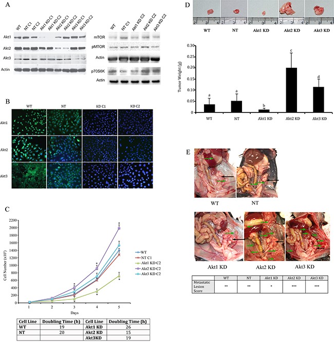
Ovarian cancer remains a significant therapeutic problem and novel, effective therapies are needed. Akt is a serine-threonine kinase that is overexpressed in numerous cancers, including ovarian. Mammalian cells express three Akt isoforms which are encoded by distinct genes. Although there are several Akt inhibitors in clinical trials, most indiscriminately target all isoforms. Current in vitro data and animal knockout experiments suggest that the Akt isoforms may have divergent roles. In this paper, we determined the isoform-specific functions of Akt in ovarian cancer cell proliferation in vitro and in ovarian cancer progression in vivo. For in vitro experiments, murine and human ovarian cancer cells were treated with Akt inhibitors and cell viability was assessed. We used two different in vivo approaches to identify the roles of Akt isoforms in ovarian cancer progression and their influence on the primary tumor and tumor microenvironment. In one experiment, wild-type C57Bl6 mice were orthotopically injected with ID8 cells with stable knockdown of Akt isoforms. In a separate experiment, mice null for Akt 1-3 were orthotopically injected with WT ID8 cells (Figure 1). Our data show that inhibition of Akt1 significantly reduced ovarian cancer cell proliferation and inhibited tumor progression in vivo. Conversely, disruption of Akt2 increased tumor growth. Inhibition of Akt3 had an intermediate phenotype, but also increased growth of ovarian cancer cells. These data suggest that there is minimal redundancy between the Akt isoforms in ovarian cancer progression. These findings have important implications in the design of Akt inhibitors for the effective treatment of ovarian cancer.

Keywords: Akt inhibitors; Akt isoforms; ovarian cancer; tumor development.


Conflict of interest statement

CONFLICTS OF INTEREST

The authors declare no conflicts of interest.

Figures

Figure 1
Figure 1. Experimental Design
Two in vivo models were utilized to determine the Akt isoform specific roles in ovarian tumor progression. In one experiment, mice were subjected to orthotopic injection of 1 × 10⁶ ID8 cells, or ID8 cells in which Akt isoforms were constitutively knocked down by stable expression of shRNAs delivered using a lentiviral vector system. ID8 cells expressing non-target shRNA were included as controls. Knockdown was confirmed with Western blot and immunofluorescence analysis. In a separate experiment, 1 × 10⁶ wild type ID8 cells were orthotopically injected under the ovarian bursa of WT, Akt 1−/−, Akt 2−/−, or Akt 3−/− mice. In each experiment, mice were either sacrificed at 60d post tumor induction (PTI), or were allowed to progress to clinical signs of morbidity for survival analysis.
Figure 2
Figure 2. Knockdown of specific Akt Isoforms has variable effects on ovarian tumor development and metastatic disease
A. Western blot analysis and B. immunofluorescence confirmed specific knockdown of Akt 1, Akt 2, and Akt 3 isoforms using lentiviral vector delivery of shRNA. C. Akt1 KD reduces, while Akt 2 KD elongates ID8 cell doubling time. 2×10⁴ ID8 cells were plated on 100mm cell culture dishes and cell number was counted using a hemocytometer every 24h for 5 days. * - statistically different from NT controls (p<0.05). Cell doubling times were quantified for each cell line and reported as the number of hours required for doubling of the cell population. D. At 60d post tumor induction, tumors were collected from mice injected with tumor cells in which the three Akt isoforms had been knocked down. Akt1 KD resulted in smaller tumors, while Akt2 knockdown led to larger tumors, with Akt3 KD resulting in an intermediate tumor size. (p<0.01). E. Akt1 knockdown also resulted in reduced metastatic disease compared to controls or other Akt isoform knockdowns. Primary tumors are shown with black arrows, while metastatic tumors are shown with green arrows. Metastatic tumors are sored as * < 3 visible tumors; ** 4-10 visible tumors; *** >, 10 visible tumors.
Figure 3
Figure 3. Akt isoform specific inhibition differentially affects tumor cell proliferation, apoptosis, and tumor vascularity
Tumors were collected and fixed at 60d post tumor induction with control cells or cells with Akt isoforms knocked down. A. Ki67 immunostaining revealed a significant reduction in tumor cell proliferation following Akt1 knockdown and increased proliferation with knockdown of Akt2. B. Conversely, Akt 1 knockdown resulted in increased apoptosis, while Akt2 knockdown decreased ovarian tumor cell death. C. Ovarian tumors induced with Akt1 KD cells had reduced microvessel density, while Akt2 KD tumors had elevated vascularity. Tumors from Akt3 KD cells had an intermediate phenotype compared to the other two KD groups. Bars with different letters are statistically different (p<0.05).
Figure 4
Figure 4. Akt isoform knockdown alters ovarian cancer cell signaling
Akt1 knockdown resulted in decreased expression of VEGFR2 and increased levels of the pro-apoptotic proto-oncogenes bax and bad in lysates from tumors collected 60d post tumor induction. Conversely, Akt2 KD elevated expression of VEGF and its receptor while suppressing expression of bax. Tumors from Akt3 KD cells had increased VEGFR2 expression and reduced bax levels. * indicates statistical difference from NT control groups.(p<0.05). Graphs represent densitometric analysis of individual tumor lysates compared to Actin loading control from a minimum of n=4 animals per group.
Figure 5
Figure 5. Tumor cell Akt isoform specific knockdown differentially impacts survival in a mouse model of epithelial ovarian cancer
Tumors were induced with WT cells, or those in which Akt1, Akt2, or Akt3 were constitutively knocked down with lentiviral vectors expressing isoform-specific shRNAs or NT shRNA. Mice were allowed to progress until clinical signs of morbidity associated with ovarian cancer. Akt2 KD mice all had to be sacrificed by 60d post tumor induction. Akt1 KD mice survived the longest of all groups, while Akt3 KD mice were all euthanized by 82d post tumor induction. Log-rank analysis showed that isoform specific knockdown of Akt significantly (p<0.05) altered survival.
Figure 6
Figure 6. Akt signaling pathways are altered in ovarian tissue collected from WT, Akt1-, Akt2-, and Akt3-null mice
A. Whole ovaries were collected from wild-type C57BL/6, Akt 1-, 2- or 3-null mice and subjected to Western blot analysis for each specific Akt isoform. B. Tumors collected from Akt 1, 2, and 3-null mice were probed for members of the AKT signaling family. C/D Tumor lysates from Akt 1, 2, and 3-null mice were evaluated for expression of members of the mTOR signaling pathway.
Figure 7
Figure 7. Ovarian tumors have a variable growth rate in Akt isoform deficient mice
Wild type ID8 cells were injected into control WT mice, or mice null for Akt1, Akt 2, or Akt 3 isoforms. A. Tumors grown in Akt1−/− mice were smaller, while those grown in Akt2−/− mice were larger than other groups. Tumors grown in Akt3−/− were not different from controls. Bars with different letters are statistically different (p<0.05). B. Akt1−/− mice had fewer metastatic abdominal tumors at 60d post tumor induction, while Akt2−/− and Akt3−/− had a higher incidence of metastases, compared to WT controls. Primary tumors are shown with black arrows, while metastatic tumors are shown with green arrows. C. Akt2−/− mice succumbed to disease earlier than the other groups (p<0.05).
Figure 8
Figure 8. Akt isoform inhibition affects ovarian cancer cell viability
WST-1 viability assay was performed on murine ID8 and human OVCAR3 and CAOV3 ovarian cancer cells following exposure of increasing doses of Akt inhibitors. The Akt1 selective inhibitor A-674563 induced the greatest reduction in viability.

Similar articles

Cited by

References

    1. Siegel RL, Miller KD, Jemal A. Cancer statistics, 2016. CA Cancer J Clin. 2016;66:7–30. - PubMed
    1. Coleman RL, Monk BJ, Sood AK, Herzog TJ. Latest research and treatment of advanced-stage epithelial ovarian cancer. Nat Rev Clin Oncol. 2013;10:211–224. - PMC - PubMed
    1. Aletti GD, Gallenberg MM, Cliby WA, Jatoi A, Hartmann LC. Current management strategies for ovarian cancer. Mayo Clin Proc. 2007;82:751–770. - PubMed
    1. Bai H, Li H, Li W, Gui T, Yang J, Cao D, Shen K. The PI3K/AKT/mTOR pathway is a potential predictor of distinct invasive and migratory capacities in human ovarian cancer cell lines. Oncotarget. 2015;6:25520–25532. doi: 10.18632/oncotarget.4550. - DOI - PMC - PubMed
    1. Noske A, Kaszubiak A, Weichert W, Sers C, Niesporek S, Koch I, Schaefer B, Sehouli J, Dietel M, Lage H, Denkert C. Specific inhibition of AKT2 by RNA interference results in reduction of ovarian cancer cell proliferation: increased expression of AKT in advanced ovarian cancer. Cancer Lett. 2007;246:190–200. - PubMed

MeSH terms