. 2016 Sep 13;7(37):60593-60608.
doi: 10.18632/oncotarget.11260.

Downregulation of homeobox gene Barx2 increases gastric cancer proliferation and metastasis and predicts poor patient outcomes

Affiliations

Downregulation of homeobox gene Barx2 increases gastric cancer proliferation and metastasis and predicts poor patient outcomes

Yushuai Mi et al. Oncotarget. .

Abstract

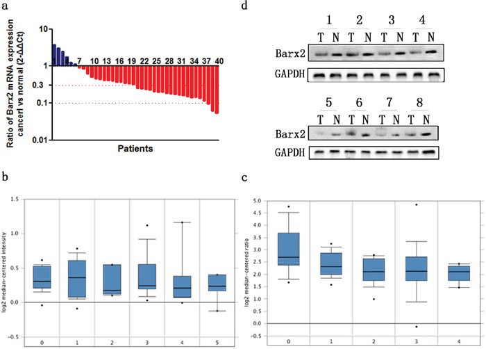
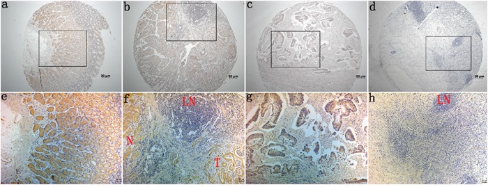
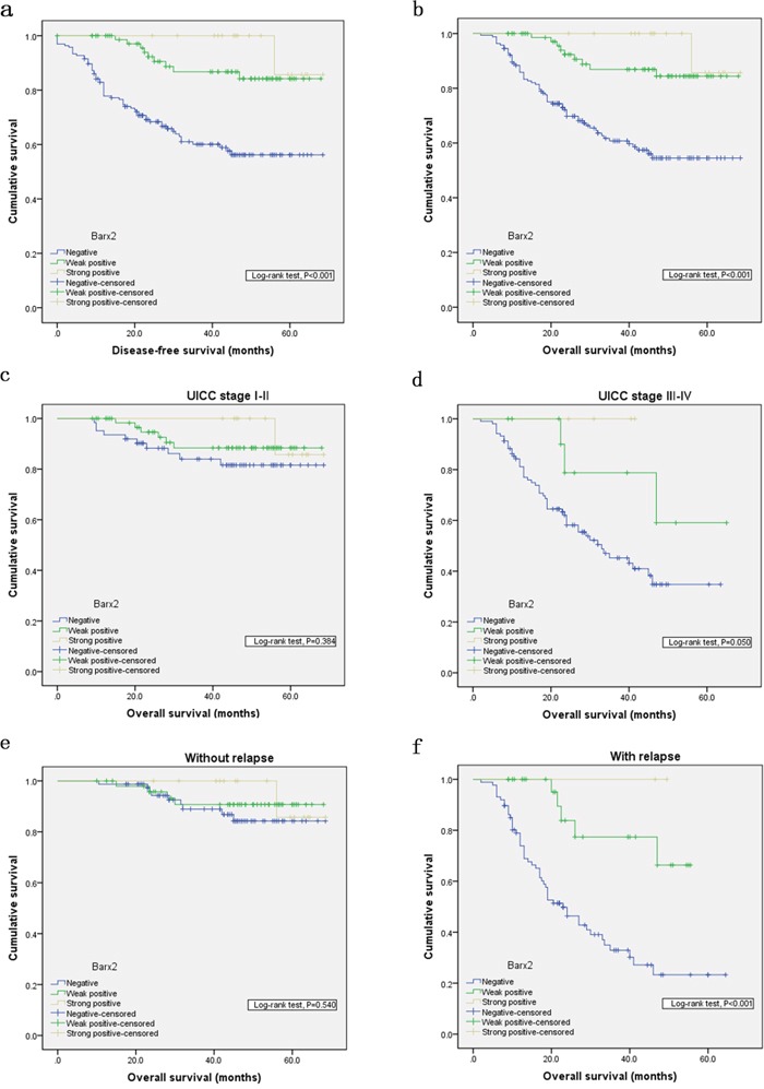
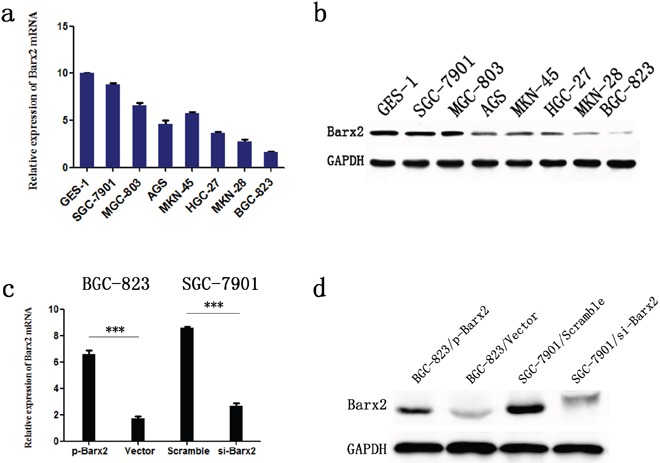
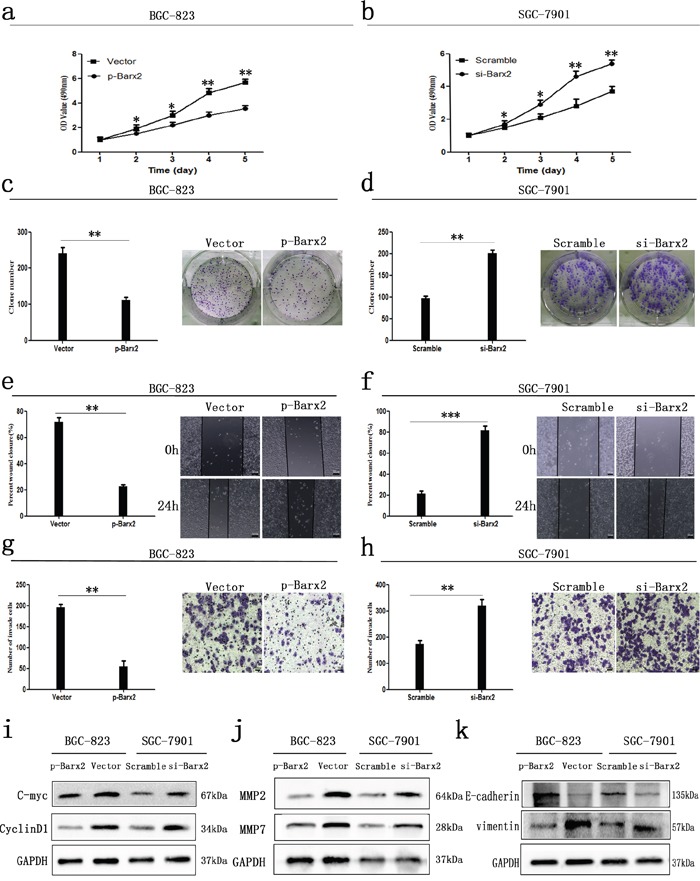
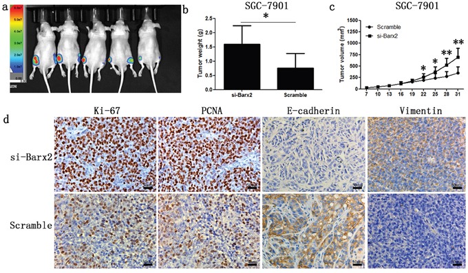
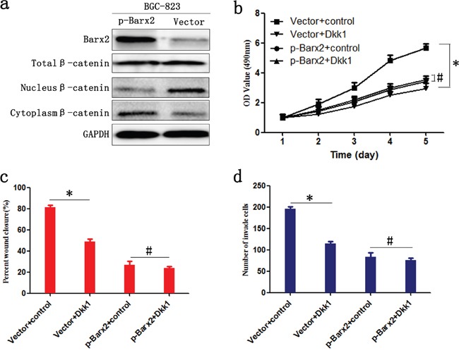
Barx2 is a Bar family homeodomain transcription factor shown to play a critical role in cell adhesion and cytoskeleton remodeling, key processes in carcinogenesis and metastasis. Using quantitative real-time PCR, Western blotting, and immunohistochemistry, we found that Barx2 is expressed at lower levels in human gastric cancer (GC) tissues than in adjacent normal mucosa. In a multivariate analysis, Barx2 expression emerged as an independent prognostic factor for disease-free and overall survival. Kaplan-Meier survival analysis showed a trend toward even shorter overall survival in the patient group with Barx2-negative tumors, independent of advanced UICC stage and tumor relapse. Using in vitro and in vivo assays, we demonstrated that under normal conditions Barx2 inhibited GC cell proliferation and invasiveness through inhibition of the Wnt/β-catenin signaling pathway. These findings indicate that reduction or loss of Barx2 dis-inhibits GC cell proliferation and invasion, and that reduction in Barx2 could serve as an independent prognostic biomarker for poor outcome in GC patients.

Keywords: Barx2; Wnt/β-catenin; gastric cancer; prognosis; progression.


Conflict of interest statement

CONFLICTS OF INTEREST

The authors confirm that there are no conflicts of interest.

Figures

Figure 1
Figure 1. The expression of Barx2 in GC tissues and paired normal mucosa
a. Quantitative real-time PCR detection of relative Barx2 expression in 40 human GC tissue specimens (T) and paired normal mucosa (N). A logarithmic 2-ΔΔCT scale was used to represent the fold change in Barx2 mRNA expression in two independent microarray datasets from Oncomine database: Cho Gastric b. and Chen Gastric c., grouped by no value (0), diffuse gastric adenocarcinoma (1), gastric adenocarcinoma (2), gastric intestinal type adenocarcinoma (3), gastric mixed adenocarcinoma (4) and gastrointestinal stromal tumor (5). d. Western blot analysis was used to detect Barx2 protein expression in 8 representative paired GC tissue samples, with GAPDH used as the loading control.
Figure 2
Figure 2. Immunohistochemical staining for Barx2 expression in normal gastric mucosa and cancerous tissues
Barx2 protein expression was significantly lower in gastric cancer tissues compared with adjacent normal mucosa, with Barx2 staining observed both in the cytoplasm and nuclei of gastric cancer cells. a, e: Strong Barx2 staining in normal gastric epithelium; b, f: Intense Barx2 staining in well-differentiated gastric cancer; c, g: Weak Barx2 staining in moderately differentiated gastric cancer; d, h: Negative Barx2 staining in poorly differentiated gastric cancer; N: Strong Barx2 staining in normal gastric epithelium; T: Intense Barx2 staining in well-differentiated gastric cancer; LN: Negative Barx2 staining in lymph node; a–d. Original magnification: 50×; e–h. Original magnification: 200×.
Figure 3
Figure 3. Kaplan-Meier analysis with a log-rank test of survival
Disease-free survival a. and overall survival b. were significantly shorter in patients with Barx2-negative tumors than in those with Barx2-positive tumors (*** P< 0.001 for both, log-rank test). Comparisons of overall survival between patients with Barx2-negative tumors and those with Barx2-positive tumors in early UICC stage (I-II) cohort and in advanced UICC stage (III-IV) cohort c-d. and in patients with or without relapse e-f. P-values were calculated by log-rank test and P<0.05 was considered significant.
Figure 4
Figure 4. Barx2 expression in cell lines
Barx2 mRNA levels a. and protein levels b. in gastric mucosa cell line (GES-1) and 7 GC cell lines. Barx2 mRNA and protein expression in BGC-823 and SGC-7901 cells transfected with Barx2 overexpression or downregulation vectors were validated using quantitative real-time PCR c. and Western blotting d. GAPDH was used to normalize mRNA and protein levels.
Figure 5
Figure 5. In vitro GC cancer cell functional assays
Overexpression or knock down of Barx2 inhibited or elevated GC cells proliferation a, b., colony formation c, d., wound healing e, f., migration and invasion ability g, h., compared with their control group, respectively (* P <0.05; ** P <0.01; *** P<0.001). e–f. Original magnification: 100×; g–h. Original magnification: 200×. i-k. Western blot analysis of cell cycle-related proteins (c-myc and CyclinD1), biomarkers of cell invasion (MMP2 and MMP7), and EMT markers (E-cadherin and vimentin) in the Barx2 overexpressing or knockdown cells compared with their control groups.
Figure 6
Figure 6. Knock-down of Barx2 promoted tumor formation ability of GC cells in nude mice
(Left flank of the 5 nude mice: Barx2 knock-down; Right flank of the 5 nude mice: vector control) a. After four weeks, xenograft weight b. and volume c. curves were compared with controls (n =5, *P <0.05, **P <0.01). d. Immunochemical staining of xenograft tumors for biomarkers of cell proliferation (Ki-67 and PCNA) and EMT markers (E-cadherin and vimentin). Original magnification: 200×.
Figure 7
Figure 7. Barx2 inhibits the proliferation, migration, and invasion of GC cells by negatively regulating the Wnt/β-catenin signaling pathway
a. WB analysis of the association between expression levels of Barx2 and β-catenin in total cellular, nuclear, and cytoplasmic fractions isolated from BGC-823 cells at 48 h after transfection with Barx2 overexpression or the control empty vector. BGC-823 cells with Barx2 overexpression or vector control were grown in media with or without 200 ng/ml inhibitor of Wnt signaling Dickkopf-related protein 1 (Dkk1); CCK-8 assays b., wound healing c. and transwell assays d. were used to examine the GC cells’ proliferation, migration, and invasion ability, respectively. (* P<0.05, # P>0.05).

References

    1. Siegel RL, Miller KD, Jemal A. Cancer statistics, 2016. CA Cancer J Clin. 2016;66:7–30. - PubMed
    1. Malvezzi M, Bonifazi M, Bertuccio P, Levi F, La Vecchia C, Decarli A, Negri E. An age-period-cohort analysis of gastric cancer mortality from 1950 to 2007 in Europe. Ann Epidemiol. 2010;20:898–905. - PubMed
    1. Torre LA, Bray F, Siegel RL, Ferlay J, Lortet-Tieulent J, Jemal A. Global cancer statistics, 2012. CA Cancer J Clin. 2015;65:87–108. - PubMed
    1. Bertuccio P, Chatenoud L, Levi F, Praud D, Ferlay J, Negri E, Malvezzi M, La Vecchia C. Recent patterns in gastric cancer: a global overview. Int J Cancer. 2009;125:666–73. - PubMed
    1. Shah MA. Update on metastatic gastric and esophageal cancers. J Clin Oncol. 2015;33:1760–9. - PubMed

MeSH terms