Review
. 2016 Aug 1;21(8):80901.
doi: 10.1117/1.JBO.21.8.080901.

Review of fluorescence guided surgery systems: identification of key performance capabilities beyond indocyanine green imaging

Affiliations
Review

Review of fluorescence guided surgery systems: identification of key performance capabilities beyond indocyanine green imaging

Alisha V DSouza et al. J Biomed Opt. .

Abstract

There is growing interest in using fluorescence imaging instruments to guide surgery, and the leading options for open-field imaging are reviewed here. While the clinical fluorescence-guided surgery (FGS) field has been focused predominantly on indocyanine green (ICG) imaging, there is accelerated development of more specific molecular tracers. These agents should help advance new indications for which FGS presents a paradigm shift in how molecular information is provided for resection decisions. There has been a steady growth in commercially marketed FGS systems, each with their own differentiated performance characteristics and specifications. A set of desirable criteria is presented to guide the evaluation of instruments, including: (i) real-time overlay of white-light and fluorescence images, (ii) operation within ambient room lighting, (iii) nanomolar-level sensitivity, (iv) quantitative capabilities, (v) simultaneous multiple fluorophore imaging, and (vi) ergonomic utility for open surgery. In this review, United States Food and Drug Administration 510(k) cleared commercial systems and some leading premarket FGS research systems were evaluated to illustrate the continual increase in this performance feature base. Generally, the systems designed for ICG-only imaging have sufficient sensitivity to ICG, but a fraction of the other desired features listed above, with both lower sensitivity and dynamic range. In comparison, the emerging research systems targeted for use with molecular agents have unique capabilities that will be essential for successful clinical imaging studies with low-concentration agents or where superior rejection of ambient light is needed. There is no perfect imaging system, but the feature differences among them are important differentiators in their utility, as outlined in the data and tables here.


Figures
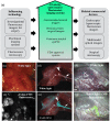
Fig. 1
Fig. 1
(a) Demonstrates the progression of systems in terms of their regulatory approval status along with parallel technologies. On the left, research-grade surgical fluorescence imagers, preclinical devices, and microscopy devices have served as contributors to the development of open-surgery fluorescence devices. On the right, the related commercial technologies, such as endoscopic imagers, multimodal imagers, and surgical microscopes, are specialized technologies that have greatly benefited from advancement in open-surgery fluorescence imagers. The central arrow illustrates the technological progression of imagers with FDA-approved systems at the trailing end, and customizable devices leading the technology development. (b)–(g) Show examples of surgical fields paired with white-light reflectance (up) and fluorescence images (bottom) shown for various applications.,, Panels (b) and (c) show white-light and fluorescence images, respectively, from the first in-human example of in situ ovarian cancer delineation using folate receptor-α targeted-fluorescent agent (reprinted by permission from Macmillan Publishers Ltd., Nature Medicine, copyright 2011). Panels (d) and (e) show white-light reflectance and white-light reflectance with pseudocolor fluorescence overlay, with Fluorescein-NP41 highlighting the peripheral nerves (reprinted by permission from Macmillan Publishers Ltd., Nature Biotechnology, copyright 2011). Panels (f) and (g) show ureters highlighted by methylene blue fluorescence, reprinted from Matsui et al., with permission from Elsevier.
Fig. 2
Fig. 2
Shows various white-light and fluorescence overlay schemes. (a) Shows a screenshot from the PerkinElmer Solaris imager during lymphatic imaging (image courtesy of PerkinElmer). The imaging windows display white light and the fluorescence overlaid on the white-light images simultaneously. User processing controls, such as ROI and display gain adjustments, are also available. (b) Shows the commonly used wavelength-based separation of collected light using dichroic mirrors and filters as seen in the Flare prototype system (reprinted from Troyan et al. with permission of Springer), Curadel Lab-Flare uses a similar setup with slightly different wavelength specifications on beam splitters and emission filters. (c) The modified Bayer filter is an alternative approach to perform simultaneous NIR detection, though this approach limits the active area for the fluorescence channel, reducing sensitivity. (d) Shows an example of simultaneous imaging and display of 700-nm (red) and 800-nm (green) fluorescence channels from the Flare prototype with the mesenteric lymph nodes highlighted by methylene blue (brackets) and a sentinel node (arrow) highlighted by ICG, reprinted from Troyan et al. with permission of Springer.
Fig. 3
Fig. 3
(a)–(d) The chemical formulas are shown along with absorption and emission spectra of the major FDA-approved fluorescent dyes such as fluorescein, PpIX, ICG, and methylene blue. (e) The normalized emission spectra are shown for common light sources used in surgery.
Fig. 4
Fig. 4
Plots of log10(Fluorophore Concentration) versus log10(Normalized Fluorescence) are shown for measurements of IRDye 800CW using FDA-approved imagers (a). Panel (b) shows IRDye 800CW measurements on imagers that are not approved for clinical use. Measurements from the LI-COR pearl impulse preclinical imager are shown for comparison. Note that large variability exists in dynamic range and detection sensitivity among FDA-approved imagers. Panel (c) shows similar plots for all systems with far-red emission imaging capability when IRDye 680RD samples were tested. A handful of imagers also performed imaging in the 700-nm channel, so IRDye 680RD was tested on these. In (d), the fitted slopes and the lower limit of detection are shown. *Fluoptics has two distinct imagers, Fluobeam700 and Fluobeam800, for imaging in the 700-nm and 800-nm emission bands, respectively. **The Li-COR Pearl imager was included simply as a standard of linearity and sensitivity achievable using an enclosed light-tight imager.
Fig. 5
Fig. 5
Images of the leading fluorescence guidance systems evaluated here, targeted for open surgery use, shown with relative approximate size comparison. The PerkinElmer Solaris, Curadel ResVet Lab-Flare, and SurgVision Explorer Air are not 510(k) cleared for human use, while the others are for ICG procedures. All have capability to image ICG in surgical trials, with differing levels of sensitivity and features. Images from left to right are from Solaris™ Open-Air Fluorescence Imaging System, Printed with permission, (c)2015-2016 PerkinElmer, Inc., all rights reserved; NOVADAQ Spy-Elite™, copyright 2016 Novadaq Technologies Inc.; Quest Spectrum™, copyright Quest Medical Imaging; Fluobeam(R), copyright 2016 Fluoptics; Hamamatsu PDE-Neo™; Lab-FLARE(R) Model R1 copyright CURADEL; Visionsense Iridium™, copyright Visionsense; SurgVision Explorer Air prototype, image courtesy of SurgVision. All images have been printed with permission from copyright holders.

References

    1. Rosenthal E. L., et al. , “Successful translation of fluorescence navigation during oncologic surgery: a consensus report,” J. Nucl. Med. 57(1), 144–150 (2016).10.2967/jnumed.115.158915 - DOI - PMC - PubMed
    1. Gioux S., Choi H. S., Frangioni J. V., “Image-guided surgery using invisible near-infrared light: fundamentals of clinical translation,” Mol. Imaging 9(5), 237–255 (2010). - PMC - PubMed
    1. Zhu B., Sevick-Muraca E. M., “A review of performance of near-infrared fluorescence imaging devices used in clinical studies,” Br. J. Radiol. 88(1045), 20140547 (2015).10.1259/bjr.20140547 - DOI - PMC - PubMed
    1. Nguyen Q. T., Tsien R. Y., “Fluorescence-guided surgery with live molecular navigation—a new cutting edge,” Nat. Rev. Cancer 13(9), 653–662 (2013).10.1038/nrc3566 - DOI - PMC - PubMed
    1. Orosco R. K., Tsien R. Y., Nguyen Q. T., “Fluorescence imaging in surgery,” IEEE Rev. Biomed. Eng. 6, 178–187 (2013).10.1109/RBME.2013.2240294 - DOI - PMC - PubMed

Publication types