

LXW7 ameliorates focal cerebral ischemia injury and attenuates inflammatory responses in activated microglia in rats
- PMID: 27533766
- PMCID: PMC4988477
- DOI: 10.1590/1414-431X20165287
LXW7 ameliorates focal cerebral ischemia injury and attenuates inflammatory responses in activated microglia in rats
Abstract
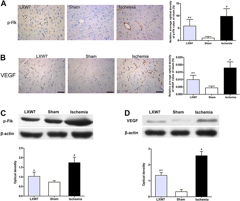
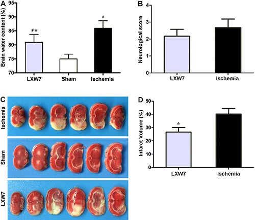
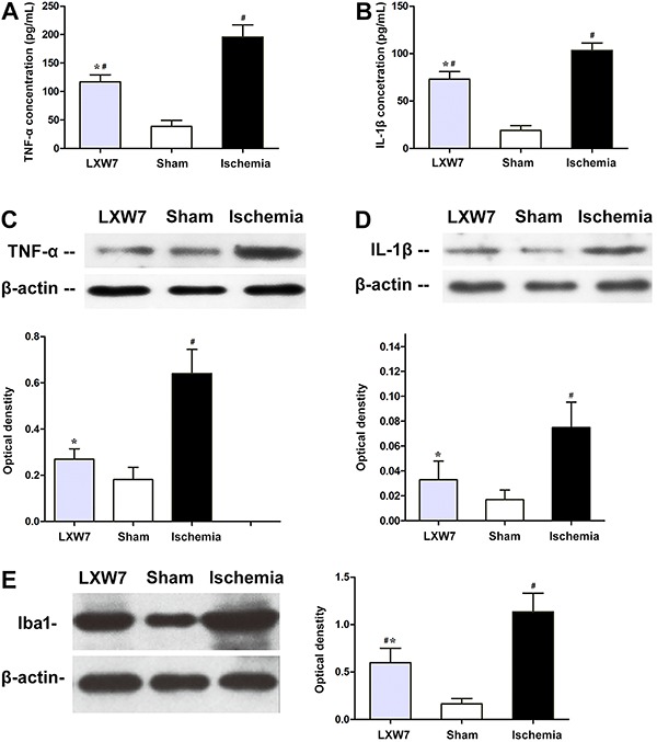
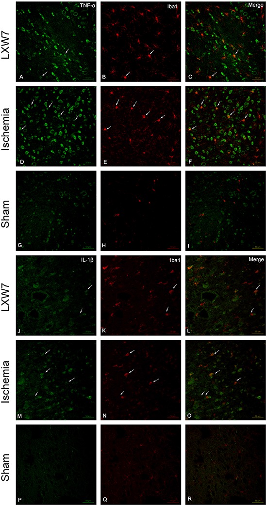
Inflammation plays a pivotal role in ischemic stroke, when activated microglia release excessive pro-inflammatory mediators. The inhibition of integrin αvβ3 improves outcomes in rat focal cerebral ischemia models. However, the mechanisms by which microglia are neuroprotective remain unclear. This study evaluated whether post-ischemic treatment with another integrin αvβ3 inhibitor, the cyclic arginine-glycine-aspartic acid (RGD) peptide-cGRGDdvc (LXW7), alleviates cerebral ischemic injury. The anti-inflammatory effect of LXW7 in activated microglia within rat focal cerebral ischemia models was examined. A total of 108 Sprague-Dawley rats (250-280 g) were subjected to middle cerebral artery occlusion (MCAO). After 2 h, the rats were given an intravenous injection of LXW7 (100 μg/kg) or phosphate-buffered saline (PBS). Neurological scores, infarct volumes, brain water content (BWC) and histology alterations were determined. The expressions of pro-inflammatory cytokines [tumor necrosis factor-alpha (TNF-α) and interleukin-1 beta (IL-1β)], and Iba1-positive activated microglia, within peri-ischemic brain tissue, were assessed with ELISA, western blot and immunofluorescence staining. Infarct volumes and BWC were significantly lower in LXW7-treated rats compared to those in the MCAO + PBS (control) group. The LXW7 treatment lowered the expression of pro-inflammatory cytokines. There was a reduction of Iba1-positive activated microglia, and the TNF-α and IL-1β expressions were attenuated. However, there was no difference in the Zea Longa scores between the ischemia and LXW7 groups. The results suggest that LXW7 protected against focal cerebral ischemia and attenuated inflammation in activated microglia. LXW7 may be neuroprotective during acute MCAO-induced brain damage and microglia-related neurodegenerative diseases.
Figures
Similar articles
-
Kellerin from Ferula sinkiangensis exerts neuroprotective effects after focal cerebral ischemia in rats by inhibiting microglia-mediated inflammatory responses.J Ethnopharmacol. 2021 Apr 6;269:113718. doi: 10.1016/j.jep.2020.113718. Epub 2020 Dec 25. J Ethnopharmacol. 2021. PMID: 33352239
-
Anti-inflammatory effects of Edaravone and Scutellarin in activated microglia in experimentally induced ischemia injury in rats and in BV-2 microglia.BMC Neurosci. 2014 Nov 22;15:125. doi: 10.1186/s12868-014-0125-3. BMC Neurosci. 2014. PMID: 25416145 Free PMC article.
-
Salvianolic Acids for Injection (SAFI) suppresses inflammatory responses in activated microglia to attenuate brain damage in focal cerebral ischemia.J Ethnopharmacol. 2017 Feb 23;198:194-204. doi: 10.1016/j.jep.2016.11.052. Epub 2017 Jan 10. J Ethnopharmacol. 2017. PMID: 28087473
-
Inflammation as a therapeutic target in acute ischemic stroke treatment.Curr Top Med Chem. 2009;9(14):1240-60. doi: 10.2174/156802609789869619. Curr Top Med Chem. 2009. PMID: 19849665 Review.
-
Neuroprotective profile of enoxaparin, a low molecular weight heparin, in in vivo models of cerebral ischemia or traumatic brain injury in rats: a review.CNS Drug Rev. 2002 Spring;8(1):1-30. doi: 10.1111/j.1527-3458.2002.tb00213.x. CNS Drug Rev. 2002. PMID: 12070524 Free PMC article. Review.
Cited by
-
Inflammatory Responses After Ischemic Stroke.Semin Immunopathol. 2022 Sep;44(5):625-648. doi: 10.1007/s00281-022-00943-7. Epub 2022 Jun 29. Semin Immunopathol. 2022. PMID: 35767089 Review.
-
Dietary teasaponin ameliorates alteration of gut microbiota and cognitive decline in diet-induced obese mice.Sci Rep. 2017 Sep 22;7(1):12203. doi: 10.1038/s41598-017-12156-2. Sci Rep. 2017. PMID: 28939875 Free PMC article.
-
Neuroprotective Effects of Chrysin Mediated by Estrogenic Receptors Following Cerebral Ischemia and Reperfusion in Male Rats.Basic Clin Neurosci. 2021 Jan-Feb;12(1):149-162. doi: 10.32598/bcn.12.1.2354.1. Epub 2021 Jan 1. Basic Clin Neurosci. 2021. PMID: 33995936 Free PMC article.
-
4R-cembranoid suppresses glial cells inflammatory phenotypes and prevents hippocampal neuronal loss in LPS-treated mice.J Neurosci Res. 2024 Apr;102(4):e25336. doi: 10.1002/jnr.25336. J Neurosci Res. 2024. PMID: 38656664 Free PMC article.
-
CeO2@PAA-LXW7 Attenuates LPS-Induced Inflammation in BV2 Microglia.Cell Mol Neurobiol. 2019 Nov;39(8):1125-1137. doi: 10.1007/s10571-019-00707-2. Epub 2019 Jun 29. Cell Mol Neurobiol. 2019. PMID: 31256326 Free PMC article.
References
MeSH terms
Substances
LinkOut - more resources
Full Text Sources
Other Literature Sources
