Review
. 2016 Nov;10(6):652-674.
doi: 10.1080/19336918.2016.1197478. Epub 2016 Aug 17.

The role of the semaphorins in cancer

Affiliations
Review

The role of the semaphorins in cancer

Gera Neufeld et al. Cell Adh Migr. 2016 Nov.

Abstract

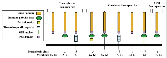
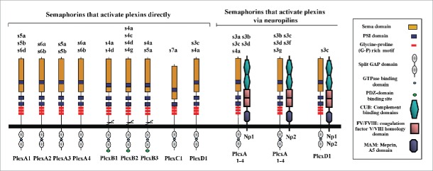
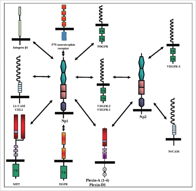
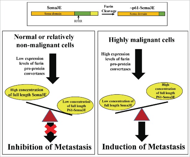
The semaphorins were initially characterized as axon guidance factors, but have subsequently been implicated also in the regulation of immune responses, angiogenesis, organ formation, and a variety of additional physiological and developmental functions. The semaphorin family contains more then 20 genes divided into 7 subfamilies, all of which contain the signature sema domain. The semaphorins transduce signals by binding to receptors belonging to the neuropilin or plexin families. Additional receptors which form complexes with these primary semaphorin receptors are also frequently involved in semaphorin signaling. Recent evidence suggests that semaphorins also fulfill important roles in the etiology of multiple forms of cancer. Some semaphorins have been found to function as bona-fide tumor suppressors and to inhibit tumor progression by various mechanisms while other semaphorins function as inducers and promoters of tumor progression.

Keywords: angiogenesis; cancer; lymphangiogenesis; semaphorins.


Figures

Figure 1.
Figure 1.
The structure of the semaphorins and their receptors. (A) The structural elements of semaphorin subclasses are shown. All feature the signature N-terminal sema domain. A conserved stretch of amino-acid residues near the C-terminal of the sema domain bears homology to the N-terminal of β-integrins and is designated as the PSI domain. Class-3 semaphorins are distinguished by a conserved basic domain at their c-termini. Class 4–7 semaphorins are membrane anchored. Class 5 semaphorins are distinguished by thrombospondin repeats. All the vertebrate semaphorins except for the class-5 and 6 semaphorins also contain an immunoglobulin like domain.
Figure 2.
Figure 2.
The interaction of the various vertebrate semaphorins with their neuropilins and plexin receptors. The different semaphorins are described using a 3 letter code in which the S stands for semaphorin, the number designates the subfamily, and the following letter designates the specific sub-family member. Thus, s3a stands for sema3A. The specific interactions between individual semaphorins and either single plexins or specific neuropilins are shown.
Figure 3.
Figure 3.
Cell surface receptors that form complexes with neuropilins. Cell surface receptors that form complexes with np1 include the vascular endothelial growth factor (VEGF-A) tyrosine-kinase receptors VEGFR-1 and VEGFR-2, the hepatocyte growth factor/scatter factor (HGF/SF) tyrosine-kinase receptor MET, and the epidermal growth factor (EGF) receptor EGFR. In addition np1 can associate with the Ig adhesion receptors L1-CAM and CHL1, with integrin-β1, with the 4 type-A plexins, and with plexin-D1. Np2 forms complexes with the 3 tyrosine-kinases receptors belonging to the VEGF receptor family, VEGFR-(1–3), with type-A plexins and plexin-D1, and with the Ig adhesion receptor NrCAM.
Figure 4.
Figure 4.
The dual role of sema3E in tumor progression. Like all class-3 semaphorins, sema3E contains a conserved cleavage site for furin like pro-protein convertases (FPPC) which when cleaved generates the active N-terminal fragment p61-Sema3E (Upper Panel). In contrast with uncleaved full length sema3E, P61-Sema3E can induce the association of the sema3E plexin-D1 receptor with ErbB2 on tumor cells, thus activating “in-trans” ErbB2 mediated signal transduction that promotes tumor metastasis. Full length sema3E has anti-metastatic and anti-angiogenic properties. However, FPPC expression is strongly up-regulated in malignant tumor cells causing most of the sema3E produced by the tumor cells to be cleaved to p61-Sema3E and as a result to promote rather than to inhibit tumor metastasis.

References

    1. Goodman CS, Kolodkin AL, Luo Y, Pueschel AW, Raper JA. Unified nomenclature for the semaphorins collapsins. Cell 1999; 97:551-2; PMID:10367884; http://dx.doi.org/10.1016/S0092-8674(00)80766-7 - DOI - PubMed
    1. Feiner L, Koppel AM, Kobayashi H, Raper JA. Secreted chick semaphorins bind recombinant neuropilin with similar affinities but bind different subsets of neurons in situ. Neuron 1997; 19:539-45; PMID:9331347; http://dx.doi.org/10.1016/S0896-6273(00)80370-0 - DOI - PubMed
    1. Love CA, Harlos K, Mavaddat N, Davis SJ, Stuart DI, Jones EY, Esnouf RM. The ligand-binding face of the semaphorins revealed by the high-resolution crystal structure of SEMA4D. Nat Struct Biol 2003; 10:843-8; PMID:12958590; http://dx.doi.org/10.1038/nsb977 - DOI - PubMed
    1. Antipenko A, Himanen JP, van Leyen K, Nardi-Dei V, Lesniak J, Barton WA, Rajashankar KR, Lu M, Hoemme C, Puschel AW, and others . Structure of the semaphorin-3A receptor binding module. Neuron 2003; 39:589-98; PMID:12925274; http://dx.doi.org/10.1016/S0896-6273(03)00502-6 - DOI - PubMed
    1. Liu H, Juo ZS, Shim AH, Focia PJ, Chen X, Garcia KC, He X. Structural basis of semaphorin-plexin recognition and viral mimicry from sema7A and A39R complexes with PlexinC1. Cell 2010; 142:749-61; PMID:20727575; http://dx.doi.org/10.1016/j.cell.2010.07.040 - DOI - PMC - PubMed

Publication types

LinkOut - more resources