. 2016 Jan 22;48(1):e205.
doi: 10.1038/emm.2015.100.

CB2 receptor activation prevents glial-derived neurotoxic mediator production, BBB leakage and peripheral immune cell infiltration and rescues dopamine neurons in the MPTP model of Parkinson's disease

Affiliations

CB2 receptor activation prevents glial-derived neurotoxic mediator production, BBB leakage and peripheral immune cell infiltration and rescues dopamine neurons in the MPTP model of Parkinson's disease

Young C Chung et al. Exp Mol Med. .

Abstract

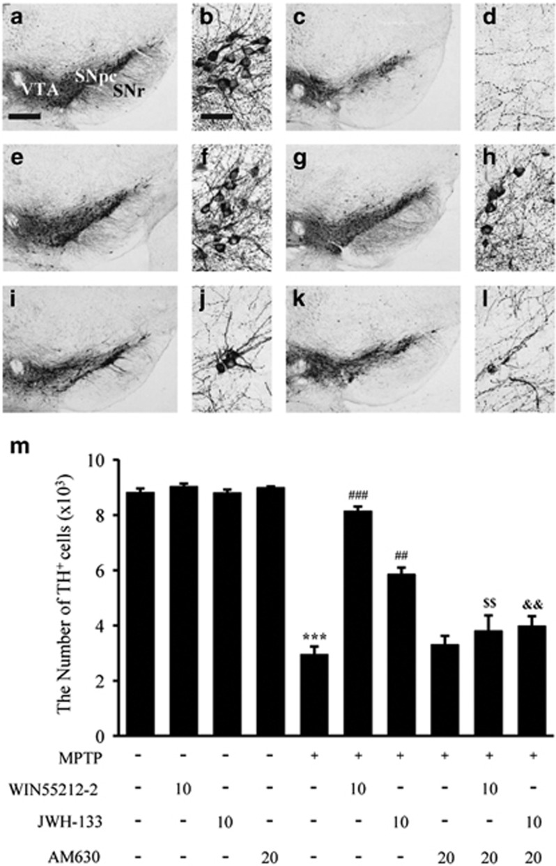
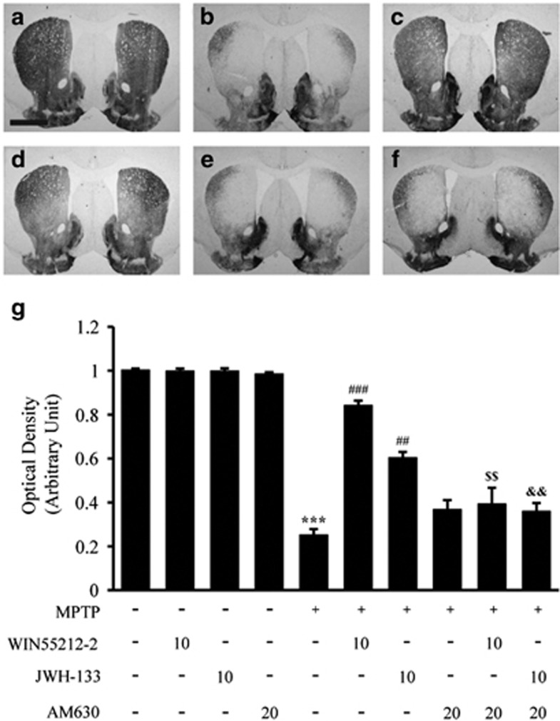
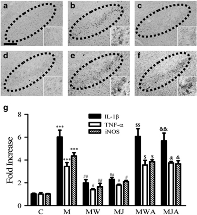
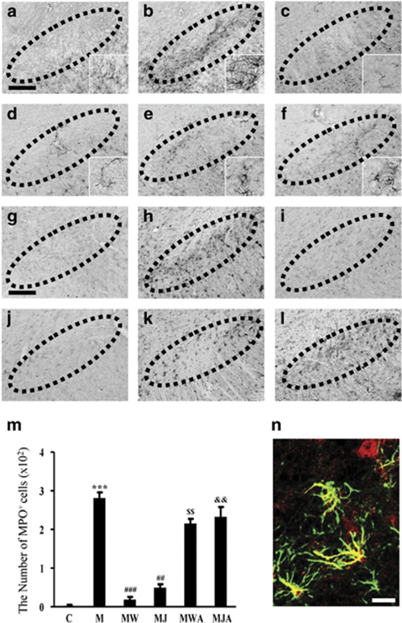
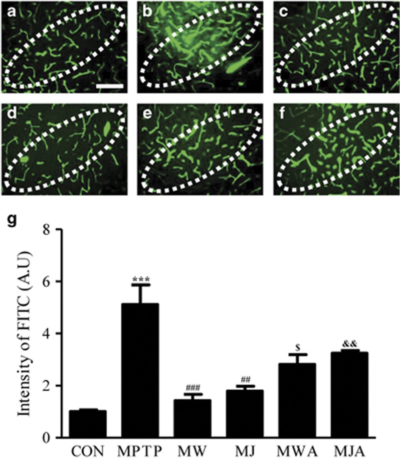
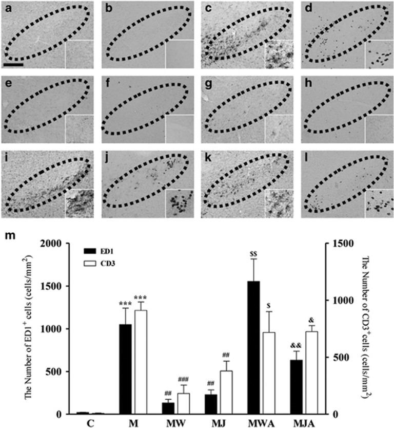
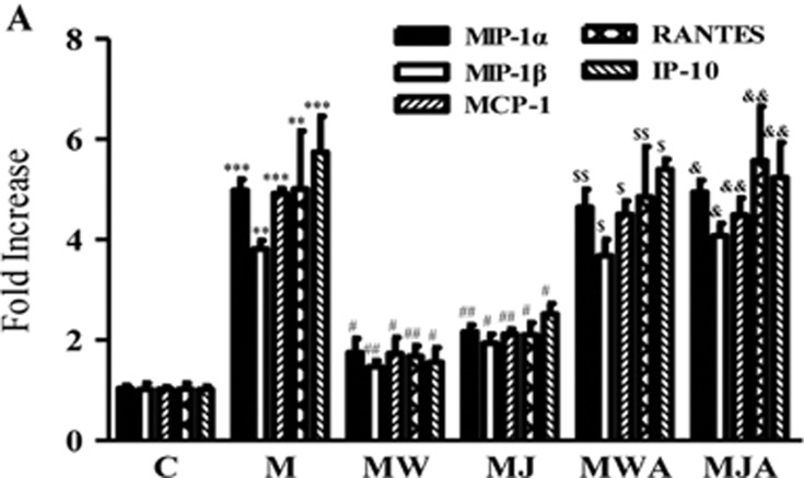
The cannabinoid (CB2) receptor type 2 has been proposed to prevent the degeneration of dopamine neurons in 1-methyl-4-phenyl-1,2,3,6-tetrahydropyridine (MPTP)-treated mice. However, the mechanisms underlying CB2 receptor-mediated neuroprotection in MPTP mice have not been elucidated. The mechanisms underlying CB2 receptor-mediated neuroprotection of dopamine neurons in the substantia nigra (SN) were evaluated in the MPTP mouse model of Parkinson's disease (PD) by immunohistochemical staining (tyrosine hydroxylase, macrophage Ag complex-1, glial fibrillary acidic protein, myeloperoxidase (MPO), and CD3 and CD68), real-time PCR and a fluorescein isothiocyanate-labeled albumin assay. Treatment with the selective CB2 receptor agonist JWH-133 (10 μg kg(-1), intraperitoneal (i.p.)) prevented MPTP-induced degeneration of dopamine neurons in the SN and of their fibers in the striatum. This JWH-133-mediated neuroprotection was associated with the suppression of blood-brain barrier (BBB) damage, astroglial MPO expression, infiltration of peripheral immune cells and production of inducible nitric oxide synthase, proinflammatory cytokines and chemokines by activated microglia. The effects of JWH-133 were mimicked by the non-selective cannabinoid receptor WIN55,212 (10 μg kg(-1), i.p.). The observed neuroprotection and inhibition of glial-mediated neurotoxic events were reversed upon treatment with the selective CB2 receptor antagonist AM630, confirming the involvement of the CB2 receptor. Our results suggest that targeting the cannabinoid system may be beneficial for the treatment of neurodegenerative diseases, such as PD, that are associated with glial activation, BBB disruption and peripheral immune cell infiltration.


Figures

Figure 1
Figure 1
The CB2 receptor protects nigral dopamine neurons from MPTP neurotoxicity in vivo. Animals that received PBS as a control (a, b); MPTP (c, d); MPTP and WIN55,212-2 (e, f); MPTP and JWH-133 (g, h); MPTP, WIN55,212-2 and AM630 (i, j); or MPTP, JWH-133 and AM630 (k, l) were killed 7 days after the last MPTP injection. Brain tissues were cut, and SN tissues were immunostained with an antibody to TH to label dopamine neurons. (b, d, f, h, j and l) higher magnifications of (a, c, e, g, i and k), respectively. (m) The numbers of TH+ neurons in the SN were counted. Five to seven animals were used for each experimental group. *P<0.001 significantly different from controls. ##P<0.01 and ###P<0.001 significantly different from MPTP. $$P<0.01 significantly different from MPTP and WIN55,212-2; and &&P<0.01 significantly different from MPTP and JWH-133 (ANOVA and Student–Neuman–Keuls analysis). SNpc, substantia nigra pars compacta; SNr, substantia nigra pars reticulata; VTA, ventral tegmental area; Scale bars: a, c, e, g, i and k, 300 μm; b, d, f, h, j and l, 50 μm.
Figure 2
Figure 2
The CB2 receptor protects striatal dopamine fibers from MPTP neurotoxicity in vivo. The STR tissues obtained from the same animals used in Figure 1 were immunostained with a TH antibody to label dopamine fibers. Control (a); MPTP (b); MPTP and WIN55,212-2 (c); MPTP and JWH-133 (d); MPTP, WIN55,212-2 and AM630 (e); and MPTP, JWH-133 and AM630 (f). (g) The optical density of TH+ fibers in the STR. ***P<0.001 significantly different from controls. ##P<0.01 and ###P<0.001 significantly different from MPTP. $$P<0.01 significantly different from MPTP and WIN55,212-2; and &&P<0.01 significantly different from MPTP and JWH-133 (ANOVA and Student–Neuman–Keuls analysis). Scale bars: a–f, 500 μm.
Figure 3
Figure 3
The CB2 receptor inhibits microglial activation and the expression of proinflammatory cytokines in the SN in vivo. Animals that received vehicle as a control (a); MPTP (b); MPTP and WIN55,212-2 (c); MPTP and JWH-133 (d); MPTP, WIN55,212-2 and AM630 (e); or MPTP, JWH-133 and AM630 (f) were killed 3 days after the last MPTP injection. Brain tissues were cut, and SN tissues were immunostained with an antibody for MAC-1 to label microglia (af). Insets show higher magnifications of a–f. Dotted lines indicate the SNpc. Scale bars: a–f, 200 μm. (g) Real-time PCR showing mRNA expression of proinflammatory mediators in the SN. Total RNA was isolated from the bilateral SN for real-time PCR 1 day after treatment with vehicle as a control (C); MPTP only (M); MPTP and WIN55,212-2 (MW); MPTP and JWH-133 (MJ); MPTP, WIN55,212-2 and AM630 (MWA); or MPTP, JWH-133 and AM630 (MJA). The CB2 receptor dramatically attenuated MPTP-induced expression of proinflammatory cytokines, including IL-1β, TNF-α and iNOS. The results represent the mean±s.e.m. of three to four separate experiments. ***P<0.001 significantly different from C. #P<0.05 and ##P<0.01, significantly different from M. $P<0.05 and $$P<0.01 significantly different from MW. &P<0.05 and &&P<0.01 significantly different from MJ (ANOVA and Student–Neuman–Keuls analysis).
Figure 4
Figure 4
The CB2 receptor attenuates MPTP-induced astroglial activation and expression of myeloperoxidase (MPO) in the SN in vivo. The SN tissues obtained from the same animals used in Figure 3 were immunostained with a GFAP antibody to label astrocytes (af) and with an MPO antibody to evaluate MPO immunoreactivity (g–l). Animals that received PBS as a control (a, g); MPTP (b, h); MPTP and WIN55,212-2 (c, i); MPTP and JWH-133 (d, j); MPTP, WIN55,212-2 and AM630 (e, k); or MPTP, JWH-133 and AM630 (f, l) were killed 3 days after the last MPTP injection. Insets show higher magnifications of a–l. Dotted lines indicate the SNpc. (m) The number of MPO-positive cells in the SN was counted. Four to five animals were used for each experimental group. C, control; M, MPTP; MW, MPTP and WIN55,212-2; MJ, MPTP and JWH-133; MWA, MPTP and WIN55,212-2 and AM630; MJA, MPTP and JWH-133 and AM630. ***P<0.001 significantly different from controls; ##P<0.01 and ###P<0.001 significantly different from MPTP only; $$P<0.01, significantly different from MW. &&P<0.01 significantly different from MJ (ANOVA and Student–Neuman–Keuls analysis). (n) Localization of MPO immunoreactivity in GFAP+ activated astrocytes in the MPTP-treated SN. The SN tissues obtained from the same animals used in b were simultaneously immunostained with antibodies against MPO and GFAP, as a marker for astrocytes. Scale bars: a–l, 200 μm.
Figure 5
Figure 5
The CB2 receptor prevents MPTP-induced BBB disruption in the SN in vivo. At 3 days after the injection of vehicle as a control or of MPTP in the absence or presence of WIN55,212-2, JWH-133 and/or AM630, FITC-labeled albumin was administered to detect brain vascular permeability. Control (a); MPTP (b); MPTP and WIN55,212-2 (c); MPTP and JWH-133 (d); MPTP, WIN55,212-2 and AM630 (e); and MPTP, JWH-133 and AM630 (f). Dotted lines indicate the SNpc. Scale bars: a–l, 100 μm. (g) Bars represent the FITC-labeled-albumin-positive area in the SNpc. Four or five animals were used for each experimental group. Actual values are normalized by the value for the PBS-injected control. CON, control; M, MPTP; MW, MPTP and WIN55,212-2; MJ, MPTP and JWH-133; MWA, MPTP and WIN55,212-2 and AM630; MJA, MPTP and JWH-133 and AM630. ***P<0.001 significantly different from controls; ##P<0.01 and ###P<0.001 significantly different from MPTP only; $P<0.05 significantly different from MW; and &&P<0.01 significantly different from MJ (ANOVA and Student–Neuman–Keuls analysis).
Figure 6
Figure 6
The CB2 receptor inhibits MPTP-induced infiltration of peripheral immune cells in the SN in vivo. The SN tissues obtained from the same animals used in Figure 5 were immunostained with an ED1 antibody to label phagocytotic macrophages and microglia (a, c, e, g, i, and k) and with a CD3 antibody to label T cells (b, d, f, h, j, and l). Animals that received PBS as a control (a, b); MPTP (c, d); MPTP and WIN55,212-2 (e, f); MPTP and JWH-133 (g, h); MPTP, WIN55,212-2 and AM630 (i, j); or MPTP, JWH-133 and AM630 (k, l) were killed 3 days after the last MPTP injection. Insets show higher magnifications of a–l. Dotted lines indicate the SNpc. Scale bars: a–l, 200 μm. (m) The number of CD3+ (white bars) or ED1+ (black bars) cells in the SN were counted. Four to five animals were used for each experimental group. C, control; M, MPTP; MW, MPTP and WIN55,212-2; MJ, MPTP and JWH-133; MWA, MPTP and WIN55,212-2 and AM630; MJA, MPTP and JWH-133 and AM630. ***P<0.01 significantly different from controls; ##P<0.01 and ###P<0.001 significantly different from MPTP only; $P<0.05 and $$P<0.01 significantly different from MW; &P<0.05 and &&P<0.01 significantly different from MJ (ANOVA and Student–Neuman–Keuls analysis).
Figure 7
Figure 7
The CB2 receptor inhibits MPTP-induced expression of chemokines in the SN in vivo. (a) Real-time PCR showing mRNA expression of chemokines in the SN. Animals that received vehicle or MPTP in the absence or presence of WIN55,212-2, JWH-133 and/or AM630 were killed 2 days later for RT-PCR analysis. The CB2 receptor dramatically reduced the MPTP-induced expression of chemokines, including MIP-1α, MIP-1β, MCP-1, RANTES and IP-10. Graphic representation of the mean±s.e.m. of three to four samples. C, control; M, MPTP; MW, MPTP and WIN55,212-2; MJ, MPTP and JWH-133; MWA, MPTP and WIN55,212-2 and AM630; MJA, MPTP and JWH-133 and AM630. **P<0.01 and ***P<0.001 significantly different from C. #P<0.05 and ##P<0.01 significantly different from M. $P<0.01 and $$P<0.05 significantly different from MW. &P<0.05 and &&P<0.01 significantly different from MJ (ANOVA and Student–Neuman–Keuls analysis).

References

    1. Savitt JM, Dawson VL, Dawson TM. Diagnosis and treatment of Parkinson disease: molecules to medicine. J Clin Invest 2006; 116: 1744–1754. - PMC - PubMed
    1. Appel SH. CD4+ T cells mediate cytotoxicity in neurodegenerative diseases. J Clin Invest 2009; 119: 13–15. - PMC - PubMed
    1. Block ML, Zecca L, Hong JS. Microglia-mediated neurotoxicity: uncovering the molecular mechanisms. Nat Rev Neurosci 2007; 8: 57–69. - PubMed
    1. Chung YC, Ko HW, Bok E, Park ES, Huh SH, Nam JH et al. The role of neuroinflammation on the pathogenesis of Parkinson's disease. BMB Rep 2010; 43: 225–232. - PubMed
    1. Hwang O. Role of oxidative stress in Parkinson's disease. Exp Neurobiol 2013; 22: 11–17. - PMC - PubMed

Publication types

MeSH terms

Substances