

Mucinous Carcinoma with Neuroendocrine Differentiation of Salivary Gland Origin
- PMID: 27534564
- PMCID: PMC5429273
- DOI: 10.1007/s12105-016-0750-5
Mucinous Carcinoma with Neuroendocrine Differentiation of Salivary Gland Origin
Abstract
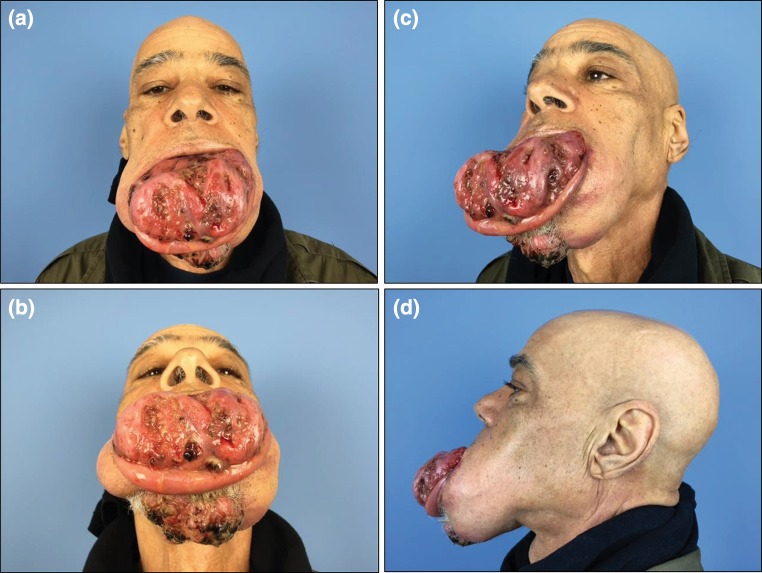
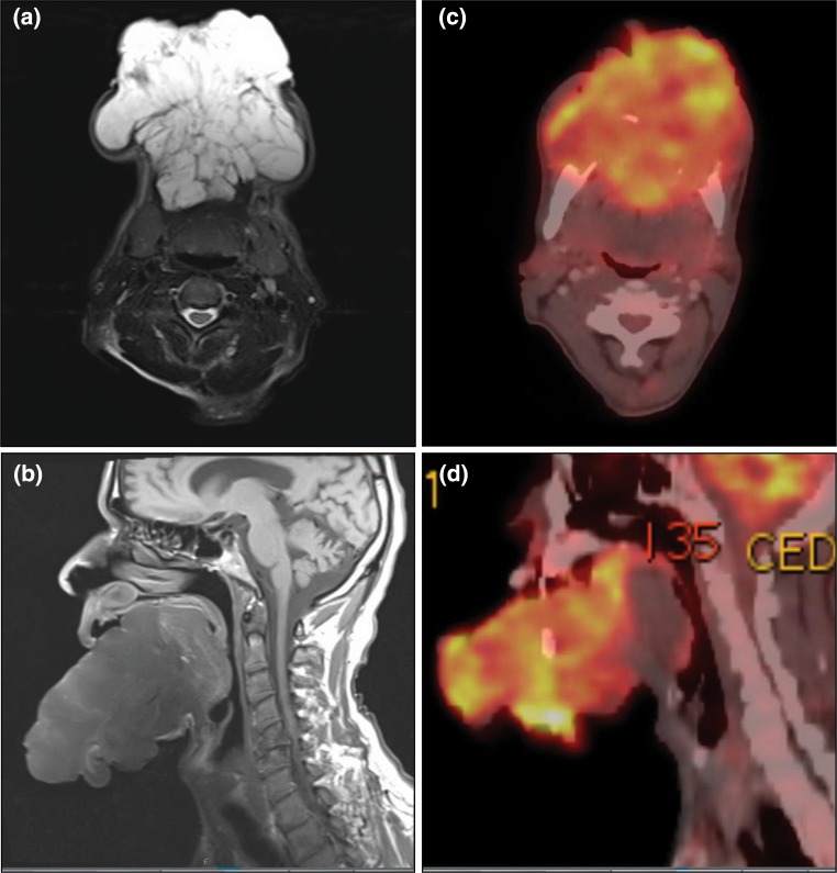
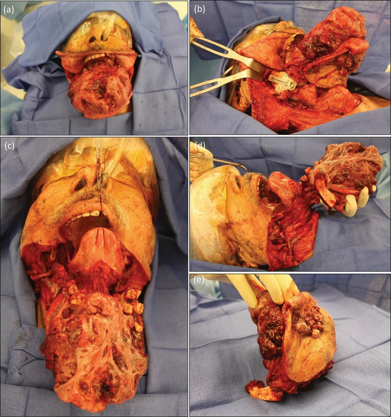
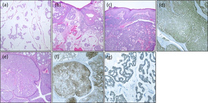
Primary mucinous adenocarcinomas of the salivary gland are rare malignancies defined by aggregates of epithelial cells suspended in large pools of extracellular mucin. We report a case of a giant mucinous adenocarcinoma of salivary gland origin, with low-grade cytoarchitectural features and neuroendocrine differentiation arising in the submental region. Grossly, the tumor measured 12.5 × 13.4 × 8.2 cm and replaced the bone and soft tissues of the anterior oral cavity. Microscopically, the neoplasm was composed of large extracellular pools of mucin, which contained papillary and acinar aggregates, and small nodules of ductal type epithelium with minimal nuclear enlargement, powdery chromatin and little pleomorphism. The nodules comprised 20 % of the tumor and showed morphologic and immunohistochemical evidence of neuroendocrine differentiation. Examination revealed histologic features comparable to mammary gland analogues in mucin predominance, ductal type morphology, expression of estrogen and progesterone receptors, and GATA-3 positivity. This is the first case reported of mucin-rich carcinoma of salivary gland origin exhibiting neuroendocrine differentiation.
Keywords: Head and neck cancer; Mucin-rich carcinoma; Mucinous adenocarcinoma; Neuroendocrine differentiation; Reconstructive surgery; Salivary cancer; Salivary duct carcinoma.
Conflict of interest statement
No conflict of interest to disclose.
Figures
Similar articles
-
Mucin-rich variant of salivary duct carcinoma: a clinicopathologic and immunohistochemical study of four cases.Am J Surg Pathol. 2003 Aug;27(8):1070-9. doi: 10.1097/00000478-200308000-00004. Am J Surg Pathol. 2003. PMID: 12883239
-
Mucinous adenocarcinoma of probable minor salivary gland origin.Oral Surg Oral Med Oral Pathol Oral Radiol Endod. 2002 Dec;94(6):738-40. doi: 10.1067/moe.2002.126698. Oral Surg Oral Med Oral Pathol Oral Radiol Endod. 2002. PMID: 12464900
-
Endocrine mucin-producing sweat gland carcinoma: twelve new cases suggest that it is a precursor of some invasive mucinous carcinomas.Am J Surg Pathol. 2005 Oct;29(10):1330-9. doi: 10.1097/01.pas.0000170348.40057.60. Am J Surg Pathol. 2005. PMID: 16160476
-
Mucin-rich salivary duct carcinoma with signet-ring cell feature ex pleomorphic adenoma of the submandibular gland: a case report of an unusual histology with immunohistochemical analysis and review of the literature.Med Mol Morphol. 2012 Dec;45(1):45-52. doi: 10.1007/s00795-011-0554-3. Epub 2012 Mar 20. Med Mol Morphol. 2012. PMID: 22431183 Review.
-
Newly Described Entities in Salivary Gland Pathology.Am J Surg Pathol. 2017 Aug;41(8):e33-e47. doi: 10.1097/PAS.0000000000000883. Am J Surg Pathol. 2017. PMID: 28614209 Review.
References
-
- World Health Organization Classification of Tumours . Pathology and genetics of head and neck tumours. Lyon: IARC Press; 2005.
-
- Ellis GL, Auclair PL. Tumors of the salivary glands, Atlas of Tumor Pathology. 3. Washington DC: Armed Forces Institute of Pathology; 1996.
-
- Brandwein-Gensler M, et al. Low-grade salivary duct carcinoma: description of 16 cases. Am J Surg Pathol. 2004;28(8):1040–1044. doi: 10.1097/01.pas.0000128662.66321.be. - DOI - PubMed
Publication types
MeSH terms
LinkOut - more resources
Full Text Sources
Other Literature Sources
Medical
