Review
. 2016 Aug;6(8):160165.
doi: 10.1098/rsob.160165.

Human leukocyte Antigen-DM polymorphisms in autoimmune diseases

Affiliations
Review

Human leukocyte Antigen-DM polymorphisms in autoimmune diseases

Miguel Alvaro-Benito et al. Open Biol. 2016 Aug.

Abstract

Classical MHC class II (MHCII) proteins present peptides for CD4(+) T-cell surveillance and are by far the most prominent risk factor for a number of autoimmune disorders. To date, many studies have shown that this link between particular MHCII alleles and disease depends on the MHCII's particular ability to bind and present certain peptides in specific physiological contexts. However, less attention has been paid to the non-classical MHCII molecule human leucocyte antigen-DM, which catalyses peptide exchange on classical MHCII proteins acting as a peptide editor. DM function impacts the presentation of both antigenic peptides in the periphery and key self-peptides during T-cell development in the thymus. In this way, DM activity directly influences the response to pathogens, as well as mechanisms of self-tolerance acquisition. While decreased DM editing of particular MHCII proteins has been proposed to be related to autoimmune disorders, no experimental evidence for different DM catalytic properties had been reported until recently. Biochemical and structural investigations, together with new animal models of loss of DM activity, have provided an attractive foundation for identifying different catalytic efficiencies for DM allotypes. Here, we revisit the current knowledge of DM function and discuss how DM function may impart autoimmunity at the organism level.

Keywords: antigen presentation; autoimmunity; human leucocyte antigen-DM; major histocompatibility complex of class II; peptidome; polymorphism.


Figures

Figure 1.
Figure 1.
Genomic organization of the MHCII locus and the link between the MHCII structure and disease susceptibility. (a) Overview of the genetic organization of the MHCII locus in human and in mouse, and numbers of natural variants of the different MHCII proteins encoded by the MHCII locus in humans. Functional genes are shown as filled boxes and dashed light blue boxes represent pseudogenes which are not translated as functional proteins (e.g. DPB2 and DPA2 in humans and I-Pa and I-Pb in mouse). TAP and LMP genes belong to MHCI genes but are located within the MHCII locus. A yellow background indicates genes encoding classical MHCII proteins and a red background indicates the genes encoding for non-classical MHCII proteins. A red cross indicates a potential hotspot for recombination. (b) Example of three-dimensional fold of classical and non-classical MHCII proteins. The ectodomains are depicted in a cartoon representation and the classical MHCII protein HLA-DR1 (PDB: 2fse) and the non-classical MHCII proteins HLA-DM (PDB: 2bc4, bottom) and HLA-DO (PDB: 4iop, from which HLA-DM has been removed) are compared. Note that while the peptide has been removed from HLA-DR1, the non-classical MHCII proteins do not bind peptides. The ectodomains are shown with the alpha subunit in green and the beta subunit in cyan. The α1, β1 and α2 and β2 domains are indicated. The most important structural differences between classical and non-classical MHCII proteins are located in the α1 and β1 domains, and can be appreciated in the top view (bottom) (c) Structural details of MHCII proteins relevant for disease; polymorphic residues are depicted as spheres: HLA-DP (3lqz), HLA-DQ (1jk8) and HLA-DR (2fse). Peptides presented by these molecules and found in the pdb files have been removed to facilitate visualization. The epitopes are indicated in each structure. (d) The same proteins as in (c) are shown, and the polymorphisms associated with disease are represented as spheres. Peptides presented by these molecules are depicted in stick representations. The P1 and P9 pockets are highlighted with a yellow and a blue circle, respectively.
Figure 2.
Figure 2.
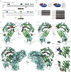
HLA-DM genes and proteins. (a) Structure for the HLA-DMA and HLA-DMB genes. ENSEMBL accession numbers are provided as well as the gene size in kbp. Exons are shown in boxes, in which the polymorphic regions are highlighted in yellow. Black lines show sequenced regions in different alleles. (b) HLA-DMA (i) and DMB (ii) allelic frequencies shown as pie charts. (c). Protein chains for DMA and DMB allelic variants are shown with the polymorphic residues highlighted. The most abundant and characterized DMA and DMB alleles (DMA*0101 and DMB*0101, respectively) are shown on top, and the polymorphic residues are shown in yellow. Any change on the amino acid level is shown in red. (d) Cartoon representation of the three-dimensional structure of HLA-DM showing the polymorphic residues as spheres. (e) Three-dimensional structure of HLA-DM (PDB: 2bc4), where residues identified in other studies as affecting enzymatic activity in B-cell lines are shown as spheres. (f) Positioning of the polymorphic residues of HLA-DM in the context of the HLA-DM–HLA-DR1 three-dimensional structure (PDB: 4fqx). (g) Zoom-in of the polymorphic residue DMα184R showing the structural rearrangement between the HLA-DM-free (i) and in complex with HLA-DR1 (ii). The three-dimensional structures are shown as cartoons and the DMαR184 (variable residue in DMA alleles) and the DMαR98 (important for the DM–DR interaction in vitro) residues are shown as spheres.
Figure 3.
Figure 3.
Known cellular functions of HLA-DM and consequences of total or partial loss of DM function. MHCII molecules are shown as black ‘V's. CLIP peptides are shown as black triangles. Antigenic peptides are shown as red triangles (lightness represents the affinity for the MHCII, with dark representing high affinity). HLA-DM is shown as a large triangle, and the colour correlates with its catalytic activity (cyan: normal; yellow: impaired) and in the case of total loss of activity a red ‘X’ is shown. (a) Peptide editor function of HLA-DM. DM function leads to the formation of highly stable peptide–MHCII complexes. MHCII molecules in complex with CLIP encounter antigenic peptides of different affinities. Under conditions of normal HLA-DM activity (yellow triangle, upper panel), CLIP is exchanged by higher affinity antigenic peptides resulting in stable peptide MHCII complexes. In the case of no HLA-DM activity, CLIP will mostly remain associated with MHCII molecules (lower panel). In the case of HLA-DM impaired activity not all CLIP will be dissociated from MHCII proteins. The arrows represent the relevant antigen processing conditions for each antigen (unfolding and/or proteolysis). (b) Chaperone function of HLA-DM. The chaperone function of DM rescues empty MHCII molecules from degradation. In the absence of HLA-DM activity, MHCII molecules with low affinity for CLIP collapse and are unable to present peptides. In conditions of normal HLA-DM activity, a large pool of the empty MHCII molecules will be rescued (upper panel). In the total absence of HLA-DM function (lower panel), the pool of rescued MHCII proteins will be lower (and therefore the total levels of MHCII proteins will also be lower). Impaired catalytic HLA-DM activity would lead to an intermediate situation. (c) HLA-DM activity in a cellular context and its impact at the peptidome level. HLA-DM and MHCII proteins (as nonameric complexes Ii3MHCII3) assemble in the ER and traffic through the Golgi. HLA-DM favours peptide exchange and acts as a chaperone mostly in MIIC (late endosomal compartments/MHCII compartments). HLA-DM is supposed to be more effective in MIIC compartments where antigens, mostly internalized, are degraded by cellular proteases (represented as scissors). The peptidome associated with MHCII proteins is represented with the same symbols as in (a,b), and the sizes indicate the relative amount of each complex. Normal DM expression levels result in a concrete peptidome (left, mostly composed by high affinity antigenic peptides) which is substantially altered in the absence of DM activity (right, where CLIP is the most abundant peptide and the presence of non-receptive MHCII molecules is higher). A partial loss of HLA-DM activity (due to catalytic impairment, centre) is expected to have consequences on the MHCII-associated peptidomes.
Figure 4.
Figure 4.
Expected impact of low DM editing activity on the MHCII-associated peptidome and its consequences for tolerance to self and peripheral presentation of self- and foreign antigens. (a) Overview of how HLA-DM affects the peptidomes presented by MHCII proteins with high affinity (black ‘V's) or low affinity (grey ‘V's) for CLIP (small black inverted triangles) in the presence of normal (upper panel, large blue inverted triangle), catalytically impaired/low (intermediate, large yellow inverted triangle) or no DM (red ‘X’) activities. Low DM activity (large yellow inverted triangle) could represent defects at transcriptional, catalytic or modulation levels. Antigenic sources contain a number of potential binding epitopes with different affinities for the particular MHCII. DM function favours the binding of high-affinity epitopes. The arrows represent the relevant antigen processing conditions for each antigen (unfolding and/or proteolysis). (b) The MHCII peptidome impacts a number of different immune processes related to T-cell development, tolerance acquisition and adaptive immune responses to pathogens. For some of the different DM editing conditions described in (a), it has been shown how DM loss affects most of these processes for both high and low MHCII affinities for CLIP alleles. T-cell numbers with respect to the normal DM editing levels are reduced when the arrows point up or increased when they point down. Although there are expected changes in the peptidome presented by MHCII proteins, it is difficult to predict the consequences in terms of T-cell numbers.

Similar articles

Cited by

References

    1. Rai E, Wakeland EK. 2011. Genetic predisposition to autoimmunity: what have we learned? Semin. Immunol. 23, 67–83. (doi:10.1016/j.smim.2011.01.015) - DOI - PubMed
    1. Wucherpfennig KW, Sethi D. 2011. T cell receptor recognition of self and foreign antigens in the induction of autoimmunity. Semin. Immunol. 23, 84–91. (doi:10.1016/j.smim.2011.01.007) - DOI - PMC - PubMed
    1. Shiina T, Hosomochi K, Kulski JK. 2009. The HLA genomic loci map: expression, interaction, diversity and disease. J. Hum. Genet. 54, 15–39. (doi:10.1038/jhg.2008.5) - DOI - PubMed
    1. Schlosstein L, Terasaki PI, Bluestone R, Pearson CM. 1973. High association of an HL-A antigen, W27, with ankylosing spondylitis. N. Engl. J. Med. 288, 704–706. (doi:10.1056/NEJM197304052881403) - DOI - PubMed
    1. Fernando MM, Stevens CR, Walsh EC, De Jager PL, Goyette P, Plenge RM, Vyse TJ, Rioux JD. 2008. Defining the role of the MHC in autoimmunity: a review and pooled analysis. PLoS Genet. 4, e1000024 (doi:10.1371/journal.pgen.1000024) - DOI - PMC - PubMed

Publication types