

Endovascular ischemic stroke models of adult rhesus monkeys: a comparison of two endovascular methods
- PMID: 27534985
- PMCID: PMC4989171
- DOI: 10.1038/srep31608
Endovascular ischemic stroke models of adult rhesus monkeys: a comparison of two endovascular methods
Abstract
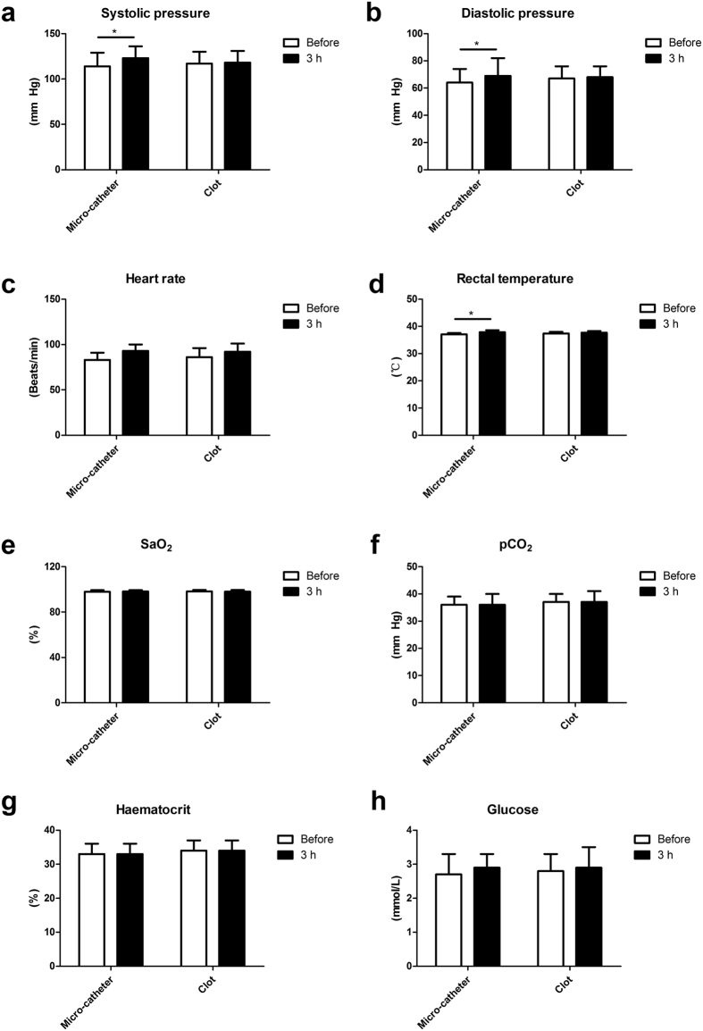
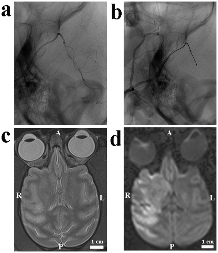
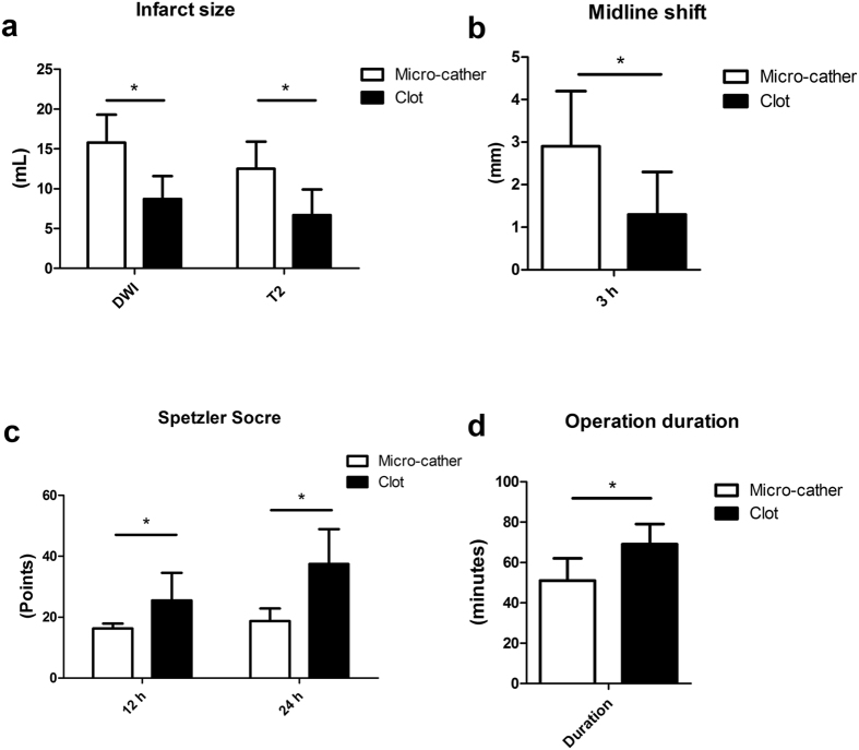
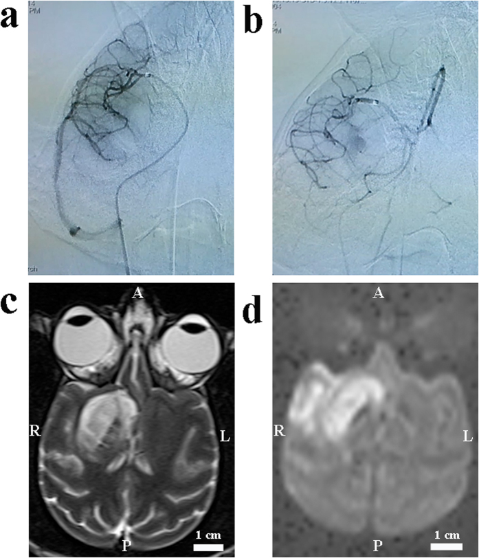
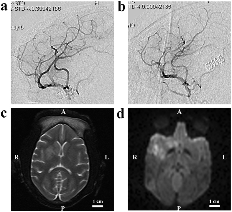
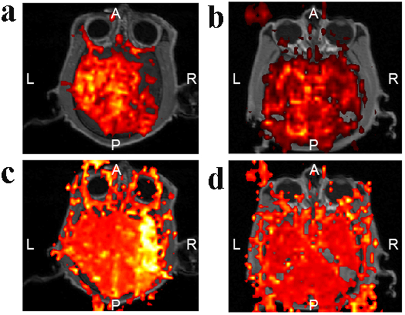
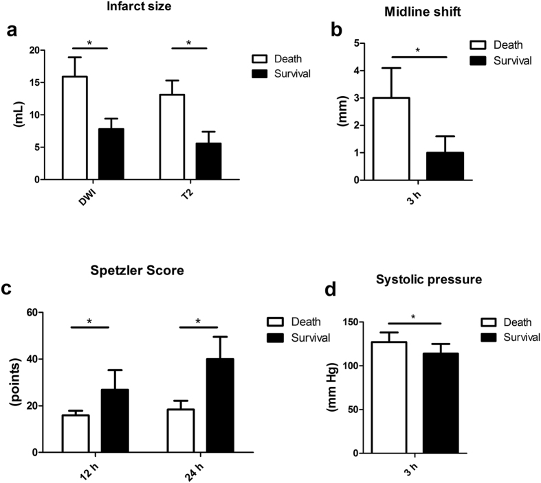
To further investigate and improve upon current stroke models in nonhuman primates, infarct size, neurologic function and survival were evaluated in two endovascular ischemic models in sixteen rhesus monkeys. The first method utilized a micro-catheter or an inflatable balloon to occlude the M1 segment in six monkeys. In the second model, an autologous clot was injected via a micro-catheter into the M1 segment in ten monkeys. MRI scanning was performed on all monkeys both at baseline and 3 hours after the onset of ischemia. Spetzler neurologic functions were assessed post-operatively, and selective perfusion deficits were confirmed by DSA and MRI in all monkeys. Animals undergoing micro-catheter or balloon occlusion demonstrated more profound hemiparesis, larger infarct sizes, lower Spetzler neurologic scores and increased mortality compared to the thrombus occlusion group. In animals injected with the clot, there was no evidence of dissolution, and the thrombus was either near the injection site (M1) or flushed into the superior division of the MCA (M2). All animals survived the M2 occlusion. M1 occlusion with thrombus generated 50% mortality. This study highlighted clinically important differences in these two models, providing a platform for further study of a translational thromboembolic model of acute ischemic stroke.
Figures
Similar articles
-
An Enhanced Model of Middle Cerebral Artery Occlusion in Nonhuman Primates Using an Endovascular Trapping Technique.AJNR Am J Neuroradiol. 2015 Dec;36(12):2354-9. doi: 10.3174/ajnr.A4448. Epub 2015 Sep 17. AJNR Am J Neuroradiol. 2015. PMID: 26381560 Free PMC article.
-
Temporal evolution of ischemic lesions in nonhuman primates: a diffusion and perfusion MRI study.PLoS One. 2015 Feb 6;10(2):e0117290. doi: 10.1371/journal.pone.0117290. eCollection 2015. PLoS One. 2015. PMID: 25659092 Free PMC article.
-
An ischemic stroke model of nonhuman primates for remote lesion studies: a behavioral and neuroimaging investigation.Restor Neurol Neurosci. 2015;33(2):131-42. doi: 10.3233/RNN-140440. Restor Neurol Neurosci. 2015. PMID: 25588459
-
A clinically relevant model of focal embolic cerebral ischemia by thrombus and thrombolysis in rhesus monkeys.Nat Protoc. 2022 Sep;17(9):2054-2084. doi: 10.1038/s41596-022-00707-5. Epub 2022 Jun 27. Nat Protoc. 2022. PMID: 35760857 Review.
-
A novel mouse model of thromboembolic stroke.J Neurosci Methods. 2015 Dec 30;256:203-11. doi: 10.1016/j.jneumeth.2015.09.013. Epub 2015 Sep 18. J Neurosci Methods. 2015. PMID: 26386284 Free PMC article. Review.
Cited by
-
CCL2 (C-C Motif Chemokine Ligand 2) Biomarker Responses in Central Versus Peripheral Compartments After Focal Cerebral Ischemia.Stroke. 2021 Nov;52(11):3670-3679. doi: 10.1161/STROKEAHA.120.032782. Epub 2021 Sep 30. Stroke. 2021. PMID: 34587791 Free PMC article.
-
A non-human primate model of stroke reproducing endovascular thrombectomy and allowing long-term imaging and neurological read-outs.J Cereb Blood Flow Metab. 2021 Apr;41(4):745-760. doi: 10.1177/0271678X20921310. Epub 2020 May 19. J Cereb Blood Flow Metab. 2021. PMID: 32428423 Free PMC article.
-
The cerebral artery in cynomolgus monkeys (Macaca fascicularis).Exp Anim. 2022 Aug 5;71(3):391-398. doi: 10.1538/expanim.22-0002. Epub 2022 Apr 20. Exp Anim. 2022. PMID: 35444076 Free PMC article.
-
Intranasal salvinorin A improves neurological outcome in rhesus monkey ischemic stroke model using autologous blood clot.J Cereb Blood Flow Metab. 2021 Apr;41(4):723-730. doi: 10.1177/0271678X20938137. Epub 2020 Jul 2. J Cereb Blood Flow Metab. 2021. PMID: 32615886 Free PMC article.
-
Short-term remote ischemic conditioning may protect monkeys after ischemic stroke.Ann Clin Transl Neurol. 2019 Jan 15;6(2):310-323. doi: 10.1002/acn3.705. eCollection 2019 Feb. Ann Clin Transl Neurol. 2019. PMID: 30847363 Free PMC article.
References
-
- Kwiecien T. D., Sy C. & Ding Y. Rodent models of ischemic stroke lack translational relevance. are baboon models the answer? Neurol Res 36, 417–422 (2014). - PubMed
-
- Stroke Therapy Academic Industry Roundtable (STAIR). Recommendations for standards regarding preclinical neuroprotective and restorative drug development. Stroke 30, 2752–2758 (1999). - PubMed
-
- Fukuda S. & delZoppo G. J. Models of focal cerebral ischemia in the nonhuman primate. ILAR J 44, 96–104 (2003). - PubMed
Publication types
MeSH terms
Grants and funding
LinkOut - more resources
Full Text Sources
Other Literature Sources
Medical
