. 2016 Oct;57(10):1831-1844.
doi: 10.1194/jlr.M069807. Epub 2016 Aug 17.

Cholesterol 7α-hydroxylase protects the liver from inflammation and fibrosis by maintaining cholesterol homeostasis

Affiliations

Cholesterol 7α-hydroxylase protects the liver from inflammation and fibrosis by maintaining cholesterol homeostasis

Hailiang Liu et al. J Lipid Res. 2016 Oct.

Abstract

Cholesterol 7α-hydroxylase (CYP7A1) plays a critical role in control of bile acid and cholesterol homeostasis. Bile acids activate farnesoid X receptor (FXR) and Takeda G protein-coupled receptor 5 (TGR5) to regulate lipid, glucose, and energy metabolism. However, the role of bile acids in hepatic inflammation and fibrosis remains unclear. In this study, we showed that adenovirus-mediated overexpression of Cyp7a1 ameliorated lipopolysaccharide (LPS)-induced inflammatory cell infiltration and pro-inflammatory cytokine production in WT and TGR5-deficient (Tgr5-/-) mice, but not in FXR-deficient (Fxr-/-) mice, suggesting that bile acid signaling through FXR protects against hepatic inflammation. Nuclear factor κ light-chain enhancer of activated B cells (NF-κB)-luciferase reporter assay showed that FXR agonists significantly inhibited TNF-α-induced NF-κB activity. Furthermore, chromatin immunoprecipitation and mammalian two-hybrid assays showed that ligand-activated FXR interacted with NF-κB and blocked recruitment of steroid receptor coactivator-1 to cytokine promoter and resulted in inhibition of NF-κB activity. Methionine/choline-deficient (MCD) diet increased hepatic inflammation, free cholesterol, oxidative stress, apoptosis, and fibrosis in CYP7A1-deficient (Cyp7a1-/-) mice compared with WT mice. Remarkably, adenovirus-mediated overexpression of Cyp7a1 effectively reduced hepatic free cholesterol and oxidative stress and reversed hepatic inflammation and fibrosis in MCD diet-fed Cyp7a1-/- mice. Current studies suggest that increased Cyp7a1 expression and bile acid synthesis ameliorate hepatic inflammation through activation of FXR, whereas reduced bile acid synthesis aggravates MCD diet-induced hepatic inflammation and fibrosis. Maintaining bile acid and cholesterol homeostasis is important for protecting against liver injury and nonalcoholic fatty liver disease.

Keywords: Takeda G protein-coupled receptor 5; bile acid; farnesoid X receptor; nuclear receptor.


Figures

Fig. 1.
Fig. 1.
Adenovirus-mediated Cyp7a1 gene transduction activated FXR signaling in liver and intestine and increased bile acid pool size. Mice were injected in the tail vein with 1 × 10⁹ pfu per mouse of either Ad-null (control) or Ad-Cyp7a1 for 7 days. A: mRNAs were isolated from liver (Left) and small intestine (Right) and mRNA levels of bile acid synthesis genes (Cyp7a1, Cyp8b1, Cyp27a1, and Cyp7b1) and FXR-induced genes [Shp and intestine fibroblast growth factor 15 (Fgf15)] were determined by qPCR. Amplification of Gapdh was used as a control. B: Bile acid contents in liver, intestine, and gallbladder of Ad-null and Ad-Cyp7a1 mice were determined, and total bile acid pool sizes were calculated. Results are shown as mean ± SE. *P < 0.05, **P < 0.01. Studentʼs t-test was used for statistical analysis (n = 12 male mice per group).
Fig. 2.
Fig. 2.
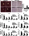
Ad-Cyp7a1 ameliorated LPS-induced hepatic inflammation via FXR, but not TGR5. WT (C57BL/6J), Fxr−/−, and Tgr5−/− mice were injected in the tail vein with 1 × 10⁹ pfu per mouse of either Ad-null or Ad-Cyp7a1. After 7 days, mice were injected ip with 20 mg/kg LPS or PBS. After 6 h, liver tissues and sera were collected for analysis. A: Ad-Cyp7a1 reduced LPS-induced hepatic inflammation. Fresh WT mouse liver tissues were kept in 10% formalin solution and 5 μM sections were stained with H&E. B: Ad-Cyp7a1 reduced macrophage infiltration in LPS-induced hepatic inflammation. Representative immunohistochemistry staining for F4/80 from WT mouse liver sections. The number of F4/80-positive cells in five microscopic fields was counted and statistically analyzed. C: Ad-Cyp7a1 reduced LPS-induced pro-inflammatory cytokines and chemokine mRNA expression in WT mice. Total RNAs from liver tissues were isolated and hepatic mRNA levels of inflammatory genes were determined by qPCR. Amplification of Gapdh was used as a control. D: Ad-Cyp7a1 reduced serum cytokines induced by LPS in WT mice. The levels of cytokines in the serum were measured by ELISA assay. Results are shown as mean ± SE. *P < 0.05, **P < 0.01 versus mice injected with Ad-null and treated with LPS. E: Ad-Cyp7a1 did not affect LPS-induced inflammatory gene mRNA expression in Fxr−/− mice. Total RNA from livers of Fxr−/− mice was isolated and hepatic mRNA levels of inflammatory genes were determined by qPCR. Amplification of Gapdh was used as control. F: Ad-Cyp7a1 did not affect serum cytokine protein levels in Fxr−/− mice. The levels of cytokines in the sera of Fxr−/− mice were measured by ELISA assay. G: Ad-Cyp7a1 reduced LPS-induced inflammatory cytokine mRNA levels in Tgr5−/− mouse liver. Total RNA from livers of Tgr5−/− mice was isolated and hepatic mRNA levels of inflammatory genes were determined by qPCR. Amplification of Gapdh was used as control. H: Ad-Cyp7a1 reduced serum cytokines in Tgr5−/− mice. The levels of cytokines in the sera of Tgr5−/− mice were measured by ELISA assay. Results are shown as mean ± SE. *P < 0.05, **P < 0.01 versus mice injected with Ad-null and treated with LPS. ANOVA was used for statistical analysis (n = 8 male mice per group).
Fig. 3.
Fig. 3.
Activation of FXR inhibited TNF-α and p65 activated NF-κB activity. A: NF-κB reporter assay showed that activation of FXR by GW4064 and OCA reduced TNF-α-induced NF-κB activity. B: NF-κB reporter assay showed that activation of FXR by GW4064 and OCA reduced p65-induced NF-κB activity. For reporter assay, HepG2 cells were cotransfected with NF-κB/luciferase reporter plasmid (pNL3.2-NF-κB-RE) and pCMV-β-gal (for normalization of β-galactosidase activity), and FXR expression plasmid (pcDNA3-hFXR), p65 expression plasmid (pCMV-p65), or pcDNA3 plasmid (control) as indicated. After 48 h, cells were treated with TNF-α (20 ng/ml), GW4064 (1 μM), or OCA (10 μM), as indicated for 6 h. Luciferase activities were measured by luciferase assay kit and luciferase activity was normalized to β-gal activity and expressed as relative luciferase units (RLU)/β-gal activity. Results shown are mean ± SE. *P < 0.05, **P < 0.01 versus vehicle (DMSO). C: Primary human hepatocytes were treated with GW4064 (1 μM) or OCA (10 μM) in the absence or presence of LPS (100 ng/ml) for 6 h. Total RNA was isolated and mRNA levels of inflammatory genes were determined by qPCR. Amplification of Gapdh was used as control. Results shown are mean ± SE. *P < 0.05, **P < 0.01 versus LPS treatment. ANOVA was used for statistical analysis.
Fig. 4.
Fig. 4.
Ad-Cyp7a1 inhibited LPS-induced NF-κB binding to cytokine gene promoters through inducing a ligand-dependent interaction between FXR and p65 and coactivator. A: ChIP assay of the NF-κB binding site showed that Ad-Cyp7a1 ameliorated LPS-induced p65 occupancy to the NF-κB binding site on Tnf-α and Il-1β gene promoters. B: ChIP assay showed that activation of FXR by OCA ameliorated LPS-induced p65 binding to the NF-κB binding site on Tnf-α and Il-1β gene promoters. For ChIP assay, WT mice were injected in the tail vein with 1 × 10⁹ pfu per mouse of either Ad-null or Ad-Cyp7a1, or treated with 30 mg/kg OCA by gavage once per day. After 7 days of adenovirus or OCA treatment, mice were injected ip with 20 mg/kg LPS or PBS (control). After 6 h, liver tissues were homogenized and the chromatin protein and DNA were cross-linked in 1% formaldehyde and sonicated to 0.2–2.0 kb fragments and mixed with either anti-p65 or anti-FXR antibody to precipitate protein/chromatin complexes. Rabbit IgG was used as negative control. DNA fragments were quantified by amplification with the primers designed to probe the NF-κB binding sites on the Tnf-α and Il-1β gene promoters. C: Mammalian two-hybrid assay showed that GW4064 stimulated FXR interaction with p65 through the FXR LBD. D: Mammalian two-hybrid assay showed that SRC-1 reduced GW4064-activated FXR and p65 interaction by competition for interaction with FXR and p65. For two-hybrid assay, HEK293T cells were cotransfected with GAL4-luciferase reporter plasmid pG5luc (or Gal4 empty as control), VP16 fusion protein plasmid, VP16-p65 and GAL4-fusion protein plasmid, GAL-FXR (full length), GAL-FXR-LBD, GAL-FXR-DBD, GAL-FXR-HINGE, and SRC-1 plasmid in the absence or presence of GW4064 (1 μM) as indicated for 48 h. Luciferase reporter activity was assayed using a luciferase assay kit. Relative luciferase activity (RLU) was measured and normalized to Renilla luciferase activity. E: NF-κB reporter assay showed that coactivator SRC-1 stimulated TNF-α-induced NF-κB activity. HepG2 cells were cotransfected with pNL3.2-NF-κB reporter plasmid and pCMV-β-gal plasmid, and pcDNA3-SRC-1 or pcDNA3 (control). After 48 h, cells were treated with GW4064 (1 μM) in the absence or presence of TNF-α (20 ng/ml) for 6 h. NF-κB reporter activity was measured by luciferase assay kit. Results shown are mean ± SE. *P < 0.05, **P < 0.01. ANOVA was used for statistical analysis.
Fig. 5.
Fig. 5.
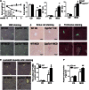
Lack of Cyp7a1 aggravated MCD diet-induced liver fibrosis. WT and Cyp7a1−/− mice were fed with chow diet or MCD diet for 3 weeks. A: MCD diet affected body weight, liver weight, and serum aspartate aminotransferase and alanine aminotransferase levels of WT and Cyp7a1−/− mice. B–E: MCD diet increased hepatic inflammation and collagen fibrosis in Cyp7a1−/− mouse livers. Fresh liver samples fixed in 10% formalin solution and 5 μM sections were stained with H&E (B), Sirius red (C), Masson’s trichrome stain (aniline blue solution) (D), and α-SMA (E). Representative immunohistochemistry stains of liver sections are shown. The number of α-SMA-positive cells in five microscopic fields was counted and statistically analyzed. F: MCD diet increased collagen hydroxyproline contents in Cyp7a1−/− mouse livers. Hydroxyproline contents in liver collagen were determined by the reaction of oxidized hydroxyproline with 4-(dimethylamino) benzaldehyde using a hydroxyproline assay kit. Results shown are mean ± SE. *P < 0.05, **P < 0.01 versus WT mice fed with chow diet. #P < 0.05, ##P < 0.01 versus WT mice fed with MCD diet. ANOVA was used for statistical analysis (n = 7–9 male mice per group).
Fig. 6.
Fig. 6.
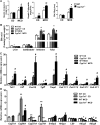
MCD diet increased hepatic lipids and decreased mRNA expression of alternative bile acid synthesis pathway genes in Cyp7a1−/− mice. WT and Cyp7a1−/− mice were fed with chow diet or MCD diet for 3 weeks. A: MCD diet increased cholesterol, triglyceride, and free fatty acid contents in Cyp7a1−/− mouse livers. Hepatic lipids were detected using lipid analysis kits. B: MCD diet did not alter bile acid pool size in Cyp7a1−/− mice. Bile acids were analyzed using a bile acid analysis kit. Total bile acid pool sizes were determined as the total amount of bile acids in liver, intestine, and gallbladder. C: MCD diet increased inflammatory cytokines and collagen gene expression in Cyp7a1−/− mouse livers. Total RNAs from liver tissues were isolated and hepatic mRNA levels were determined by qPCR. Amplification of Gapdh was used as control. D: MCD diet decreased mRNA levels of alternative bile acid synthesis pathway genes in Cyp7a1−/− mouse livers. Quantitative real-time PCR was used to analyze mRNA expression levels of bile acid synthesis and cholesterol synthesis and transport genes. Total RNAs from liver tissues were isolated and hepatic mRNA levels were determined by qPCR. Amplification of Gapdh was used as control. Results shown are mean ± SE. *P < 0.05, **P < 0.01 versus WT mice fed with chow diet. #P < 0.05, ##P < 0.01 versus WT mice fed with MCD diet. ANOVA was used for statistical analysis (n = 7–9 male mice per group).
Fig. 7.
Fig. 7.
MCD diet increased free cholesterol, oxidative stress, and apoptosis, and aggravated hepatic fibrosis in Cyp7a1−/− mice. WT and Cyp7a1−/− mice were fed with chow diet or MCD diet for 3 weeks. A: MCD diet increased free cholesterol in Cyp7a1−/− mouse livers. Free cholesterol contents in the liver were detected using a cholesterol assay kit. B: MCD diet increased free cholesterol in Cyp7a1−/− mouse livers. Fresh liver tissues were kept in 10% formalin solution and 5 μM sections were stained with filipin and determined by fluorescence microscope. Fluorescence density was analyzed by Image J software. C: MCD diet increased oxidative stress in Cyp7a1−/− mouse livers. Liver tissues were homogenized and oxidative stress was assayed using a TBARS assay kit. SOD activity was determined by a SOD kit. D: MCD diet increased hepatic apoptosis in Cyp7a1−/− mouse livers. Representative TUNEL apoptosis staining of liver sections from WT and Cyp7a1−/− mice are shown. The number of TUNEL-positive cells in five microscopic fields was counted and statistically analyzed. E: MCD diet increased apoptosis proteins in Cyp7a1−/− mouse livers. Liver tissues were homogenized and cell death-related proteins were determined by Western blot. β-Actin was used as control. Results shown are mean ± SE. *P < 0.05, versus WT mice fed with chow diet. #P < 0.05, versus WT mice fed with MCD diet. ANOVA was used for statistical analysis (n = 7–9 male mice per group).
Fig. 8.
Fig. 8.
Ad-Cyp7a1 reduced MCD diet-induced hepatic steatosis and fibrosis in Cyp7a1−/− mice. Cyp7a1−/− mice were fed with MCD diet for 3 weeks. On week 2, mice were injected in the tail vein with 1 × 10⁹ pfu per mouse of either Ad-null or Ad-Cyp7a1. One week later, mice were euthanized and liver tissues were collected for analysis. A: Ad-Cyp7a1 reduced MCD diet-induced inflammatory cytokines and collagen gene mRNA expression in Cyp7a1−/− mouse livers. Total RNAs from liver tissues were isolated and mRNA levels were determined by qPCR. B: Ad-Cyp7a1 reduced α-SMA in Cyp7a1−/− mice. C: Ad-Cyp7a1 reduced collagen and fibrosis in Cyp7a1−/− mouse livers. Representative H&E, Sirius red, and Masson’s trichrome stains are shown. D: Ad-Cyp7a1 reduced free cholesterol contents, TBARS levels, and apoptosis in Cyp7a1−/− mouse livers. All assays were performed as described in the Fig. 7 legend. Results a shown are mean ± SE. *P < 0.05 versus Ad-null Cyp7a1−/− mice fed with MCD diet. Studentʼs t-test was used for statistical analysis (n = 5–7 male mice per group).

Similar articles

Cited by

References

    1. Li T., and Chiang J. Y.. 2014. Bile acid signaling in metabolic disease and drug therapy. Pharmacol. Rev. 66: 948–983. - PMC - PubMed
    1. Chiang J. Y. 2009. Bile acids: regulation of synthesis. J. Lipid Res. 50: 1955–1966. - PMC - PubMed
    1. Trauner M., Fickert P., and Stauber R. E.. 1999. Inflammation-induced cholestasis. J. Gastroenterol. Hepatol. 14: 946–959. - PubMed
    1. Luedde T., and Schwabe R. F.. 2011. NF-kappaB in the liver-linking injury, fibrosis and hepatocellular carcinoma. Nat. Rev. Gastroenterol. Hepatol. 8: 108–118. - PMC - PubMed
    1. Greve J. W., Gouma D. J., and Buurman W. A.. 1989. Bile acids inhibit endotoxin-induced release of tumor necrosis factor by monocytes: an in vitro study. Hepatology. 10: 454–458. - PubMed

Publication types

MeSH terms

LinkOut - more resources