. 2017 Jan 24;26(1):145-156.
doi: 10.3727/096368916X692861. Epub 2016 Aug 16.

Pivotal Role of Brain-Derived Neurotrophic Factor Secreted by Mesenchymal Stem Cells in Severe Intraventricular Hemorrhage in Newborn Rats

Pivotal Role of Brain-Derived Neurotrophic Factor Secreted by Mesenchymal Stem Cells in Severe Intraventricular Hemorrhage in Newborn Rats

So Yoon Ahn et al. Cell Transplant. .

Abstract

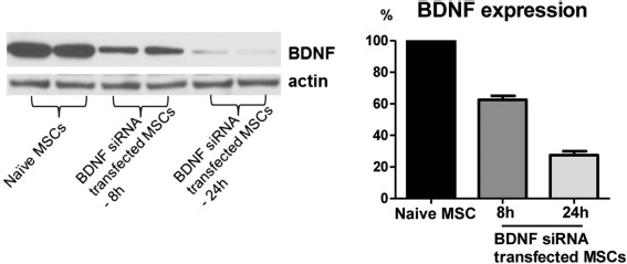
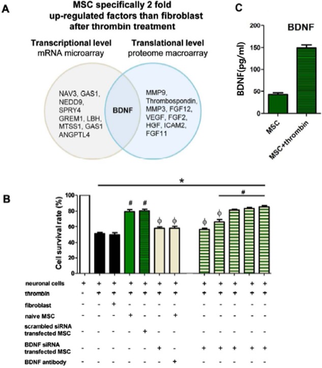
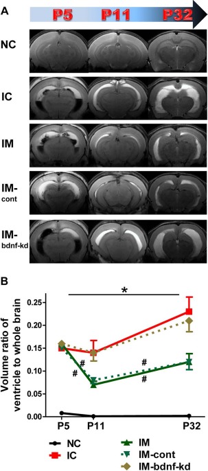
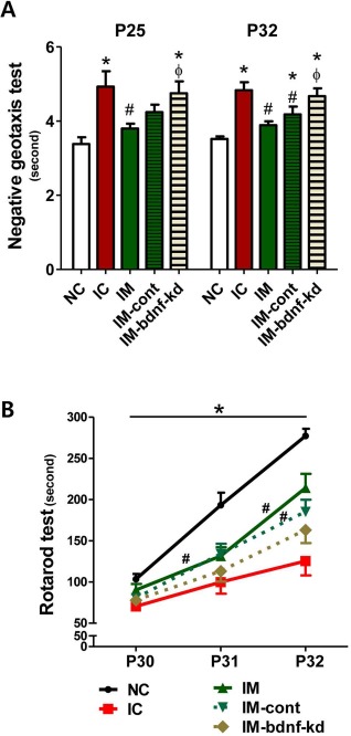
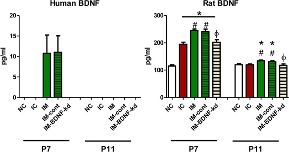
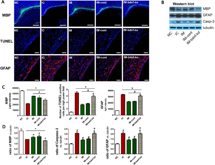
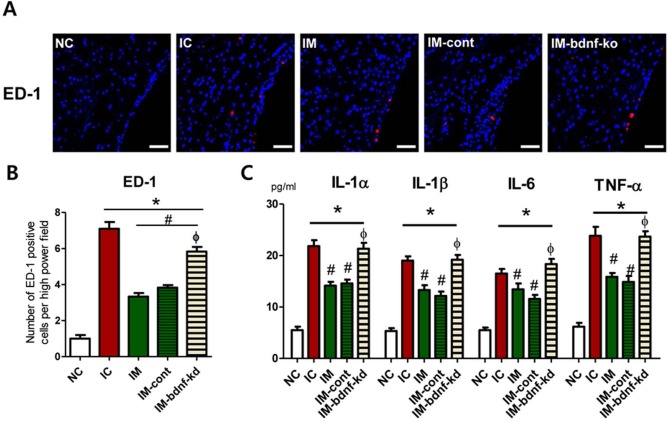
Mesenchymal stem cell (MSC) transplantation protects against neonatal severe intraventricular hemorrhage (IVH)-induced brain injury by a paracrine rather than regenerative mechanism; however, the paracrine factors involved and their roles have not yet been delineated. This study aimed to identify the paracrine mediator(s) and to determine their role in mediating the therapeutic effects of MSCs in severe IVH. We first identified significant upregulation of brain-derived neurotrophic factor (BDNF) in MSCs compared with fibroblasts, in both DNA and antibody microarrays, after thrombin exposure. We then knocked down BDNF in MSCs by transfection with small interfering (si)RNA specific for human BDNF. The therapeutic effects of MSCs with or without BDNF knockdown were evaluated in vitro in rat neuronal cells challenged with thrombin, and in vivo in newborn Sprague-Dawley rats by injecting 200 μl of blood on postnatal day 4 (P4), and transplanting MSCs (1 × 10⁵ cells) intraventricularly on P6. siRNA-induced BDNF knockdown abolished the in vitro benefits of MSCs on thrombin-induced neuronal cell death. BDNF knockdown also abolished the in vivo protective effects against severe IVH-induced brain injuries such as the attenuation of posthemorrhagic hydrocephalus, impaired behavioral test performance, increased astrogliosis, increased number of TUNEL cells, ED-1+ cells, and inflammatory cytokines, and reduced myelin basic protein expression. Our data indicate that BDNF secreted by transplanted MSCs is one of the critical paracrine factors that play a seminal role in attenuating severe IVH-induced brain injuries in newborn rats.


Figures

Figure 1.
Figure 1.
Knock down of brain-derived neurotrophic factor (BDNF) in the mesenchymal stem cells (MSCs). Western blot assay for BDNF expression from the naive MSCs and BDNF small interfering (si)RNA-transfected MSCs after 8 and 24 h of transfection. To confirm the successful knockdown of BDNF, the BDNF level was measured in the culture medium of BDNF siRNA-transfected MSCs. BDNF knockdown MSCs showed substantially reduced BDNF production at 8 h and 24 h after transfection.
Figure 2.
Figure 2.
Gene expression analysis of upregulation in MSCs and in vitro protective effects of BDNF secreted from MSCs. (A) Gene expression analysis of upregulation in MSCs compared with fibroblasts in microarray and macroarray. Note that the BDNF gene was commonly upregulated in microarray and macroarray. (B) In vitro expression level of BDNF in MSCs. (C) Cell survival rate in each group was evaluated by 3-(4,5-dimethylthiazol-2-yl)-2,5-diphenyl tetrazolium (MTT) assay. Data are expressed as mean ± standard error of the mean (SEM). *p < 0.05 versus neuronal cells without any intervention, #p < 0.05 versus neuronal cells treated with thrombin, Φp < 0.05 versus neuronal cells treated with thrombin and naive MSCs. Abbreviations: (si)RNA, small interfering; VEGF, vascular endothelial growth factor.
Figure 3.
Figure 3.
Knock down of BDNF in MSCs abolished the therapeutic effects of MSCs in attenuating ventricular dilatation and its progression after severe intraventricular hemorrhage (IVH). (A) Representative serial brain magnetic resonance images (MRIs) from each group 1, 7, and 28 days after inducing IVH (P5/P11/P32). (B) Ventricle-to-whole brain volume ratio as measured by MRI. Data are expressed as mean ± SEM. Abbreviations: NC, normal control rats; IC, IVH control rats; IM, IVH with transplantation of human umbilical cord blood-derived mesenchymal stem cells (hUCB-MSCs); IM-cont, IVH with transplantation of scrambled small interfering (si)RNA-transfected hUCB-MSCs; IM-bdnf-kd, transplantation of BDNF siRNA-transfected hUCB-MSCs. *p < 0.05 versus NC, #p < 0.05 versus IC.
Figure 4.
Figure 4.
Knock down of BDNF in MSCs abolished the therapeutic effects of MSCs in improving behavioral function after severe IVH. Sensorimotor functional outcomes on the negative geotaxis test on P25 and P32 (A) and rotarod test on P30–32 (B). Data are expressed as mean ± SEM. Abbreviations: NC, normal control rats; IC, IVH control rats; IM, IVH with transplantation of human umbilical cord blood-derived mesenchymal stem cells (hUCB-MSCs); IM-cont, IVH with transplantation of scrambled small interfering (si)RNA-transfected hUCB-MSCs; IM-bdnf-kd, transplantation of BDNF siRNA-transfected hUCB-MSCs. *p < 0.05 versus NC, #p < 0.05 versus IC, Fp < 0.05 versus IM.
Figure 5.
Figure 5.
Human and rat BDNF expressions in the brain tissue. On the second and fifth day after human MSC transplantation (P5), human BDNF and rat BDNF were measured, respectively, in rat brains from each group. Abbreviations: NC, normal control rats; IC, IVH control rats; IM, IVH with transplantation of human umbilical cord blood-derived mesenchymal stem cells (hUCB-MSCs); IM-cont, IVH with transplantation of scrambled small interfering (si)RNA-transfected hUCB-MSCs; IM-bdnf-kd, transplantation of BDNF siRNA-transfected hUCB-MSCs. *p < 0.05 versus NC, #p < 0.05 versus IC, Φp < 0.05 versus IM.
Figure 6.
Figure 6.
Knock down of BDNF in MSCs abolished the therapeutic effects of MSCs in improving brain myelination and in attenuating cell death and reactive gliosis after severe IVH. (A) Representative immunofluorescence photomicrographs of the periventricular area with staining for myelin basic protein (MBP) (green), TUNEL (green), glial fibrillary acidic protein (GFAP) (red), and 4′,6-diamidino-2-pheylindole (DAPI) (blue) (original magnification: 200×, scale bars: 50 μm for MBP and original magnification: 85×, scale bars: 500 μm for TUNEL and GFAP). (B) Western blot assay of MBP, GFAP, and caspase 3 with tubulin in the brain tissue homogenates on P32. Brain MBP expression, cell death, and reactive gliosis evidenced by light intensity of MBP immunofluorescence, average number of TUNEL+ cells, and light intensity of GFAP staining, respectively, in the periventricular area (C) and those indicated by the ratio of MBP, caspase 3, and GFAP to tubulin with Western assay, respectively, in the brain tissue homogenates (D) on P32. Data are expressed as mean ± SEM. Abbreviations: NC, normal control rats; IC, IVH control rats; IM, IVH with transplantation of human umbilical cord blood-derived mesenchymal stem cells (hUCB-MSCs); IM-cont, IVH with transplantation of scrambled small interfering (si)RNA-transfected hUCB-MSCs; IM-bdnf-kd, transplantation of BDNF siRNA-transfected hUCB-MSCs. *p<0.05 versus NC, #p<0.05 versus IC, Φp <0.05 versus IM.
Figure 7.
Figure 7.
Knock down of BDNF in MSCs abolished the therapeutic effects of MSCs in downregulating brain inflammation after severe IVH. (A) Representative immunofluorescence photomicrographs of the periventricular area with staining for active macrophages (ED-1, red) and DAPI (blue) (original magnification: 200×, scale bars: 25 μm). (B) Average number of ED-1+ cells, which indicate active macrophages in the periventricular area on P32. (C) Levels of inflammatory cytokines including interleukin (IL)-1α, IL-β, IL-6, and tumor necrosis factor (TNF)-α in the brain tissue homogenates on P32. Data are expressed as mean ± SEM. NC, normal control rats; IC, IVH control rats; IM, IVH with transplantation of hUCB-MSCs; IM-cont, IVH with transplantation of scrambled small interfering (si)RNA-transfected hUCB-MSCs; IM-bdnf-kd, transplantation of BDNF siRNA-transfected hUCB-MSCs. *p < 0.05 versus NC, #p < 0.05 versus IC, Φp < 0.05 versus IM.

Similar articles

Cited by

References

    1. Vohr B.R., Wright L.L., Dusick A.M., Mele L., Verter J., Steichen J.J., Simon N.P., Wilson D.C., Broyles S., Bauer C.R., Delaney-Black V., Yolton K.A., Fleisher B.E., Papile L.A., Kaplan M.D.. Neurodevelopmental and functional outcomes of extremely low birth weight infants in the National Institute of Child Health and Human Development Neonatal Research Network, 1993–1994. Pediatrics 2000;105(6): 1216–26. - PubMed
    1. Ahn S.Y., Chang Y.S., Sung D.K., Sung S.I., Yoo H.S., Lee J.H., Oh W.I., Park W.S.. Mesenchymal stem cells prevent hydrocephalus after severe intraventricular hemorrhage. Stroke 2013; 44(2): 497–504. - PubMed
    1. Park W.S., Sung S.I., Ahn S.Y., Sung D.K., Im G.H., Yoo H.S., Choi S.J., Chang Y.S.. Optimal timing of mesenchymal stem cells therapy for neonatal intraventricular hemorrhage. Cell Transplant. 2016; 25(6): 1131–44. - PubMed
    1. Ahn S.Y., Chang Y.S., Sung D.K., Sung S.I., Yoo H.S., Im G.H., Choi S.J., Park W.S.. Optimal route for mesenchymal stem cells transplantation after severe intraventricular hemorrhage in newborn rats. PLoS One 2015; 10(7): e0132919. - PMC - PubMed
    1. Chang Y.S., Ahn S.Y., Jeon H.B., Sung D.K., Kim E.S., Sung S.I., Yoo H.S., Choi S.J., Oh W.I., Park W.S.. Critical role of VEGF secreted by mesenchymal stem cells in hyperoxic lung injury. Am J Respir Cell Mol Biol. 2014; 51(3): 391–9. - PubMed

MeSH terms