Double primary hepatic cancer (hepatocellular carcinoma and intrahepatic cholangiocarcinoma) originating from hepatic progenitor cell: a case report and review of the literature
- PMID: 27535234
- PMCID: PMC4989533
- DOI: 10.1186/s12957-016-0974-6
Double primary hepatic cancer (hepatocellular carcinoma and intrahepatic cholangiocarcinoma) originating from hepatic progenitor cell: a case report and review of the literature
Abstract
Background: Synchronous development of primary hepatocellular carcinoma (HCC) and intrahepatic cholangiocarcinoma (ICC) in different sites of the liver have rarely been reported before. The purpose of this study is to investigate the clinicopathological characteristics of synchronous double cancer of HCC and ICC.
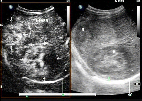
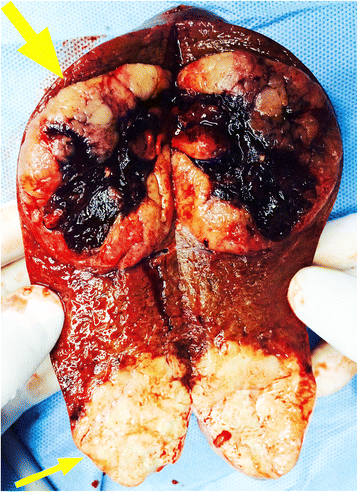
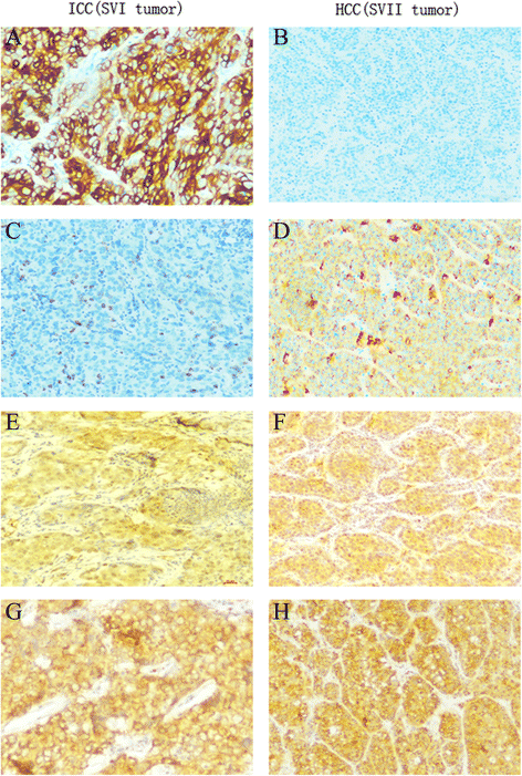
Case presentation: A 56-year-old Chinese man without obvious liver cirrhosis was preoperation diagnosed with multiple HCC in segments VI (SVI) and VII (SVII) by the abdominal computed tomography (CT) and contrast-enhanced ultrasonography (CEUS). We performed hepatic resection of both segments. The tumors in SVI and SVII were pathologically diagnosed as ICC and HCC, respectively. Immunohistochemically, the HCC in SVII was positive for HepPar-1 and negative for CK19, while the ICC in SVI tumor was positive for CK19 and negative for HepPar-1. Interestingly, the immunohistochemical results also showed that the classic hepatic progenitor cell (HPCs) markers CD34 and CD117 were both positive of the two tumors. The patient still survived and at a 1-year follow-up did not show evidence of metastasis or new recurrent lesions. We speculate that the two masses may have originated from HPCs based on the findings of this patient.
Conclusions: Synchronous development of HCC and ICC is very rare with unique clinical and pathological features. The correct preoperative diagnosis of double hepatic cancer of HCC and ICC is difficult. Hepatitis B virus (HBV) and hepatitis C virus (HCV) infection were both the independent risk factor to the development of double liver cancer. Hepatic resection is the preferred and most effective treatment choice. The prognosis of synchronous occurrence of double hepatic cancer was poorer than for either HCC or ICC, and the origin of it needs further study.
Keywords: Double hepatic cancer; Hepatic progenitor cell; Hepatocellular carcinoma; Intrahepatic cholangiocarcinoma; Prognosis.
Figures


Similar articles
-
Synchronous double cancers of primary hepatocellular carcinoma and intrahepatic cholangiocarcinoma: a case report and review of the literature.World J Surg Oncol. 2014 Nov 10;12:337. doi: 10.1186/1477-7819-12-337. World J Surg Oncol. 2014. PMID: 25385169 Free PMC article. Review.
-
Synchronous double primary hepatocellular carcinoma and intrahepatic cholangiocarcinoma: A case report and review of the literature.Medicine (Baltimore). 2021 Nov 19;100(46):e27349. doi: 10.1097/MD.0000000000027349. Medicine (Baltimore). 2021. PMID: 34797273 Free PMC article. Review.
-
Comparison of combined hepatocellular and cholangiocarcinoma with hepatocellular carcinoma and intrahepatic cholangiocarcinoma.Surg Today. 2006;36(10):892-7. doi: 10.1007/s00595-006-3276-8. Surg Today. 2006. PMID: 16998683
-
Double cancer - hepatocellular carcinoma and intrahepatic cholangiocarcinoma with a spindle-cell variant.J Hepatobiliary Pancreat Surg. 1999;6(4):422-6. doi: 10.1007/s005340050144. J Hepatobiliary Pancreat Surg. 1999. PMID: 10664295
-
Double primary liver cancer (intrahepatic cholangiocarcinoma and hepatocellular carcinoma) in a patient with hepatitis C virus-related cirrhosis.J Hepatobiliary Pancreat Surg. 2007;14(2):204-9. doi: 10.1007/s00534-006-1134-0. Epub 2007 Mar 27. J Hepatobiliary Pancreat Surg. 2007. PMID: 17384916
Cited by
-
Hepatocellular carcinoma with biliary and neuroendocrine differentiation: A case report.World J Clin Oncol. 2021 Apr 24;12(4):262-271. doi: 10.5306/wjco.v12.i4.262. World J Clin Oncol. 2021. PMID: 33959479 Free PMC article.
-
Synchronous double primary hepatocellular carcinoma and intrahepatic cholangiocarcinoma in a single patient with chronic hepatitis B: two case reports and literature review.Front Oncol. 2025 Jun 16;15:1507454. doi: 10.3389/fonc.2025.1507454. eCollection 2025. Front Oncol. 2025. PMID: 40589631 Free PMC article.
-
Diagnosis of liver tumors by multimodal ultrasound imaging.Medicine (Baltimore). 2020 Aug 7;99(32):e21652. doi: 10.1097/MD.0000000000021652. Medicine (Baltimore). 2020. PMID: 32769936 Free PMC article.
-
Characteristics, diagnosis, treatment and prognosis of double primary hepatic cancer: experience based on a series of 12 cases.Am J Transl Res. 2024 Aug 15;16(8):4234-4245. doi: 10.62347/PIWA3282. eCollection 2024. Am J Transl Res. 2024. PMID: 39262740 Free PMC article.
-
Synchronous Hepatocellular Carcinoma and Cholangiocellular Carcinoma on 18F-FDG PET/CT.Mol Imaging Radionucl Ther. 2018 Oct 9;27(3):136-137. doi: 10.4274/mirt.93723. Mol Imaging Radionucl Ther. 2018. PMID: 30317851 Free PMC article.
References
-
- Inaba K, Suzuki S, Sakaguchi T, Kobayasi Y, Takehara Y, Miura K, Baba S, Nakamura S, Konno H. Double primary liver cancer (intrahepatic cholangiocarcinoma and hepatocellular carcinoma) in a patient with hepatitis C virus-related cirrhosis. J Hepatobiliary Pancreat Surg. 2007;14:204–209. doi: 10.1007/s00534-006-1134-0. - DOI - PubMed
-
- Jung KS, Chun KH, Choi GH, Jeon HM, Shin HS, Park YN, Park JY. Synchronous development of intrahepatic cholangiocarcinoma and hepatocellular carcinoma in different sites of the liver with chronic B-viral hepatitis: two case reports. BMC Res Notes. 2013;6:520. doi: 10.1186/1756-0500-6-520. - DOI - PMC - PubMed
Publication types
MeSH terms
LinkOut - more resources
Full Text Sources
Other Literature Sources
Medical

