. 2016 Oct 1;116(4):1946-1955.
doi: 10.1152/jn.00085.2016. Epub 2016 Aug 17.

The inferior olive is essential for long-term maintenance of a simple motor skill

Affiliations

The inferior olive is essential for long-term maintenance of a simple motor skill

Xiang Yang Chen et al. J Neurophysiol. .

Abstract

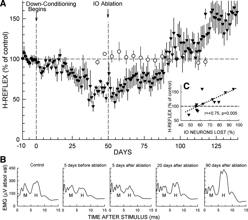
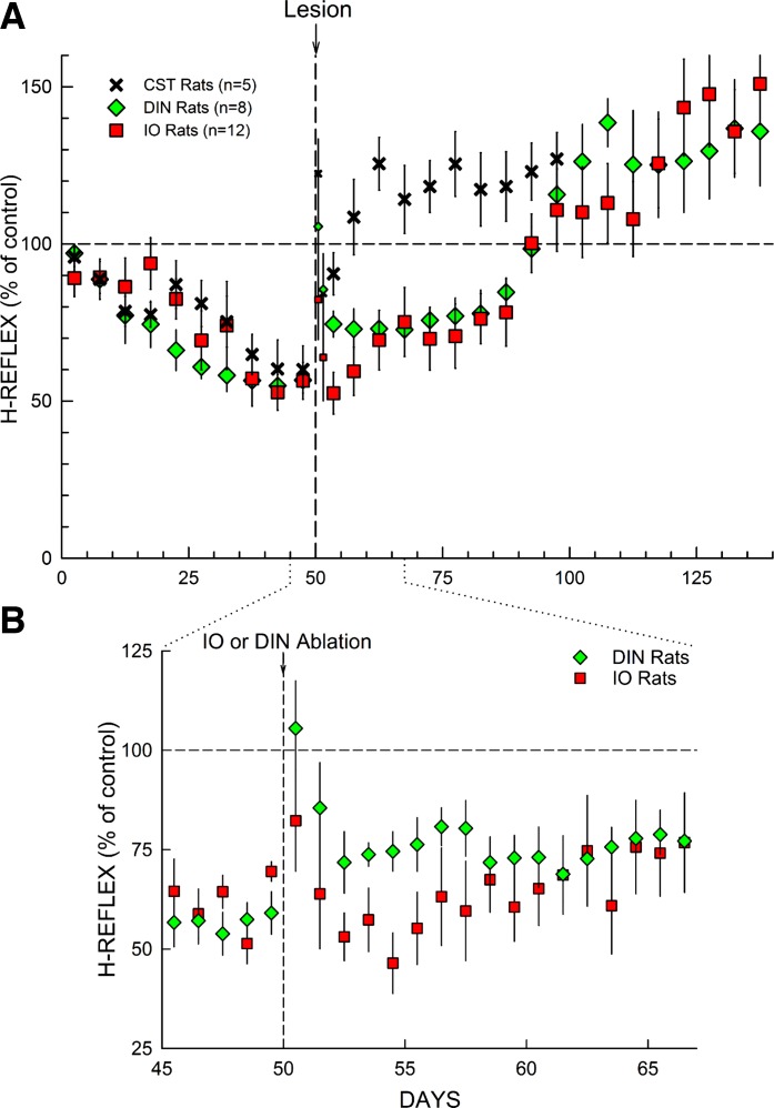
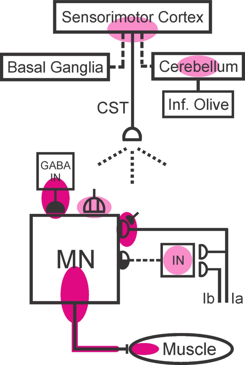
The inferior olive (IO) is essential for operant down-conditioning of the rat soleus H-reflex, a simple motor skill. To evaluate the role of the IO in long-term maintenance of this skill, the H-reflex was down-conditioned over 50 days, the IO was chemically ablated, and down-conditioning continued for up to 102 more days. H-reflex size just before IO ablation averaged 62(±2 SE)% of its initial value (P < 0.001 vs. initial). After IO ablation, H-reflex size rose to 75-80% over ∼10 days, remained there for ∼30 days, rose over 10 days to above its initial value, and averaged 140(±14)% for the final 10 days of study (P < 0.01 vs. initial). This two-stage loss of down-conditioning maintenance correlated with IO neuronal loss (r = 0.75, P < 0.01) and was similar to the loss of down-conditioning that follows ablation of the cerebellar output nuclei dentate and interpositus. In control (i.e., unconditioned) rats, IO ablation has no long-term effect on H-reflex size. These results indicate that the IO is essential for long-term maintenance of a down-conditioned H-reflex. With previous data, they support the hypothesis that IO and cortical inputs to cerebellum combine to produce cerebellar plasticity that produces sensorimotor cortex plasticity that produces spinal cord plasticity that produces the smaller H-reflex. H-reflex down-conditioning appears to depend on a hierarchy of plasticity that may be guided by the IO and begin in the cerebellum. Similar hierarchies may underlie other motor learning.

Keywords: H-reflex; cerebellum; learning; memory; operant conditioning; plasticity; sensorimotor cortex; spinal cord.


Figures

Fig. 1.
Fig. 1.
A: Research design. At least 20 days after implantation surgery, rats were exposed to the control mode for 20 days and then to the H-reflex down-conditioning mode for 50 days. The inferior olive (IO) was then ablated. After the ablation, exposure to the down-conditioning mode continued for 56–102 more days. The last 10 control-mode days, the 10 down-conditioning days just before IO ablation (i.e., days 41–50 of conditioning), and the last 10 days of continued down-conditioning after IO ablation (heavy horizontal lines) provided the data used to assess the effect of down-conditioning on the soleus H-reflex and the final impact of IO ablation on the maintenance of soleus H-reflex down-conditioning. B: IO ablation. Cresyl-violet stained photomicrographs showing the IO nucleus complex and IO neurons in a naive control (NC) rat (B1 and B3) and in an IO-ablated rat (IO rat; B2 and B4). (B3 and B4 show the areas outlined in B1 and B2, respectively.) DAO, dorsal accessory olive; DM, dorsomedial group; MAO, medial accessory olive; PO, principal olive. Scale bars: 400 μm in B1 and B2, 40 μm in B3 and B4. The loss of most IO neurons in the IO-ablated rat is evident. C: VGLUT2 immunoreactivity (VGLUT2-IR) in cerebellar cortex. C1 and C2: photomicrographs of VGLUT2-IR-labeled cerebellar sections showing VGLUT2-IR labeling in cerebellar cortex from a NC rat (C1) and an IO rat (C2). The molecular (M), Purkinje (P), and granular (G) layers are indicated. VGLUT2-IR in the molecular layer is much weaker in the IO rat (C2). Scale bar: 100 μm. C3: correlation in IO rats between cerebellar VGLUT2-IR intensity in the molecular layer and remaining IO neurons (in % of NC neuron number). The strong correlation (r = 0.80, P < 0.01) is consistent with the fact that VGLUT2-IR labeling in the molecular layer is selective for excitatory olivocerebellar climbing fiber terminals on Purkinje cell dendrites (Fremeau et al. 2001; Kaneko et al. 2002).
Fig. 2.
Fig. 2.
Top: Representative low (left)- and high (right)-magnification transverse sections of L5 spinal cord showing labeled motoneurons from an IO rat and an NC rat. Bottom: average numbers of motoneurons per section in L3, L4, L5, and L6 spinal cord (left) and average L3–L6 motoneuron area (right) in IO rats and NC rats (±SE). Motoneuron number and size did not differ between IO and NC rats (P > 0.15 by t-test).
Fig. 3.
Fig. 3.
Effects of IO ablation on H-reflex size in down-conditioned rats and in unconditioned rats. A: ▼: average daily H-reflex (±SE) (in % of initial size) for the 12 IO rats for the final 10 days in control mode and for up to 140 days of exposure to the down-conditioning mode. As shown, the IO was ablated after the first 50 days of down-conditioning. All 12 rats were then studied through day 106 (56 days after ablation); 11 were studied through day 112; and 7 were studied through day 140 (90 days after ablation). H-reflex size averaged 62(±2)% of initial value for the 10 days immediately before IO ablation (P < 0.001). It rose to ∼75% in the next 10 days, remained there for ∼30 days, and then over the next 10 days rose to above its initial value and remained high [140(±14)% for each rat's final 10 days of data collection (P = 0.005 vs. initial)]. Background EMG amplitude and M-response size were stable throughout. ○: 5-day average H-reflex (±SE) size (in % of initial size) for 7 rats exposed to the control-mode before and for 50–70 days after IO ablation. All 7 rats were followed for 50 days after ablation, and 5 were followed for 70 days. Background EMG amplitude and M-response size were stable throughout. In these unconditioned rats, IO ablation had no detectable long-term effect on H-reflex size [data from Chen et al. (2016) plus additional more recent data]. B: average daily poststimulus EMG activity from a representative IO rat for: a control-mode day; 5 days prior to IO ablation; 5 days after IO ablation; 20 days after IO ablation; and 90 days after IO ablation. The early and delayed increases following IO ablation are evident. Background EMG amplitude (i.e., the amplitude at time 0) and M-response size are stable throughout. C: average H-reflex size for days 101–110 (51–60 days after IO ablation) for each IO rat vs. the percent of IO ablated [i.e., 100 − (no. of IO neurons remaining in % of average number in NC rats)]. H-reflex size is strongly correlated with IO neuronal loss, implying that the IO plays a key role in the preservation of the H-reflex decrease produced by down-conditioning.
Fig. 4.
Fig. 4.
A: effects of different lesions on maintenance of H-reflex down-conditioning. Average H-reflex sizes (±SE) for each 5-day period for corticospinal tract (CST)-transected rats (n = 5), DIN-ablated rats (n = 8), and IO-ablated rats (n = 12) for the first 50 days of down-conditioning before the lesion and for the next 50–100 days after the lesion [CST data from Chen and Wolpaw (2002); DIN data from Wolpaw and Chen (2006); IO data from present study]. For the 5 days immediately after lesion, H-reflex sizes are shown for the 1st day and the 2nd day (smaller symbols) and for the next 3 days together. All 3 lesions show a transient increase in the first 1–2 days (see footnote 1). After this brief nonspecific effect dissipates, all 3 lesions result eventually in an H-reflex larger than its initial control size. The H-reflex down-conditioning mode remains in effect throughout. B: this expansion from A shows average daily H-reflex values for the IO and DIN rats for the days immediately before and after IO or DIN ablation. In the DIN rats, the first stage of the H-reflex increase occurs within the first 2 days after DIN ablation; in the IO rats, it develops over the initial 10 days after IO ablation. [The brief nonspecific increase in the first 1–2 postablation days is evident for both ablations (footnote 1).]
Fig. 5.
Fig. 5.
Present knowledge of the multisite brain and spinal cord plasticity underlying H-reflex conditioning. Shaded ovals indicate the spinal and supraspinal sites of definite or probable plasticity associated with operant conditioning of the spinal stretch reflex (SSR) or its electrical analog, the H-reflex. MN, the motoneuron; CST, the main corticospinal tract; IN, a spinal interneuron; GABA IN, a GABAergic spinal interneuron. Open synaptic terminals are excitatory, solid ones are inhibitory, half-open ones could be either, and the subdivided one is a cluster of C terminals. Dashed pathways imply the possibility of intervening spinal interneurons. The monosynaptic and probably oligosynaptic SSR/H-reflex pathway from Ia and Ib afferents to the motoneuron is shown. Definite (red shading) or probable (pink shading) sites of plasticity include: the motoneuron membrane (i.e., firing threshold and axonal conduction velocity); motor unit properties; GABAergic interneurons in the ventral horn; GABAergic inhibitory terminals, C terminals, and Ia afferent terminals on the motoneuron; terminals conveying disynaptic group I inhibition or excitation to the motoneuron; sensorimotor cortex (SMC); and cerebellum. The essential roles of the corticospinal tract (which originates largely in SMC), cerebellar and basal ganglia output to cortex, and inferior olive input to cerebellum are indicated. The spinal cord plasticity that is directly responsible for H-reflex conditioning appears to be induced and maintained by cortical plasticity that may depend for its long-term survival on cerebellar plasticity that in turn depends for its survival on input from the inferior olive (see text). [Updated from Wolpaw (2010) with permission.]

Similar articles

Cited by

References

    1. Andre P, Arrogi P. Hipnic modulation of cerebellar information processing: implications for the cerebro-cerebellar dialogue. Cerebellum 2: 84–95, 2003. - PubMed
    1. Aoki H, Sugihara I. Morphology of single olivocerebellar axons in the denervation-reinnervation model produced by subtotal lesion of the rat inferior olive. Brain Res 1449: 24–37, 2012. - PubMed
    1. Balaban CD. Central neurotoxic effects of intraperitoneal administered 3-acetylpyridine, harmaline and nicotinamide in Sprague-Dawley and Long-Evans rats: a critical review of 3-acetyl-pyridine neurotoxicity. Brain Res Rev 9: 21–42, 1985. - PubMed
    1. Bardin JM, Batini C, Billard JM, Buisseret-Delmas C, Conrath-Verrier M, Corvaja N. Cerebellar output regulation by the climbing and mossy fibers with and without the inferior olive. J Comp Neurol 213: 464–477, 1983. - PubMed
    1. Benedetti F, Montarolo PG, Rabacchi S. Inferior olive lesion induces long-lasting functional modification in the Purkinje cells. Exp Brain Res 55: 368–371, 1984. - PubMed

Publication types

LinkOut - more resources