. 2016 Aug 18:7:12564.
doi: 10.1038/ncomms12564.

Mechanosensing by the α6-integrin confers an invasive fibroblast phenotype and mediates lung fibrosis

Affiliations

Mechanosensing by the α6-integrin confers an invasive fibroblast phenotype and mediates lung fibrosis

Huaping Chen et al. Nat Commun. .

Abstract

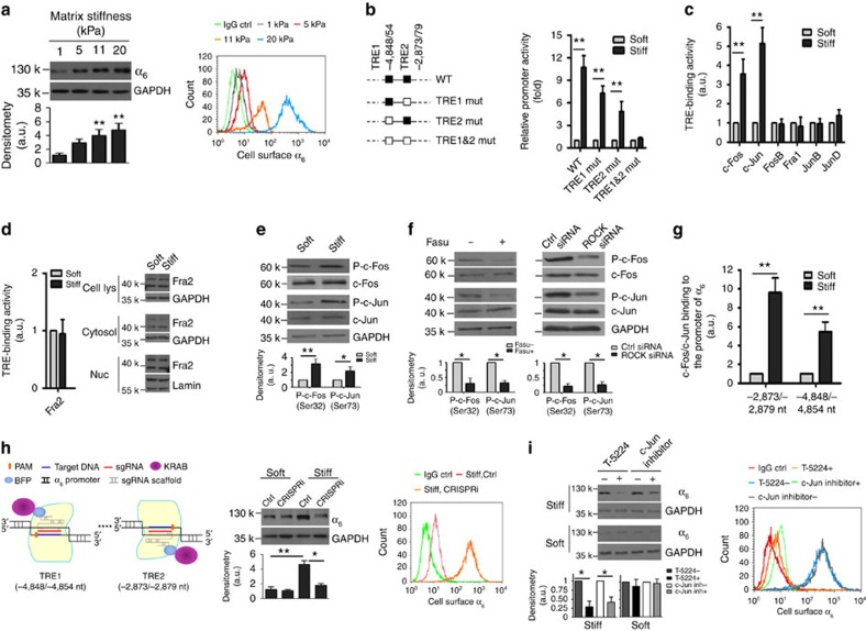
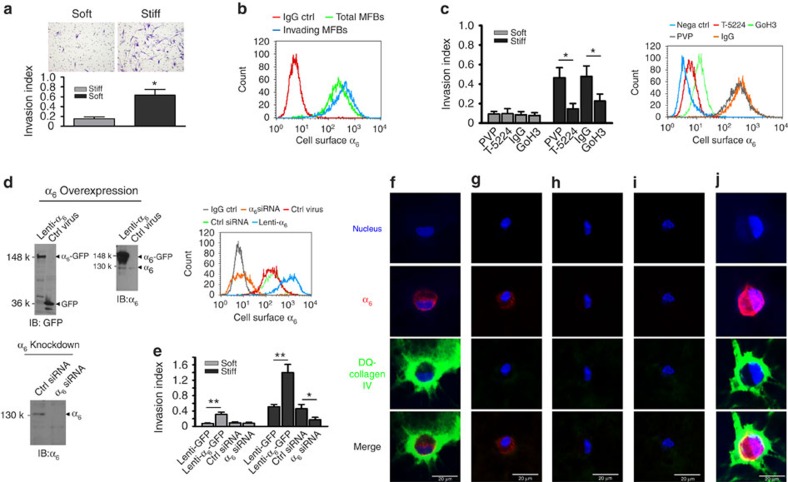
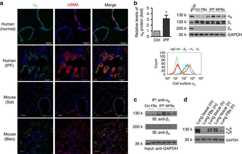
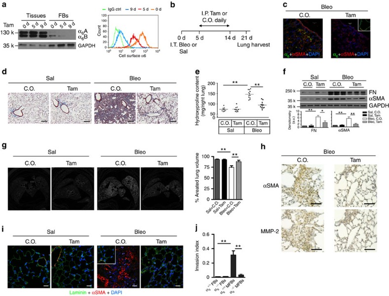
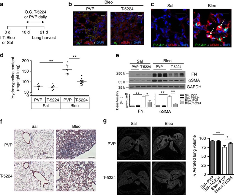
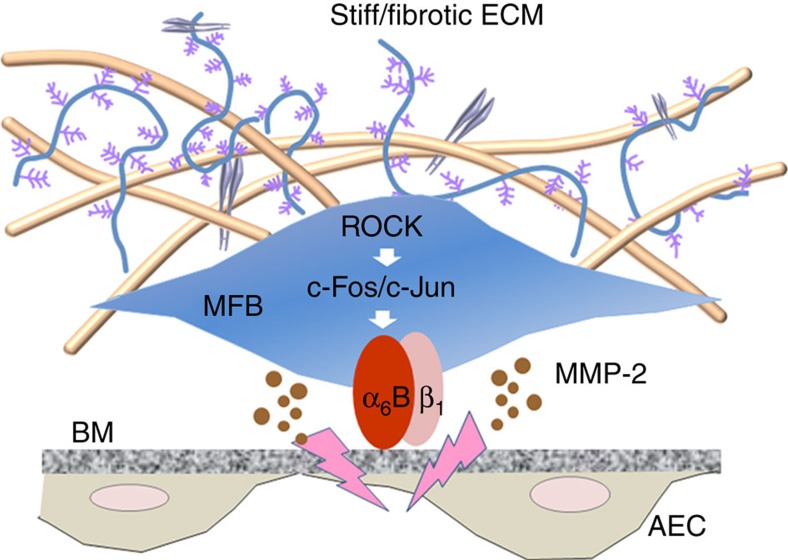
Matrix stiffening is a prominent feature of pulmonary fibrosis. In this study, we demonstrate that matrix stiffness regulates the ability of fibrotic lung myofibroblasts to invade the basement membrane (BM). We identify α6-integrin as a mechanosensing integrin subunit that mediates matrix stiffness-regulated myofibroblast invasion. Increasing α6-expression, specifically the B isoform (α6B), couples β1-integrin to mediate MMP-2-dependent pericellular proteolysis of BM collagen IV, leading to myofibroblast invasion. Human idiopathic pulmonary fibrosis lung myofibroblasts express high levels of α6-integrin in vitro and in vivo. Genetic ablation of α6 in collagen-expressing mesenchymal cells or pharmacological blockade of matrix stiffness-regulated α6-expression protects mice against bleomycin injury-induced experimental lung fibrosis. These findings suggest that α6-integrin is a matrix stiffness-regulated mechanosensitive molecule which confers an invasive fibroblast phenotype and mediates experimental lung fibrosis. Targeting this mechanosensing α6(β1)-integrin offers a novel anti-fibrotic strategy against lung fibrosis.


Figures

Figure 1
Figure 1. Stiff matrix upregulates α6-expression by ROCK-dependent activation of c-Fos/c-Jun transcription complex.
(a) IPF lung myofibroblasts were cultured on PA hydrogels with increasing stiffness (1, 5, 11 and 20 kPa). Levels of α6-protein were determined by immunoblot and flow cytometry, respectively. In flow cytometry, non-immune rat IgG2a, κ was used as isotype IgG control. (b) Schematic shows the WT and mutated human α6-promoters. Promoter activities were determined by luciferase assay. (c) Nuclear extracts from myofibroblasts cultured on soft and stiff matrix were incubated with immobilized oligonucleotides containing TREs. The TRE-binding activities of six AP-1 components as indicated were quantified by colorimetric enzyme-linked immunosorbant assay (ELISA). (d) The TRE-binding activity of Fra2 in nuclear extracts was quantified by colorimetric ELISA. Levels of Fra2 protein in cell lyates, cytoplasmic and nuclear fractions were determined by immunoblot. (e) Protein levels of phospho and total c-Fos and c-Jun under soft versus stiff matrix conditions were determined by immunoblot. (f) Effects of ROCK inhibitor Fasudil (Fasu) and ROCK-specific siRNAs on stiff matrix-induced phosphorylation of c-Fos and c-Jun. (g) The binding of c-Fos/c-Jun complex to the α6-promoter under soft versus stiff matrix conditions was measured by quantitative chromatin immunoprecipitation. (h) Schematic shows sgRNA-mediated targeted expression of KRAB transcription repressor at the distal TRE1 and the proximal TRE2 regions in human α6-promoter. Effects of CRISPRi-based disruption of c-Fos/c-Jun-dependent promoter activation on stiff matrix-induced α6-expression were evaluated by immunoblot and flow cytometry analyses. Control (Ctrl) indicates cells transfected with empty vector. (i) Effects of c-Fos/c-Jun inhibitors (T-5224 and c-Jun peptides) on matrix stiffness-regulated α6-expression were evaluated by immunoblot and flow cytometry. Results are the means ±s.d. of at least three separate experiments; *P<0.05; **P<0.01; one-way analysis of variance. a.u., arbitrary units.
Figure 2
Figure 2. α6 Mediates matrix stiffness-dependent lung myofibroblast invasion into the BM.
(a) The ability of IPF myofibroblasts cultured on soft versus stiff matrix to invade the BM was evaluated by Matrigel invasion assay. (b) α6-Expression on the cell surface of invading myofibroblasts versus total (myo)fibroblasts was evaluated by flow cytometry. (c) Effects of NKI-GoH3 and T-5224 on stiffness-regulated myofibroblast invasion into the BM. α6-Expression on the cell surface was evaluated by flow cytometry using FITC-labelled GoH3. PVP, a vehicle for T-5224; IgG, FITC-labelled isotype control IgG for NKI-GoH3; Nega ctrl, plain cells with no treatments and no incubation with FITC-labelled GoH3/IgG. (d) Overexpression of α6-GFP fusion protein by lentivirus and knockdown of α6 by siRNA in cell lysates were determined by immunoblot and flow cytometry. (e) Effects of overexpression or knockdown of α6 on stiffness-regulated myofibroblast invasion into the BM. (fj) α6-expression (red) and proteolytic activation of DQ-collagen IV (green) in the absence (f) or presence of NKI-GoH3 (g), T-5224 (h), α6-siRNA (i) and Lenti-α6 (j) were determined by confocal immunofluorescent microscopy. Nuclei (blue) were stained by DAPI. Results are the means±s.d. of at least three separate experiments; *P<0.05, **P<0.01; one-way analysis of variance. Scale bar, 20 μm.
Figure 3
Figure 3. Lung myofibroblasts demonstrate increased α6-expression.
(a) Frozen lung tissue sections obtained from failed normal human donors, patients with IPF, saline-treated mice and bleomycin-treated mice were double-stained for α6 (green) and αSMA (red). Nuclei were stained by DAPI (blue). Confocal immunofluorescent images were overlaid to show α6-expression in αSMA-positive lung myofibroblasts. Scale bar, 50 μm; scale bar, 20 μm for mouse with bleo images. (b) Comparison for α6-expression in lung (myo)fibroblasts isolated from patients with IPF (n=10) and non-ILD control human subjects (n=6) by immunoblot; Relative levels of α6-protein normalized to GAPDH expression. Results are the means±s.d. Representative blots for α6-expression as well as β1- and β4-expression were shown. A549 cells were used as positive control for β4-expression in immunoblot analysis. Relative levels of α6-, β1- and β4-expression on the cell surface of IPF lung myofibroblasts were analysed by flow cytometry. (c) Detection of α6β1- and α6β4-complexes in IPF lung myofibroblasts by immunoprecipitation and immunoblot. (d) Identification of α6A and α6B expression in human and mouse lung tissues and fibroblasts by immunoblot; *P<0.05, one-way analysis of variance.
Figure 4
Figure 4. Fibroblast-specific deletion of α6 protects mice against bleomycin injury-induced experimental lung fibrosis.
(a) Time-dependent deletion of α6-expression in lung fibroblasts in conditional α6−/− mice following tamoxifen (Tam) administration. Levels of α6-protein in cell lysates and on the cell surface were determined by immunoblot and flow cytometry. (b) Schematic shows the design of animal experiments. (c) Frozen lung tissue sections from bleomycin-treated mice were double-stained for α6 (green) and αSMA (red). Nuclei were stained by DAPI (blue). Confocal immunofluorescent images were overlaid to show α6-expression in αSMA-positive lung myofibroblasts. Epithelial α6-expression in tam-treated mice was shown in the inset. Scale bar, 20 μm. (d) Representative images for trichrome staining of collagens in paraffin-embedded lung tissue sections. Scale bar, 150 μm. (e) Quantification of hydroxyproline contents in right lungs of mice from four mouse groups: Sal+C.O., Sal+Tam, Bleo+C.O. and Bleo+Tam. (f) Quantification of fibronectin (FN) and αSMA protein expression in left lungs by immunoblot. Shown are representative blots. (g) Shown are representative images for ex vivo mid-lung transaxial μCT scans. The average percentages of aerated lung volumes of mice in four groups (n=5 per group) are shown in the bar graph. (h) Immunohistochemical staining of two adjacent lung sections shows Mmp-2 expression in the areas of αSMA-expressing lung myofibroblasts. Nuclei were stained by hematoxylin (blue). Scale bar, 100 μm. (i) Frozen lung tissue sections were stained for laminin (green) (a component of the BMs) and αSMA (red). Nuclei were stained by DAPI (blue). Inset shows laminin and αSMA staining in the relatively normal area of the same lung section. Scale bar, 20 μm. (j) Lung (myo)fibroblasts (FB and MFB) were isolated from mice in four groups. The ability of (M)FBs to invade the BM matrices was determined by invasion assay. Results are the means±s.d. of three separate experiments, each performed in triplicates; *P<0.05 and **P<0.01; one-way analysis of variance. Bleo, bleomycin; C.O., corn oil; Sal, saline.
Figure 5
Figure 5. Pharmacological inhibition of c-Fos/c-Jun protects mice against bleomycin injury-induced experimental lung fibrosis.
(a) Animal experimental design. (b) Overlaid confocal immunofluorescent images show α6-expression (green) in αSMA-positive lung myofibroblasts (red) in mice with treatments as indicated. Nuclei were stained by DAPI (blue). Scale bar, 20 μm. (c) Overlaid confocal immunofluorescent images show phospho c-Jun (green) in the nuclei of αSMA-positive lung myofibroblasts (red) (arrows) in mice treated with saline or bleomycin. Nuclei were stained by DAPI (blue). Scale bar, 20 μm. (d) Quantification of hydroxyproline contents in right lungs of C57BL6 mice in four groups: Sal+PVP, Sal+T-5224, Bleo+PVP and Bleo+T-5224. Results are the means ±s.d. (e) Quantification of FN and αSMA protein expression in left lungs by immunoblot. Shown are representative blots. (f) Representative images for trichrome staining of collagens in paraffin-embedded lung tissue sections. Scale bar, 150 μm. (g) Shown are representative images for ex vivo mid-lung transaxial μCT scans. The average percentages of aerated lung volumes are shown in the bar graph (n=5 mice per group). Results are the means±s.d.; *P<0.05 and **P<0.01; one-way analysis of variance. O.G., oral gavage.
Figure 6
Figure 6. A model for mechanosensing α6 in the regulation of lung myofibroblast invasion into the BM.
Stiff/fibrotic matrix upregulates α6-expression by ROCK-dependent activation of c-Fos/c-Jun transcription complex. Interactions between α6-integrins, specifically α61-integrins, and the BM bring lung myofibroblasts into the close proximity to the BM. This facilitates MMP-2-mediated pericellular proteolysis of BM component collagen IV, leading to lung myofibroblast invasion.

References

    1. Liu F. et al.. Feedback amplification of fibrosis through matrix stiffening and COX-2 suppression. J. Cell Biol. 190, 693–706 (2010). - PMC - PubMed
    1. Booth A. J. et al.. Acellular normal and fibrotic human lung matrices as a culture system for in vitro investigation. Am. J. Respir. Crit. Care. Med. 186, 866–876 (2012). - PMC - PubMed
    1. Rahaman S. O. et al.. TRPV4 mediates myofibroblast differentiation and pulmonary fibrosis in mice. J. Clin. Invest. 124, 5225–5238 (2014). - PMC - PubMed
    1. Zhou Y. et al.. Inhibition of mechanosensitive signaling in myofibroblasts ameliorates experimental pulmonary fibrosis. J. Clin. Invest. 123, 1096–1108 (2013). - PMC - PubMed
    1. Wipff P. J., Rifkin D. B., Meister J. J. & Hinz B. Myofibroblast contraction activates latent TGF-beta1 from the extracellular matrix. J. Cell Biol. 179, 1311–1323 (2007). - PMC - PubMed

Publication types

MeSH terms