

Serial analysis of 3D H-1 MRSI for patients with newly diagnosed GBM treated with combination therapy that includes bevacizumab
- PMID: 27535746
- PMCID: PMC5069332
- DOI: 10.1007/s11060-016-2229-3
Serial analysis of 3D H-1 MRSI for patients with newly diagnosed GBM treated with combination therapy that includes bevacizumab
Abstract
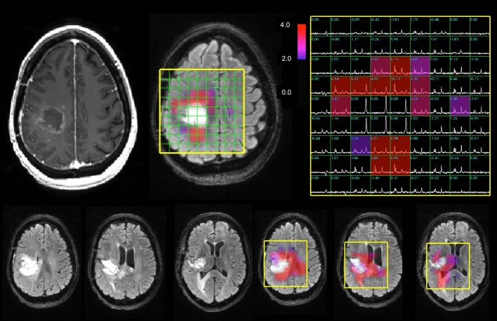
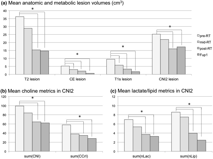
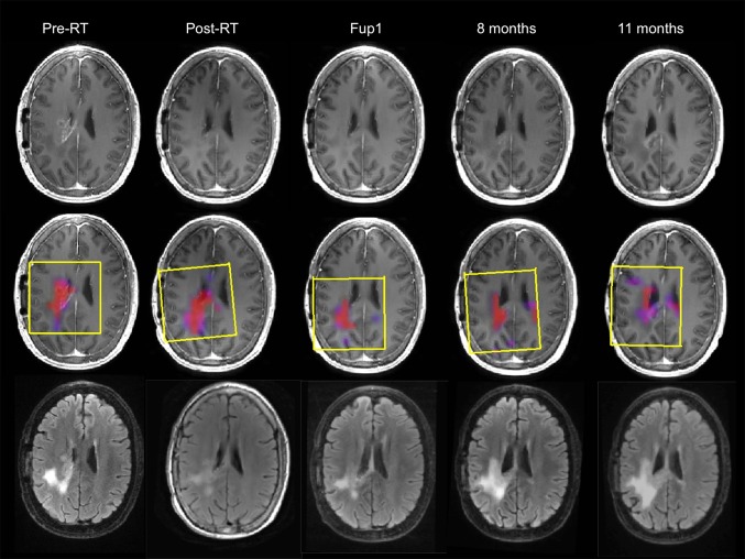
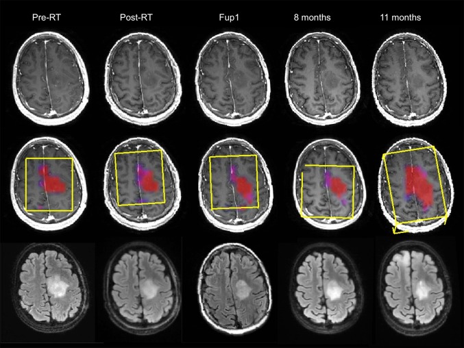
Interpretation of changes in the T1- and T2-weighted MR images from patients with newly diagnosed glioblastoma (GBM) treated with standard of care in conjunction with anti-angiogenic agents is complicated by pseudoprogression and pseudoresponse. The hypothesis being tested in this study was that 3D H-1 magnetic resonance spectroscopic imaging (MRSI) provides estimates of levels of choline, creatine, N-acetylaspartate (NAA), lactate and lipid that change in response to treatment and that metrics describing these characteristics are associated with survival. Thirty-one patients with newly diagnosed GBM and being treated with radiation therapy (RT), temozolomide, erlotinib and bevacizumab were recruited to receive serial MR scans that included 3-D lactate edited MRSI at baseline, mid-RT, post-RT and at specific follow-up time points. The data were processed to provide estimates of metrics representing changes in metabolite levels relative to normal appearing brain. Cox proportional hazards analysis was applied to examine the relationship of these parameters with progression free survival (PFS) and overall survival (OS). There were significant reductions in parameters that describe relative levels of choline to NAA and creatine, indicating that the treatment caused a decrease in tumor cellularity. Changes in the levels of lactate and lipid relative to the NAA from contralateral brain were consistent with vascular normalization. Metabolic parameters from the first serial follow-up scan were associated with PFS and OS, when accounting for age and extent of resection. Integrating metabolic parameters into the assessment of patients with newly diagnosed GBM receiving therapies that include anti-angiogenic agents may be helpful for tracking changes in tumor burden, resolving ambiguities in anatomic images caused by non-specific treatment effects and for predicting outcome.
Keywords: Anti-angiogenic therapy; MRSI; Metabolic imaging; Newly diagnosed glioblastoma; Survival.
Conflict of interest statement
The authors declare that they have no conflict of interest.
Figures
Similar articles
-
Association of early changes in 1H MRSI parameters with survival for patients with newly diagnosed glioblastoma receiving a multimodality treatment regimen.Neuro Oncol. 2017 Mar 1;19(3):430-439. doi: 10.1093/neuonc/now159. Neuro Oncol. 2017. PMID: 27576874 Free PMC article.
-
3-Dimensional magnetic resonance spectroscopic imaging at 3 Tesla for early response assessment of glioblastoma patients during external beam radiation therapy.Int J Radiat Oncol Biol Phys. 2014 Sep 1;90(1):181-9. doi: 10.1016/j.ijrobp.2014.05.014. Epub 2014 Jun 28. Int J Radiat Oncol Biol Phys. 2014. PMID: 24986746 Free PMC article.
-
Comparison of ADC metrics and their association with outcome for patients with newly diagnosed glioblastoma being treated with radiation therapy, temozolomide, erlotinib and bevacizumab.J Neurooncol. 2015 Jan;121(2):331-9. doi: 10.1007/s11060-014-1636-6. Epub 2014 Oct 29. J Neurooncol. 2015. PMID: 25351579 Free PMC article.
-
[Pseudoprogression or pseudoresponse: a challenge for the diagnostic imaging in Glioblastoma multiforme].Wien Med Wochenschr. 2011 Jan;161(1-2):13-9. doi: 10.1007/s10354-010-0860-8. Wien Med Wochenschr. 2011. PMID: 21312094 Review. German.
-
Magnetic resonance spectroscopy imaging of the newborn brain--a technical review.Semin Perinatol. 2010 Feb;34(1):20-7. doi: 10.1053/j.semperi.2009.10.003. Semin Perinatol. 2010. PMID: 20109969 Free PMC article. Review.
Cited by
-
Clinical Applications of Magnetic Resonance Spectroscopy in Brain Tumors: From Diagnosis to Treatment.Radiol Clin North Am. 2021 May;59(3):349-362. doi: 10.1016/j.rcl.2021.01.004. Epub 2021 Mar 23. Radiol Clin North Am. 2021. PMID: 33926682 Free PMC article. Review.
-
Magnetic resonance spectroscopy for the study of cns malignancies.Prog Nucl Magn Reson Spectrosc. 2021 Feb;122:23-41. doi: 10.1016/j.pnmrs.2020.11.001. Epub 2020 Dec 2. Prog Nucl Magn Reson Spectrosc. 2021. PMID: 33632416 Free PMC article.
-
Quantitative Magnetic Resonance Imaging for Biological Image-Guided Adaptive Radiotherapy.Front Oncol. 2021 Jan 29;10:615643. doi: 10.3389/fonc.2020.615643. eCollection 2020. Front Oncol. 2021. PMID: 33585242 Free PMC article. Review.
-
Early changes in glioblastoma metabolism measured by MR spectroscopic imaging during combination of anti-angiogenic cediranib and chemoradiation therapy are associated with survival.NPJ Precis Oncol. 2017;1:20. doi: 10.1038/s41698-017-0020-3. Epub 2017 Jun 12. NPJ Precis Oncol. 2017. PMID: 29202103 Free PMC article.
-
Deep learning super-resolution magnetic resonance spectroscopic imaging of brain metabolism and mutant isocitrate dehydrogenase glioma.Neurooncol Adv. 2022 May 24;4(1):vdac071. doi: 10.1093/noajnl/vdac071. eCollection 2022 Jan-Dec. Neurooncol Adv. 2022. PMID: 35911635 Free PMC article.
References
-
- Stupp R, Hegi ME, Mason WP, van den Bent MJ, Taphoorn MJ, Janzer RC, Ludwin SK, Allgeier A, Fisher B, Belanger K, Hau P, Brandes AA, Gijtenbeek J, Marosi C, Vecht CJ, Mokhtari K, Wesseling P, Villa S, Eisenhauer E, Gorlia T, Weller M, Lacombe D, Cairncross JG, Mirimanoff RO, European Organisation for Research and Treatment of Cancer Brain Tumour and Radiation Oncology Groups; National Cancer Institute of Canada Clinical Trials Group Effects of radiotherapy with concomitant and adjuvant temozolomide versus radiotherapy alone on survival in glioblastoma in a randomised phase III study: 5-year analysis of the EORTC-NCIC trial. Lancet Oncol. 2009;10(5):459–466. doi: 10.1016/S1470-2045(09)70025-7. - DOI - PubMed
MeSH terms
Substances
Grants and funding
LinkOut - more resources
Full Text Sources
Other Literature Sources
Medical
