. 2016 Aug 18:6:31877.
doi: 10.1038/srep31877.

MASP-3 is the exclusive pro-factor D activator in resting blood: the lectin and the alternative complement pathways are fundamentally linked

Affiliations

MASP-3 is the exclusive pro-factor D activator in resting blood: the lectin and the alternative complement pathways are fundamentally linked

József Dobó et al. Sci Rep. .

Abstract

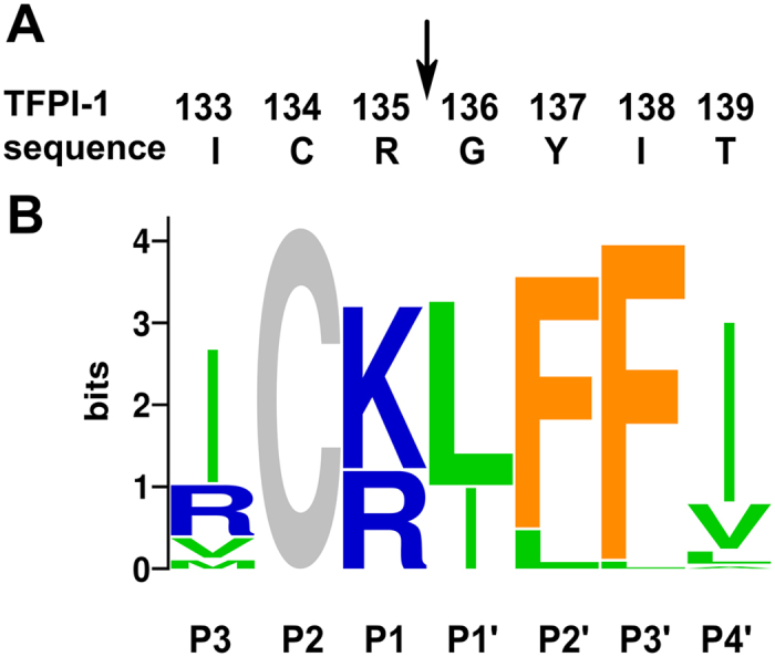
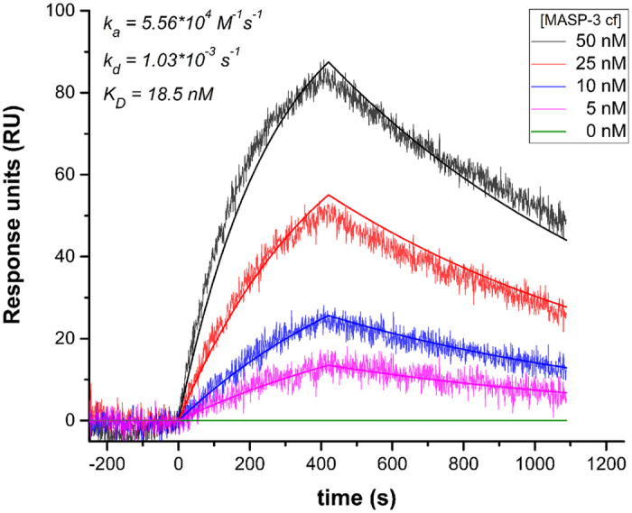
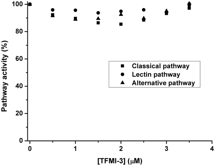
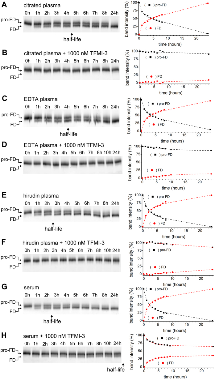
MASP-3 was discovered 15 years ago as the third mannan-binding lectin (MBL)-associated serine protease of the complement lectin pathway. Lacking any verified substrate its role remained ambiguous. MASP-3 was shown to compete with a key lectin pathway enzyme MASP-2 for MBL binding, and was therefore considered to be a negative complement regulator. Later, knock-out mice experiments suggested that MASP-1 and/or MASP-3 play important roles in complement pro-factor D (pro-FD) maturation. However, studies on a MASP-1/MASP-3-deficient human patient produced contradicting results. In normal resting blood unperturbed by ongoing coagulation or complement activation, factor D is present predominantly in its active form, suggesting that resting blood contains at least one pro-FD activating proteinase that is not a direct initiator of coagulation or complement activation. We have recently showed that all three MASPs can activate pro-FD in vitro. In resting blood, however, using our previously evolved MASP-1 and MASP-2 inhibitors we proved that neither MASP-1 nor MASP-2 activates pro-FD. Other plasma proteinases, particularly MASP-3, remained candidates for that function. For this study we evolved a specific MASP-3 inhibitor and unambiguously proved that activated MASP-3 is the exclusive pro-FD activator in resting blood, which demonstrates a fundamental link between the lectin and alternative pathways.


Figures

Figure 1
Figure 1. Development of the MASP-3 specific inhibitor, TFMI-3.
(A) The TFPI-1 domain 2 (TFPI-D2) sequence around the P1 (135) position. The numbering corresponds to the full length protein sequence from the UniProt database. (B) Codon-bias normalized sequence logo of MASP-3-selected unique clones from the TFPI-D2 phage library. Position heights represent the degree of conservation. The nonrandomized Cys residue shows the maximum height. Letter heights indicate normalized amino acid frequencies. Colors reflect chemical properties.
Figure 2
Figure 2. Interaction of TFMI-3 and MASP-3 determined by Surface Plasmon Resonance.
MASP-3cf was injected over TFMI-3_HA immobilized on anti-HA antibody-coated GLM sensor chip as described in “Experimental Procedures”. Result of one of the three independent runs is shown as sensograms corresponding to five parallel analyte injections of different MASP-3cf concentrations. Smooth lines represent global fit of the experimental data to a 1:1 Langmuir model.
Figure 3
Figure 3. Lack of inhibitory effect of TFMI-3 on the three complement pathways.
TFMI-3 was applied in 0–3.5 μM in standard complement activation assays (Wieslab). No significant inhibition of any of the three pathways, CP (■), LP (●) or AP (▲) was observed in normal human serum. Typical curves from 3 parallel assays are shown where each data point represents the mean of 3 repeats. SD bars are not shown to allow distinction of the symbols.
Figure 4
Figure 4. Inhibition of pro-FD activation by TFMI-3.
Conversion of Cy3-labeled pro-FD to Cy3-FD (referred to as activation) in different types of normal human blood preparations was detected by reducing SDS-PAGE followed by fluorimetric scanning and densitometry. Representative gels out of at least 2 parallels are shown on the left side. On the right side of each panel densitometric analysis of the pro-FD and the FD bands of the same gel is shown. The intersection point of the dashed trend lines gives the half-life of pro-FD. Calculated half-lives are found in s 3. (A) Activation of Cy3-pro-FD in citrated normal human plasma. (B) Lack of significant activation of Cy3-pro-FD in citrated normal human plasma in the presence of 1000 nM TFMI-3. (C) Activation of Cy3-pro-FD in normal human EDTA plasma. (D) Lack of significant activation of Cy3-pro-FD in normal human EDTA plasma in the presence of 1000 nM TFMI-3. (E) Activation of Cy3-pro-FD in hirudin-treated normal human plasma. (F) Lack of significant activation of Cy3-pro-FD in normal human hirudin plasma in the presence of 1000 nM TFMI-3. (G) Activation of Cy3-pro-FD in normal human serum. (H) Delayed activation of Cy3-labelled pro-FD in normal human serum in the presence of 1000 nM TFMI-3.

Similar articles

Cited by

References

    1. Merle N. S., Church S. E., Fremeaux-Bacchi V. & Roumenina L. T. Complement system part I - molecular mechanisms of activation and regulation. Front. Immunol. 6 (2015). - PMC - PubMed
    1. Ricklin D., Hajishengallis G., Yang K. & Lambris J. D. Complement: a key system for immune surveillance and homeostasis. Nat. Immunol. 11, 785–797 (2010). - PMC - PubMed
    1. Gaboriaud C., Teillet F., Gregory L. A., Thielens N. M. & Arlaud G. J. Assembly of C1 and the MBL- and ficolin-MASP complexes: Structural insights. Immunobiology 212, 279–288 (2007). - PubMed
    1. Harboe M. & Mollnes T. E. The alternative complement pathway revisited. J. Cell. Mol. Med. 12, 1074–1084 (2008). - PMC - PubMed
    1. Forneris F. et al. Structures of C3b in Complex with Factors B and D Give Insight into Complement Convertase Formation. Science 330, 1816–1820 (2010). - PMC - PubMed

Publication types

MeSH terms

Substances