. 2016 Aug 17;36(33):8562-73.
doi: 10.1523/JNEUROSCI.1059-16.2016.

Dysregulation of Neuronal Ca2+ Channel Linked to Heightened Sympathetic Phenotype in Prohypertensive States

Affiliations

Dysregulation of Neuronal Ca2+ Channel Linked to Heightened Sympathetic Phenotype in Prohypertensive States

Hege E Larsen et al. J Neurosci. .

Abstract

Hypertension is associated with impaired nitric oxide (NO)-cyclic nucleotide (CN)-coupled intracellular calcium (Ca(2+)) homeostasis that enhances cardiac sympathetic neurotransmission. Because neuronal membrane Ca(2+) currents are reduced by NO-activated S-nitrosylation, we tested whether CNs affect membrane channel conductance directly in neurons isolated from the stellate ganglia of spontaneously hypertensive rats (SHRs) and their normotensive controls. Using voltage-clamp and cAMP-protein kinase A (PKA) FRET sensors, we hypothesized that impaired CN regulation provides a direct link to abnormal signaling of neuronal calcium channels in the SHR and that targeting cGMP can restore the channel phenotype. We found significantly larger whole-cell Ca(2+) currents from diseased neurons that were largely mediated by the N-type Ca(2+) channel (Cav2.2). Elevating cGMP restored the SHR Ca(2+) current to levels seen in normal neurons that were not affected by cGMP. cGMP also decreased cAMP levels and PKA activity in diseased neurons. In contrast, cAMP-PKA activity was increased in normal neurons, suggesting differential switching in phosphodiesterase (PDE) activity. PDE2A inhibition enhanced the Ca(2+) current in normal neurons to a conductance similar to that seen in SHR neurons, whereas the inhibitor slightly decreased the current in diseased neurons. Pharmacological evidence supported a switching from cGMP acting via PDE3 in control neurons to PDE2A in SHR neurons in the modulation of the Ca(2+) current. Our data suggest that a disturbance in the regulation of PDE-coupled CNs linked to N-type Ca(2+) channels is an early hallmark of the prohypertensive phenotype associated with intracellular Ca(2+) impairment underpinning sympathetic dysautonomia.

Significance statement: Here, we identify dysregulation of cyclic-nucleotide (CN)-linked neuronal Ca(2+) channel activity that could provide the trigger for the enhanced sympathetic neurotransmission observed in the prohypertensive state. Furthermore, we provide evidence that increasing cGMP rescues the channel phenotype and restores ion channel activity to levels seen in normal neurons. We also observed CN cross-talk in sympathetic neurons that may be related to a differential switching in phosphodiesterase activity. The presence of these early molecular changes in asymptomatic, prohypertensive animals could facilitate the identification of novel therapeutic targets with which to modulate intracellular Ca(2+) Turning down the gain of sympathetic hyperresponsiveness in cardiovascular disease associated with sympathetic dysautonomia would have significant therapeutic utility.

Keywords: autonomic nervous system; cGMP; dysautonomia; hypertension; N-type Ca2+ channel; sympathetic.


Figures

Figure 1.
Figure 1.
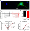
The whole-cell Ca2+ current is larger in the prohypertensive SHR. Whole-cell voltage clamp was performed on the cardiac sympathetic stellate ganglion innervating the heart to investigate the whole-cell Ca2+ properties of 4-week-old prohypertensive SHR and normotensive control rats; 50 ms, 10 mV voltage steps from −50 to +50 were applied to the cell before the resulting current was measured. Immunofluoresence showed TH positivity, confirming sympathetic phenotype of the neurons (A). B, Representative voltage-clamp traces of control and SHR neurons showing significantly larger currents in the SHR neurons. C, Peak current (−10 mV) was −127.5 ± 5.94 pA/pF (n = 10) in the SHR and −108.0 ± 6.80 pA/pF (n = 10, p < 0.045) in the control. D, Current–voltage relationship showing significantly larger currents in the SHR compared with the control at multiple voltages. E, There were no changes in the voltage dependency of the channel, as shown by the activation curve, suggesting that the biophysical properties of the channels are unaffected. Data are represented as the mean ± SEM.
Figure 2.
Figure 2.
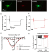
1 μm conotoxin GVIA blocks 75% and 83% of the current, suggesting that the N-type Ca2+ channel is the major contributor to ionic conductance. A, Immunofluorescence showing TH positivity and cell surface expression of the N-type Ca2+ channel. The nuclear stain DAPI is included on the overlay image. Representative peak (−10 mV) voltage-clamp traces of control (B) and SHR (C) in normal conditions (solid lines) and after treatment with conotoxin GVIA (dashed lines). D, Current–voltage relationship showing uniform reduction in both the SHR and control neurons at multiple voltages. The peak current remains at −10 mV and is not significantly different between the control and SHR. E, Bar chart of the peak currents (−10 mV) showing a 75% (control) and 83% (SHR) reduction of the N-type Ca2+ current down to levels that were not significantly different between the strains (−26.88 ± 1.7 pA/pF, n = 6 and −22.04 ± 1.60 pA/pF, p = 0.07 n = 5). Dashed lines represent the mean of the control (black) and SHR (red) control data. Data are represented as the mean ± SEM.
Figure 3.
Figure 3.
Restoring the cGMP levels rescues the channel phenotype seen in the SHR. Representative peak voltage–clamp traces at −10 mV of control (A) and SHR (B) neurons showing a decrease in the SHR current after application of 100 μm of a membrane-permeable cGMP analog 8b-cGMP. Current–voltage relationship of control (C) and SHR (D) neurons shows a normalization of the SHR current down to control levels at all voltages. The peak current remains at −10 mV and is not significantly different from the controls. The drug does not significantly alter the control currents. E, Bar chart of the peak currents (−10 mV) showing a significant reduction and normalization of the SHR current (−127.5 ± 5.94 pA/pF, n = 10 to −105.2 ± 7.79 pA/pF, n = 7, p = 0.035) down to control levels (−108.0 ± 6.80 pA/pF, n = 10, p = 0.79). Dashed lines represent the mean of the control (black) and SHR (red) control data. F, Activation plot showing no significant difference between the SHR and control or the drug groups, suggesting that the biophysical properties of the channels are unchanged. Data are represented as the mean ± SEM.
Figure 4.
Figure 4.
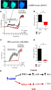
Differential cGMP–cAMP cross-talk in the SHR. In the control cells, 8b-cGMP application results in an increase in cAMP levels, but a reduction in the SHR neurons (11.68 ± 1.54% vs −4.82 ± 2.02%, p < 0.0001, unpaired t test, n = 14–16; A, B). As a result, the PKA activity in the control neurons was found to be significantly higher than that of the SHR (19.15 ± 3.51% vs 1.09 ± 0.57%, p < 0.0001, Mann–Whitney test, n = 6–8; C, D). These results point toward PDE2A dominance in the SHR, but a greater regulation by a cGMP inhibited PDE in the control cells (E).
Figure 5.
Figure 5.
PDE2A blockade suggests differential involvement of PDEs in the control and SHR neurons. Current–voltage relationships of the control (A) and SHR (B) neurons after the application of 1 μm BAY 60–7550, a PDE2A-selective inhibitor. BAY 60–7550 significantly increased the peak control channel currents (−108.0 ± 6.803 pA/pF to −138.7 ± 9.610 pA/pF, n = 9–10, p = 0.0169), but showed a slight, nonsignificant decrease on the SHR currents (−127.5 ± 5.937 pA/pF to −118.0 ± 6.673 pA/pF, p = 0.052 n = 10 and 9). After PDE2A inhibition, the control currents were trending toward being larger than the SHR, but this was not quite significant (138.7 ± 9.610 pA/pF to −118.0 ± 6.673 pA/pF, p = 0.052; C).
Figure 6.
Figure 6.
Evidence for the functional expression of PDE2 and PDE3 in the sympathetic neurons of the SHR and WKY. To assess the functional expression of PDE2 and PDE3, FRET imaging of cAMP levels (H187) in response to well characterized PDE-selective blockers was performed. Pharmacological inhibition of PDE2A (1 μm Bay 60–7550; A) and PDE3 (10 μm milrinone; B) increased the cAMP levels in both the control and SHR neurons. This was found to be nonsignificant between the two strains (n = 8–16; PDE2: 19.69 ± 5.16%, n = 10 vs 36.55 ± 21.47%, n = 8, p = 0.94. PDE3: milrinone; 22.98 ± 6.81%, n = 10 vs 31.14 ± 6.25% n = 16, p = 0.41. cilostamide; 28.15 ± 15.45%, n = 5 vs 33.23 ± 8.18% n = 8, p = 0.58, Mann–Whitney test). The presence of PDE2A in sympathetic stellate neurons is known, but this provides the first evidence for the functional expression of PDE3 in these cells.
Figure 7.
Figure 7.
cAMP–PKA signaling pathway is functional in SHR neurons. To investigate the ability of the cells to generate cAMP and PKA, activity was compared between the SHRs and controls using FRET imaging. Cells were infected with the H30 Epac and AKAR sensors and the changes in fluorescence emission intensity of CFP and YFP was monitored (A). No significant differences were found in forskolin-induced cAMP generation (B) or PKA activity (C) in the sympathetic neurons from the prohypertensive SHR or controls. Comparisons were made using the Mann–Whitney test.
Figure 8.
Figure 8.
Sympathetic neurotransmission. The arrival of an action potential in the synaptic nerve terminal leads to Ca2+ entry via voltage-gated Ca2+ channels. This triggers exocytosis of noradrenaline into the synaptic clefts, which acts on the β1 adrenoreceptor, altering myocyte behavior. In hypertension, nNOS expression and the β1 subunit of sGC expression is down, resulting in lower levels of cGMP, which, coupled with enhanced PDE2A activity, results in increased neurotransmission. Pharmacological elevation of cGMP reverses the channel phenotype, but also leads to changes in cAMP levels and PKA activity as a result of interactions with PDEs. The differential responses of the control and SHR neurons to cGMP elevation suggests that cAMP levels are controlled by both PDE2A and PDE3 subtypes in the control neurons, but that the prohypertensive phenotype is associated with PDE2A dominance.

Comment in

Similar articles

Cited by

References

    1. Bannister RA, Meza U, Adams BA. Phosphorylation-dependent regulation of voltage-gated Ca2+ channels. In: Zamponi G., editor. Voltage-gated calcium channels. Boston: Springer; 2005. pp. 168–182.
    1. Bar-Yehuda D, Korngreen A. Space-clamp problems when voltage clamping neurons expressing voltage-gated conductances. J Neurophysiol. 2008;99:1127–1136. doi: 10.1152/jn.01232.2007. - DOI - PubMed
    1. Bender AT, Beavo JA. Cyclic nucleotide phosphodiesterases: molecular regulation to clinical use. Pharmacol Rev. 2006;58:488–520. doi: 10.1124/pr.58.3.5. - DOI - PubMed
    1. Catterall WA. Structure and regulation of voltage-gated Ca2+ channels. Annu Rev Cell Dev Biol. 2000;16:521–555. doi: 10.1146/annurev.cellbio.16.1.521. - DOI - PubMed
    1. Catterall WA. Voltage-gated calcium channels. Cold Spring Harb Perspect Biol. 2011;3:a003947. doi: 10.1101/cshperspect.a003947. - DOI - PMC - PubMed

Publication types

MeSH terms