. 2016 Aug 17;36(33):8641-52.
doi: 10.1523/JNEUROSCI.0630-16.2016.

The Brain-Enriched MicroRNA miR-9-3p Regulates Synaptic Plasticity and Memory

Affiliations

The Brain-Enriched MicroRNA miR-9-3p Regulates Synaptic Plasticity and Memory

Su-Eon Sim et al. J Neurosci. .

Abstract

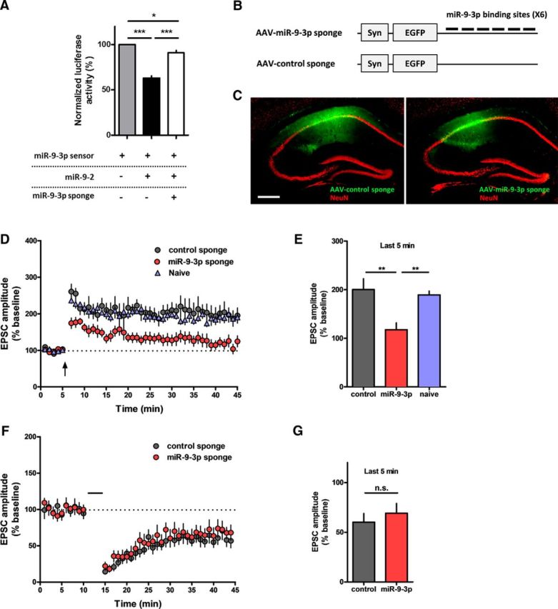
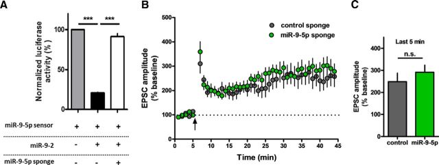
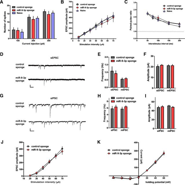
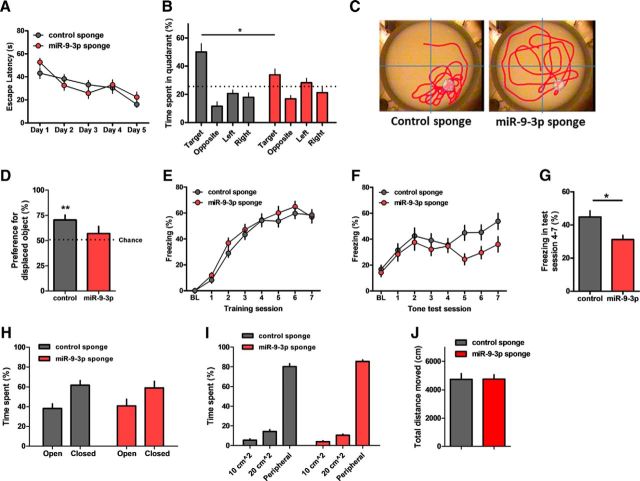
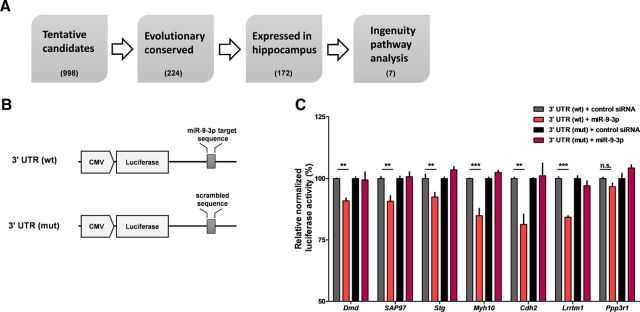
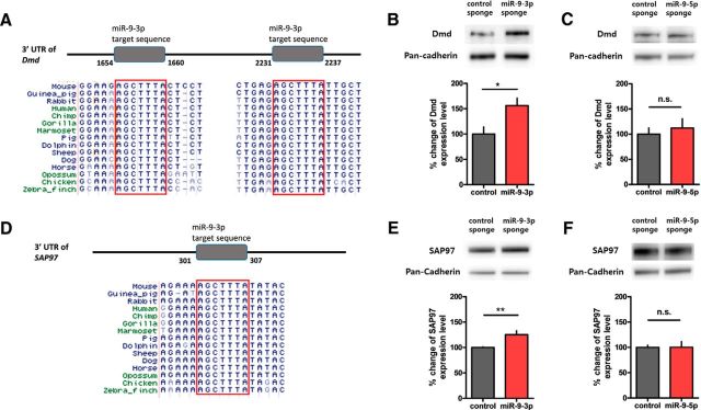
MicroRNAs (miRNAs) are small, noncoding RNAs that posttranscriptionally regulate gene expression in many tissues. Although a number of brain-enriched miRNAs have been identified, only a few specific miRNAs have been revealed as critical regulators of synaptic plasticity, learning, and memory. miR-9-5p/3p are brain-enriched miRNAs known to regulate development and their changes have been implicated in several neurological disorders, yet their role in mature neurons in mice is largely unknown. Here, we report that inhibition of miR-9-3p, but not miR-9-5p, impaired hippocampal long-term potentiation (LTP) without affecting basal synaptic transmission. Moreover, inhibition of miR-9-3p in the hippocampus resulted in learning and memory deficits. Furthermore, miR-9-3p inhibition increased the expression of the LTP-related genes Dmd and SAP97, the expression levels of which are negatively correlated with LTP. These results suggest that miR-9-3p-mediated gene regulation plays important roles in synaptic plasticity and hippocampus-dependent memory.

Significance statement: Despite the abundant expression of the brain-specific microRNA miR-9-5p/3p in both proliferating and postmitotic neurons, most functional studies have focused on their role in neuronal development. Here, we examined the role of miR-9-5p/3p in adult brain and found that miR-9-3p, but not miR-9-5p, has a critical role in hippocampal synaptic plasticity and memory. Moreover, we identified in vivo binding targets of miR-9-3p that are involved in the regulation of long-term potentiation. Our study provides the very first evidence for the critical role of miR-9-3p in synaptic plasticity and memory in the adult mouse.

Keywords: hippocampus; long-term potentiation; memory; microRNA.


Figures

Figure 1.
Figure 1.
AAV-miR-9-3p sponge-mediated miR-9-3p inhibition blocks hippocampal LTP. A, Luciferase assay showing that the miR-9-3p sponge specifically suppresses the activity of miR-9-3p (*p < 0.05, ***p < 0.001, n = 5 per group, Tukey's multiple-comparisons test after significant 1-way ANOVA). The expression of miR-9-2, a genetic locus encoding miR-9-5p/3p, inhibited the expression of the miR-9-3p sensor, which rescued by cotransfection of the miR-9-3p sponge. B, Schematic diagrams of AAV-miR-9-3p-sponge. Six bulged miR-9-3p-binding sites were contained in the 3′ UTR of the EGFP gene. C, Representative fluorescence images showing EGFP expression in hippocampus 4 weeks after injection of AAV-miR-sponge. Scale bar, 300 μm. D, Theta-burst LTP recordings were significantly impaired in miR-9-3p sponge-expressing neurons. E, Summary graph representing the average EPSC amplitudes of the last 5 min of recording in naive, control sponge, and miR-9-3p sponge groups (last 5 min of recording, control, n = 6 cells from 3 mice, miR-9-3p, n = 9 cells from 6 mice, naive, n = 7 cells from 5 mice, **p < 0.01, Tukey's multiple-comparisons test after 1-way ANOVA). F, Hippocampal LTD was comparable between control and miR-9-5p sponge-expressing neurons. G, Summary graph representing the average EPSC amplitudes of the last 5 min recording in control sponge and miR-9-3p sponge groups (last 5 min of recording, control, n = 9 cells from 4 mice, miR-9-3p, n = 7 cells from 3 mice, unpaired two-tailed t test, t(14) = 0.6813, p = 0.5068). Data are mean ± SEM.
Figure 2.
Figure 2.
AAV-miR-9-5p sponge-mediated miR-9-5p inhibition has no effect on hippocampal LTP. A, Luciferase assay showing that the miR-9-5p sponge specifically suppresses the activity of miR-9-5p (***p < 0.001, n = 4 per group, Tukey's multiple-comparisons test after significant 1-way ANOVA). B, Theta-burst LTP was comparable between control and miR-9-5p sponge. C, Summary graph represents the average EPSC amplitudes of the last 5 min recording in control and miR-9-5p sponge groups (control, n = 12 cells from 7 mice, miR-9-5p, n = 14 cells from 6 mice, unpaired 2-tailed t test, t(24) = 0.8454, p = 0.4062). Data are mean ± SEM.
Figure 3.
Figure 3.
AAV-miR sponge-mediated inhibition of miR-9-3p activity has no effects on basal synaptic properties and NMDAR-mediated synaptic transmission. A, Number of action potentials generated by current injection (n = 6 for each group). B, Input–output relationship (n = 7, naive; n = 8, control sponge; n = 6, miR-9-3p sponge). C, Paired-pulse ratio at the indicated interstimulus intervals (n = 6 for each group). DF, sEPSCs and mEPSCs from CA1 pyramidal neurons expressing control sponge or miR-9-3p sponge. D, Representative sEPSC recording traces. Scale bars, 25 pA and 200 ms. Graphs show sEPSC (n = 6, control sponge; n = 11, miR-9-3p sponge) and mEPSC (n = 7, control sponge; n = 9, miR-9-3p sponge) frequency (E) and amplitude (F). GI, sIPSCs and mIPSCs from CA1 pyramidal neurons expressing control sponge or miR-9-3p sponge. G, Representative sIPSC recording traces. Scale bars, 50 pA and 200 ms. Graphs show sIPSC (n = 10, control sponge; n = 9, miR-9-3p sponge) and mIPSC (n = 11, control sponge; n = 6, miR-9-3p sponge) frequency (H) and amplitude (I). J, Input–output relationship of NMDAR-mediated EPSCs (n = 8, control sponge; n = 11, miR-9-3p sponge). K, NMDAR-mediated I–V plot (n = 11, control sponge; n = 12, miR-9-3p sponge). Data are mean ± SEM.
Figure 4.
Figure 4.
Inhibition of miR-9-3p activity impairs hippocampus-dependent memories. AC, AAV-miR-9-3p sponge-expressing mice showed impaired spatial memory in the Morris water maze test (n = 9, AAV-control sponge; n = 10, AAV-miR-9-3p sponge). Escape latencies to the platform were similar in control and miR-9-3p sponge groups (repeated-measures 2-way ANOVA, the effect of sponge, F(1,68) = 0.15, p = 0.7062) (A). Probe test examining time spent in the target quadrant on day 6 showed significant spatial memory deficits in miR-9-3p sponge group (2-way ANOVA, sponge × quadrant, F(3,68) = 4.44, p = 0.0066; Bonferroni's post tests, *p < 0.05) (B). C, Representative images showing the swimming traces of mice during the probe trial. Dashed circle indicates the location of removed platform. D, Object location memory was significantly impaired in the miR-9-3p sponge group (n = 8, AAV-control sponge; n = 9, AAV-miR-9-3p sponge, 1-sample paired t test, **p < 0.01). The percentage preference for the displaced objects in the testing session is reported and the dotted line indicates chance (50%) preference. EG, Trace fear conditioning was impaired in the miR-9-3p sponge group (n = 20, AAV-control sponge; n = 19, AAV-miR-9-3p sponge). The average freezing duration during training sessions was similar in control and miR-9-3p sponge groups (repeated-measures 2-way ANOVA, sponge × day, F(7,259) = 0.72, p = 0.6560) (E). Average freezing duration during testing sessions (repeated-measures 2-way ANOVA, sponge × day, F(7,259) = 2.02, p = 0.0526) (F). Average freezing duration during testing sessions 4–7 (unpaired 2-tailed t test, *p < 0.05) (G). HJ, Basal anxiety level and locomotor activity were intact in both AAV-sponge groups. The elevated zero maze test (n = 10, control sponge; n = 11, miR-9-3p sponge) (H). Open-field test (n = 10, control sponge, n = 11, miR-9-3p sponge) (I, J). Data are mean ± SEM.
Figure 5.
Figure 5.
miR-9-3p regulates the expression of LTP-related genes. A, Flow chart of bioinformatic analyses. The TargetScan algorithm predicted tentative target genes of miR-9-3p. Further analyses were performed based on evolutionary conservation of miR-9-3p target sequence and reliable expression in hippocampus. Finally, seven candidate target genes were identified via Ingenuity Pathway Analysis. Numbers in parentheses indicate the number of counted target genes via each bioinformatics analysis. B, Schematic diagrams of the reporter constructs used for the luciferase assays. C, miR-9-3p suppresses the expression of the luciferase reporters containing the 3′ UTRs of selected LTP-related genes [n = 4–6 for each group, 2-way ANOVA, miR-9-3p × mutation; Dmd, F(1,20) = 5.91, p = 0.0245; SAP97, F(1,20) = 9.59, p = 0.0057; Stg, F(1,20) = 14.97, p = 0.001; Myh10, F(1,20) = 31.39, p < 0.0001; Cdh2, F(1,12) = 8.95, p = 0.0112; Lrrtm1, F(1,12) = 27.5, p = 0.0002; Ppp3r1, F(1,12) = 7.06, p = 0.0209; Bonferroni's post tests, 3′ UTR (wt) + control siRNA versus 3′ UTR (wt) + miR-9-3p, **p < 0.01, ***p < 0.001]. Data are mean ± SEM.
Figure 6.
Figure 6.
Dmd and SAP97 are novel targets of miR-9-3p. A, Bioinformatic analyses identified highly conserved miR-9-3p target sequences within Dmd 3′ UTR. B, The Dmd protein level was significantly increased in the hippocampi overexpressing miR-9-3p sponge (n = 7, AAV-control sponge; n = 8, AAV-miR-9-3p sponge, unpaired 2-tailed t test, *p < 0.05). C, The Dmd protein level was not changed by miR-9-5p inhibition (n = 4, AAV-control sponge; n = 4, AAV-miR-9-5p sponge, unpaired 2-tailed t test). D, Bioinformatic analyses identified highly conserved miR-9-3p target sequence within SAP97 3′ UTR. E, The SAP97 protein level was significantly increased in hippocampi overexpressing the miR-9-3p sponge (n = 5, AAV-control sponge; n = 4, AAV-miR-9-3p sponge, unpaired 2-tailed t test, **p < 0.01). F, The SAP97 protein level was not changed by miR-9-5p inhibition (n = 4, AAV-control sponge; n = 4, AAV-miR-9-5p sponge, unpaired 2-tailed t test). Data are mean ± SEM.

Similar articles

Cited by

References

    1. Bartel DP. MicroRNAs: genomics, biogenesis, mechanism, and function. Cell. 2004;116:281–297. doi: 10.1016/S0092-8674(04)00045-5. - DOI - PubMed
    1. Bartel DP. MicroRNAs: target recognition and regulatory functions. Cell. 2009;136:215–233. doi: 10.1016/j.cell.2009.01.002. - DOI - PMC - PubMed
    1. Bliss TV, Collingridge GL. A synaptic model of memory: long-term potentiation in the hippocampus. Nature. 1993;361:31–39. doi: 10.1038/361031a0. - DOI - PubMed
    1. Bozdagi O, Shan W, Tanaka H, Benson DL, Huntley GW. Increasing numbers of synaptic puncta during late-phase LTP: N-cadherin is synthesized, recruited to synaptic sites, and required for potentiation. Neuron. 2000;28:245–259. doi: 10.1016/S0896-6273(00)00100-8. - DOI - PubMed
    1. Cai C, Coleman SK, Niemi K, Keinänen K. Selective binding of synapse-associated protein 97 to GluR-A α-amino-5-hydroxy-3-methyl-4-isoxazole propionate receptor subunit is determined by a novel sequence motif. J Biol Chem. 2002;277:31484–31490. doi: 10.1074/jbc.M204354200. - DOI - PubMed

MeSH terms