. 2016 Aug 17;36(33):8653-67.
doi: 10.1523/JNEUROSCI.1429-16.2016.

Neutral Sphingomyelinase-2 Deficiency Ameliorates Alzheimer's Disease Pathology and Improves Cognition in the 5XFAD Mouse

Affiliations

Neutral Sphingomyelinase-2 Deficiency Ameliorates Alzheimer's Disease Pathology and Improves Cognition in the 5XFAD Mouse

Michael B Dinkins et al. J Neurosci. .

Abstract

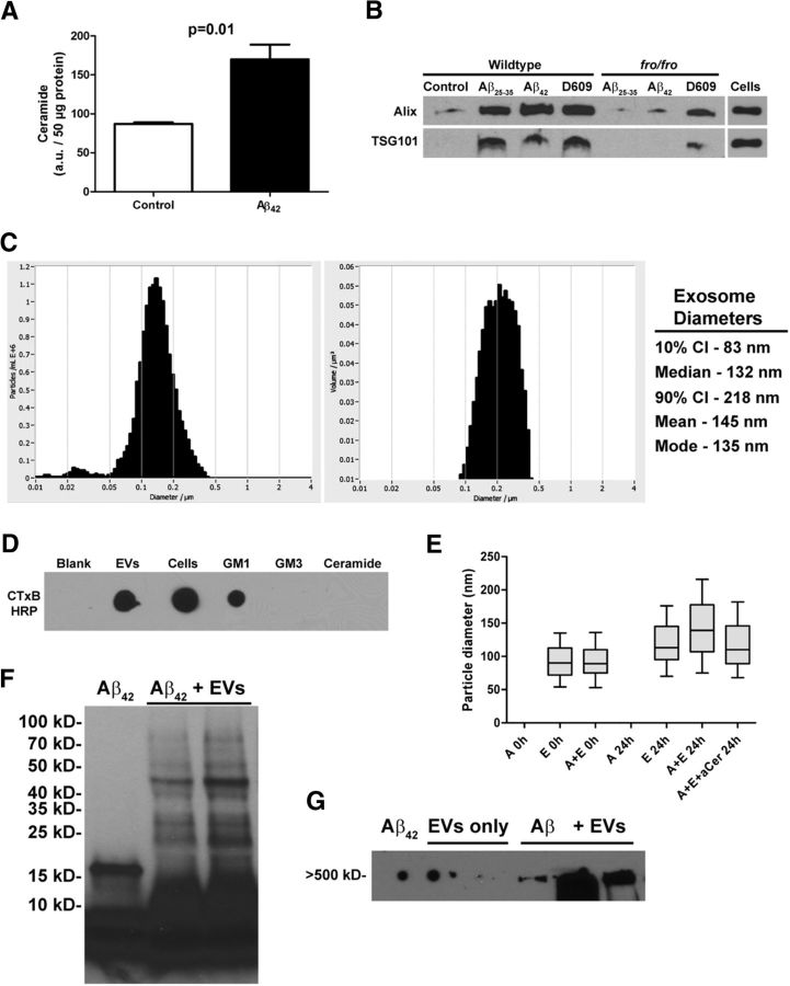
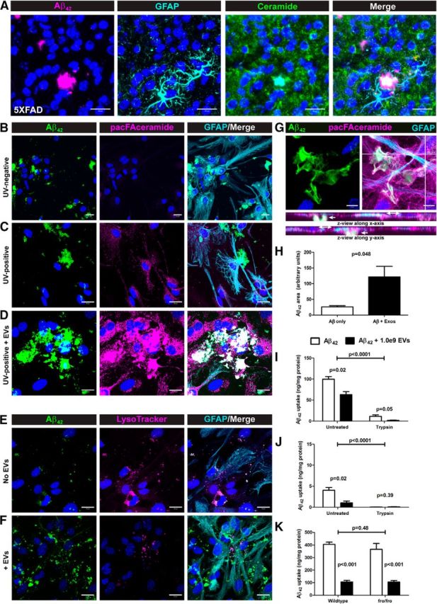
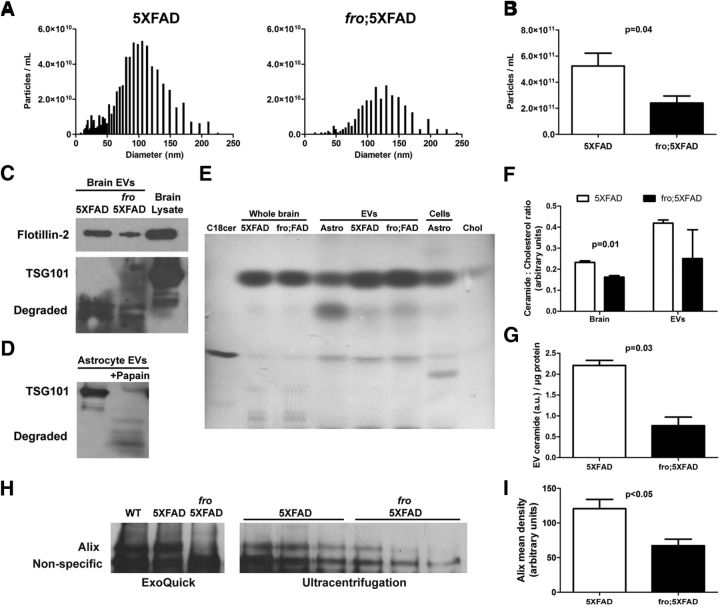
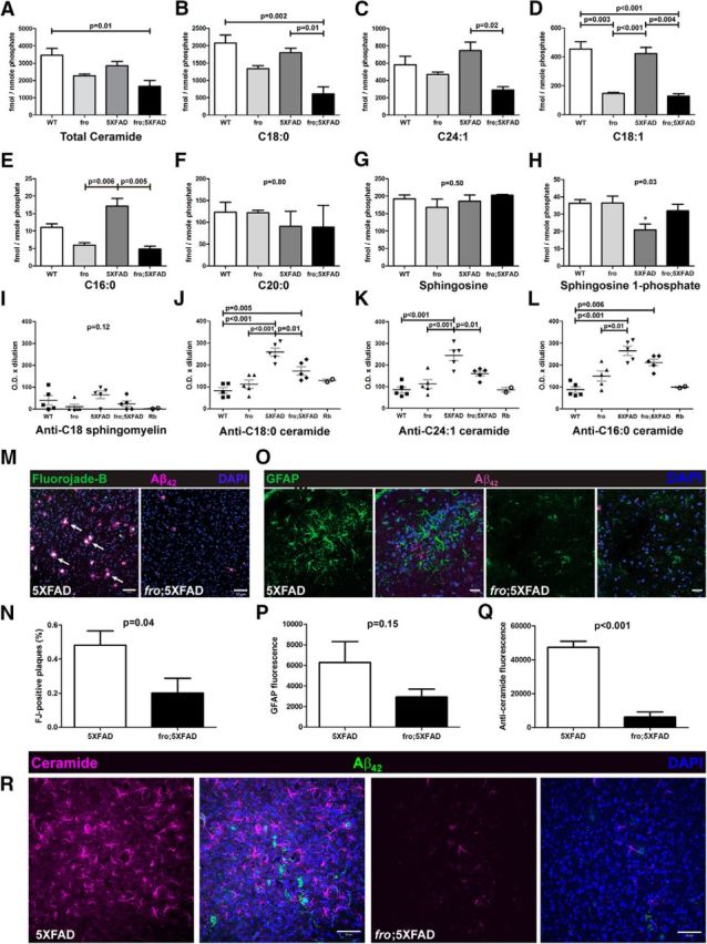
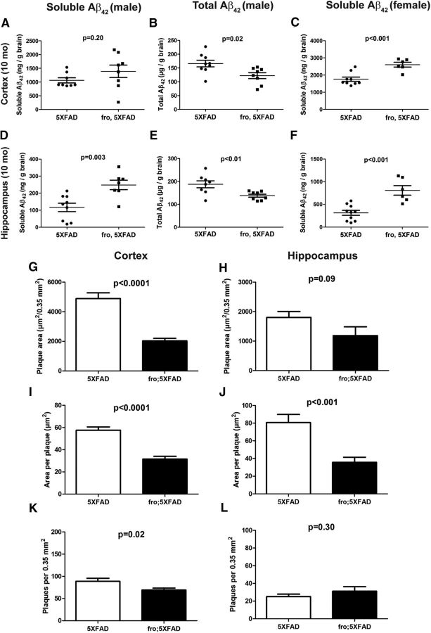
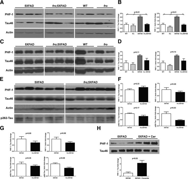
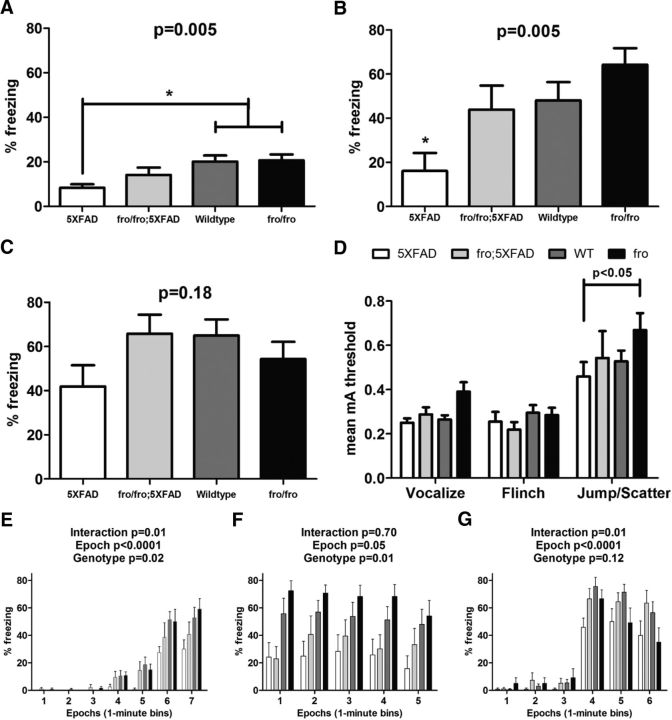
Recent evidence implicates exosomes in the aggregation of Aβ and spreading of tau in Alzheimer's disease. In neural cells, exosome formation can be blocked by inhibition or silencing of neutral sphingomyelinase-2 (nSMase2). We generated genetically nSMase2-deficient 5XFAD mice (fro;5XFAD) to assess AD-related pathology in a mouse model with consistently reduced ceramide generation. We conducted in vitro assays to assess Aβ42 aggregation and glial clearance with and without exosomes isolated by ultracentrifugation and determined exosome-induced amyloid aggregation by particle counting. We analyzed brain exosome content, amyloid plaque formation, neuronal degeneration, sphingolipid, Aβ42 and phospho-tau levels, and memory-related behaviors in 5XFAD versus fro;5XFAD mice using contextual and cued fear conditioning. Astrocyte-derived exosomes accelerated aggregation of Aβ42 and blocked glial clearance of Aβ42 in vitro Aβ42 aggregates were colocalized with extracellular ceramide in vitro using a bifunctional ceramide analog preloaded into exosomes and in vivo using anticeramide IgG, implicating ceramide-enriched exosomes in plaque formation. Compared with 5XFAD mice, the fro;5XFAD mice had reduced brain exosomes, ceramide levels, serum anticeramide IgG, glial activation, total Aβ42 and plaque burden, tau phosphorylation, and improved cognition in a fear-conditioned learning task. Ceramide-enriched exosomes appear to exacerbate AD-related brain pathology by promoting the aggregation of Aβ. Reduction of exosome secretion by nSMase2 loss of function improves pathology and cognition in the 5XFAD mouse model.

Significance statement: We present for the first time evidence, using Alzheimer's disease (AD) model mice deficient in neural exosome secretion due to lack of neutral sphingomyelinase-2 function, that ceramide-enriched exosomes exacerbate AD-related pathologies and cognitive deficits. Our results provide rationale to pursue a means of inhibiting exosome secretion as a potential therapy for individuals at risk for developing AD.

Keywords: 5XFAD; Alzheimer's; ceramide; exosomes; fear conditioning; sphingomyelinase.


Figures

Figure 1.
Figure 1.
Astrocyte-derived exosomes accelerate aggregation of Aβ42. A, Quantification of ceramide by densitometry of high-performance thin-layer chromatography after 24 h exposure of primary astrocytes to 1 μm42 (n = 3). B, Western blot analysis of exosome markers Alix and TSG101 from WT and fro/fro astrocytes after differential ultracentrifugation of conditioned medium. Lanes represent pellets from equivalent amount of medium. C, Representative histograms showing ZetaView-measured (left) particle concentration (y-axis) versus diameter (x-axis) and (right) volume (y-axis) versus diameter of astrocyte exosomes with descriptive statistics listed on the right. D, Dot-blot comparison of astrocyte- and exosome-derived lipids and standards probed with cholera toxin B subunit to detect GM1 ganglioside. E, ZetaView-measured particle diameter of exosomes alone or in the presence of Aβ42 with and without anticeramide IgG. Box plots show median diameter, with bars indicating the 10th to 90th percentile range (n = 3). F, G, Western blot analyses of Aβ42 after incubation alone or in the presence of exosomes showing various aggregation states. Lines above the blot images indicate multiple samples run side by side (in F, n = 2 samples of Aβ+ EVs; in G, 2 lanes of EVs only and 3 lanes of Aβ+ EVs). In G, the spots on the left side of the blot image are due to nonspecific background.
Figure 2.
Figure 2.
Ceramide-enriched exosomes accelerate Aβ deposition at the cell surface and reduce glial Aβ clearance in vitro. A, Confocal micrographs of brain tissue from 10-month-old 5XFAD mice labeled for Aβ42, ceramide and GFAP as indicated showing ceramide localization at the core of Aβ42 plaques. BD, Confocal projections of cultured astrocytes exposed to HiLyte488-Aβ42 after preloading with pacFA-ceramide (B, C) or upon concomitant addition of pacFA-ceramide-loaded EVs (D); pacFA-ceramide was labeled by Click reaction with Alexa Fluor 647-azide followed by GFAP immunolabeling. E, F, Confocal projections of cultured astrocytes exposed to HiLyte488-Aβ42 in the absence (E) or presence (F) of EVs and labeled with LysoTracker Red DND-99 followed by GFAP immunolabeling. G, Confocal micrograph of cultured astrocytes labeled pacFA-ceramide (Alexa Fluor 647) and immunolabeled for GFAP and Aβ42 with orthogonal views to indicate extracellular and intracellular planes. Scale bars: AF, 20 μm; G, 10 μm. H, Quantification of Aβ42 aggregate size (area) from 20× views (data not shown) of confocal micrographs of astrocytes exposed to HiLyte488-Aβ42 in the absence and presence of EVs (n = 3, Student's t test). IK, Aβ42 ELISA data (uptake) after Aβ in vitro clearance assays with mixed glial cultures (I, K) and astrocytes (K) (n = 3 each, 2-way ANOVA with Bonferroni post hoc test). All quantitative data shown are means ± SEM.
Figure 3.
Figure 3.
Reduced extracellular vesicles in fro;5XFAD mice. A, Representative frequency distribution histograms showing ZetaView-measured EV particle concentration (y-axis) versus diameter (x-axis) from 5XFAD (left) and fro;5XFAD (right) mouse brains. B, Quantification of EV particles from 5XFAD and fro;5XFAD brains (n = 4; Student's t test). C, D, Western blots of EVs isolated from 5XFAD and fro;5XFAD brains (C) and astrocytes after digestion with papain (D). E, Representative thin-layer chromatograph of lipids extracted from whole brains and brain EVs from 5XFAD and fro;5XFAD mice and astrocytes and astrocyte EVs. Ceramide and cholesterol are used as standards. F, Quantification of the ceramide:cholesterol ratios from 5XFAD and fro;5XFAD brain and brain EVs as shown in E (n = 3; Student's t test). G, Quantification of ceramide from thin-layer chromatographs of 5XFAD and fro;5XFAD brain EVs normalized to protein (n = 2; Student's t test). H, Western blot of exosome marker Alix from 5XFAD and fro;5XFAD sera after ExoQuick isolation (left) or ultracentrifugation (right). I, Quantification of Alix band intensity in H (n = 3; Student's t test). All quantitative data shown are means ± SEM.
Figure 4.
Figure 4.
Reduced total brain ceramides, serum anticeramide IgG, degenerating neurons, and astrocyte activation in fro;5XFAD mice. AH, Liquid chromatography and dual-mass spectrometry analysis of indicated ceramides and sphingosines in WT, fro, 5XFAD, and fro;5XFAD mice brain (n = 3 each; one-way ANOVA with Bonferroni post hoc test). IL, Quantification of lipid ELISAs to determine relative serum titers of indicated anticeramide IgG in WT, fro, 5XFAD, and fro;5XFAD mice (n = 5 each; one-way ANOVA with Bonferroni's post hoc test). M, Confocal micrographs of brain tissue from 10-month-old 5XFAD and fro;5XFAD mice labeled with FJB to label degenerating neurons and anti-Aβ42. N, Quantification of FJB from M (n = 8; Student's t test). O, Confocal micrographs of brain tissue from 10-month-old 5XFAD and fro;5XFAD mice immunolabeled with anti-GFAP and anti-Aβ42. P, Quantification of GFAP immunolabeling from O (n = 6; Student's t test). Q, Quantification of anticeramide immunolabeling from R (n = 3; Student's t test). R, Confocal micrographs of brain tissue from 10-month-old 5XFAD and fro;5XFAD mice immunolabeled with anticeramide and anti-Aβ42. Scale bars: M, R, 50 μm; O, 20 μm. All quantitative data shown indicate means ± SEM.
Figure 5.
Figure 5.
Total Aβ42 and plaque loads are reduced in fro;5XFAD males, whereas soluble Aβ42 is increased. AF, Dot plots of Aβ42 ELISA data from brains of 5XFAD and fro;5XFAD mice at 10 months (sex and soluble vs total Aβ indicated in figure). The number (n) of mice in each group is indicated by the number of dots in each group (e.g., n = 8 for 5XFAD in A). GL, Graphs showing total plaque area (G, H), average plaque size (I, J), and plaque counts (K, L) per 0.35 mm2 (n = 5 male mice for each group; 4 images from each region: retrosplenial, somatosensory, motor, and auditory; 5 mice × 4 regions × 4 images = 80 images). All data shown are presented means ± SEM and were analyzed by Student's t test.
Figure 6.
Figure 6.
p-tau ratios are reduced in fro;5XFAD and increased in 5XFAD mice treated with ceramide. A, C, E, Western analyses of 5XFAD and fro;5XFAD brain at 3 (A), 5 (C), and 10 (E) months of age. Homogenates were probed with antibodies against pS396/404-tau (PHF-1), p262-tau, tau (Tau46), and actin. B, D, F, Quantification of p-tau to tau or p-tau to actin ratios from blots in A (n = 3; one-way ANOVA with Bonferroni post hoc test), C (n = 3; one-way ANOVA with Bonferroni post hoc test), and E (n = 5; Student's t test), respectively. In G, the data for hippocampi are presented, but blots are not shown (n = 5; Student's t test). H, Western blot analysis of p-tau (PHF-1) and tau in cortices of 5XFAD mice injected with Freund's adjuvant with and without C18:0 ceramide and quantification of band intensity in H (bottom; n = 3; Student's t test). Quantitative data are presented as means ± SEM. All samples within a comparison group were run on the same gel and cropped for the figure.
Figure 7.
Figure 7.
Male fro;5XFAD mice show improved fear-conditioned memory at 8 months. AC, Graphs showing means ± SEM of freezing behavior associated with training (A) and both contextual (B) and cued (C) fear-conditioned learning in WT (n = 13), fro (n = 9), 5XFAD (n = 9), and fro;5XFAD (n = 7) mice analyzed by one-way ANOVA with Bonferroni's post hoc test. D, Graph showing the mean (±SEM) threshold (reported in milliamp units) required to elicit vocalization, flinching, and jumping in response to the presentation electric stock stimuli (range = 0.1–1.0 mA). EG, Graphs showing means ± SEM of freezing behavior presented as the percentage of time mice were frozen in epochs of 1 min bins during training (A), contextual testing (B), and cued testing (C). Data were analyzed by two-way ANOVA with Bonferroni post hoc test.

Similar articles

Cited by

References

    1. Alessenko AV, Bugrova AE, Dudnik LB. Connection of lipid peroxide oxidation with the sphingomyelin pathway in the development of Alzheimer's disease. Biochem Soc Trans. 2004;32:144–146. doi: 10.1042/bst0320144. - DOI - PubMed
    1. An K, Klyubin I, Kim Y, Jung JH, Mably AJ, O'Dowd ST, Lynch T, Kanmert D, Lemere CA, Finan GM, Park JW, Kim TW, Walsh DM, Rowan MJ, Kim JH. Exosomes neutralize synaptic-plasticity-disrupting activity of Abeta assemblies in vivo. Mol Brain. 2013;6:47. doi: 10.1186/1756-6606-6-47. - DOI - PMC - PubMed
    1. Asai H, Ikezu S, Tsunoda S, Medalla M, Luebke J, Haydar T, Wolozin B, Butovsky O, Kügler S, Ikezu T. Depletion of microglia and inhibition of exosome synthesis halt tau propagation. Nat Neurosci. 2015;18:1584–1593. doi: 10.1038/nn.4132. - DOI - PMC - PubMed
    1. Aubin I, Adams CP, Opsahl S, Septier D, Bishop CE, Auge N, Salvayre R, Negre-Salvayre A, Goldberg M, Guénet JL, Poirier C. A deletion in the gene encoding sphingomyelin phosphodiesterase 3 (Smpd3) results in osteogenesis and dentinogenesis imperfecta in the mouse. Nat Genet. 2005;37:803–805. doi: 10.1038/ng1603. - DOI - PubMed
    1. Baleriola J, Walker CA, Jean YY, Crary JF, Troy CM, Nagy PL, Hengst U. Axonally synthesized ATF4 transmits a neurodegenerative signal across brain regions. Cell. 2014;158:1159–1172. doi: 10.1016/j.cell.2014.07.001. - DOI - PMC - PubMed

Publication types

MeSH terms