

A review of the role of ultrasound biomicroscopy in glaucoma associated with rare diseases of the anterior segment
- PMID: 27536058
- PMCID: PMC4975163
- DOI: 10.2147/OPTH.S112166
A review of the role of ultrasound biomicroscopy in glaucoma associated with rare diseases of the anterior segment
Abstract
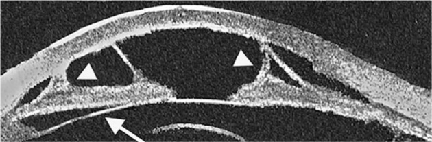
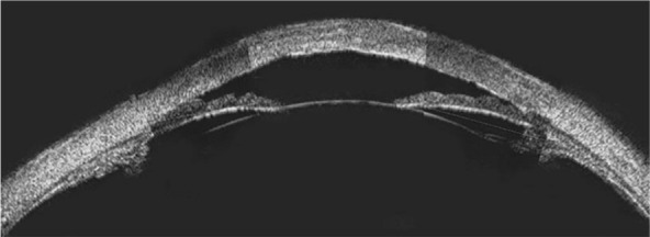
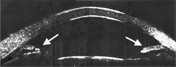
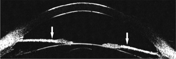
Ultrasound biomicroscopy is a non-invasive imaging technique, which allows high-resolution evaluation of the anatomical features of the anterior segment of the eye regardless of optical media transparency. This technique provides diagnostically significant information in vivo for the cornea, anterior chamber, chamber angle, iris, posterior chamber, zonules, ciliary body, and lens, and is of great value in assessment of the mechanisms of glaucoma onset. The purpose of this paper is to review the use of ultrasound biomicroscopy in the diagnosis and management of rare diseases of the anterior segment such as mesodermal dysgenesis of the neural crest, iridocorneal endothelial syndrome, phakomatoses, and metabolic disorders.
Keywords: glaucoma; iridocorneal syndrome; metabolic disorders; neural crest; phakomatoses; rare diseases; ultrasound biomicroscopy.
Figures
References
-
- Pavlin CJ, Foster FS. Ultrasound biomicroscopy of the eye. New York: Springer & Verlag; 1995.
-
- Mannino G, Papale A, De Bella F. Biomicroscopia ad ultrasuoni [Ultrasound biomicroscopy] Fabiano Editore; 2004. Italian.
-
- Pavlin CJ, Haraseiwicz K, Sherar MD, Foster FS. Clinical use of ultrasound biomicroscopy. Ophthalmology. 1991;98(3):287–295. - PubMed
-
- Kiryu J, Park M, Kobayashi H, Kondo T. Ultrasound biomicroscopy of the anterior segment of newborn infants. J Pediatr Ophthalmol Strabismus. 1998;35:320–322. - PubMed
Publication types
LinkOut - more resources
Full Text Sources
Other Literature Sources
