. 2016 Aug 3:7:1194.
doi: 10.3389/fmicb.2016.01194. eCollection 2016.

Heterokont Predator Develorapax marinus gen. et sp. nov. - A Model of the Ochrophyte Ancestor

Affiliations

Heterokont Predator Develorapax marinus gen. et sp. nov. - A Model of the Ochrophyte Ancestor

Vladimir V Aleoshin et al. Front Microbiol. .

Abstract

Heterotrophic lineages of Heterokonta (or stramenopiles), in contrast to a single monophyletic group of autotrophs, Ochrophyta, form several clades that independently branch off the heterokont stem lineage. The nearest neighbors of Ochrophyta in the phylogenetic tree appear to be almost exclusively bacterivorous, whereas the hypothesis of plastid acquisition by the ancestors of the ochrophyte lineage suggests an ability to engulf eukaryotic alga. In line with this hypothesis, the heterotrophic predator at the base of the ochrophyte lineage may be regarded as a model for the ochrophyte ancestor. Here, we present a new genus and species of marine free-living heterotrophic heterokont Develorapax marinus, which falls into an isolated heterokont cluster, along with the marine flagellate Developayella elegans, and is able to engulf eukaryotic cells. Together with environmental sequences D. marinus and D. elegans form a class-level clade Developea nom. nov. represented by species adapted to different environmental conditions and with a wide geographical distribution. The position of Developea among Heterokonta in large-scale phylogenetic tree is discussed. We propose that members of the Developea clade represent a model for transition from bacterivory to a predatory feeding mode by selection for larger prey. Presumably, such transition in the grazing strategy is possible in the presence of bacterial biofilms or aggregates expected in eutrophic environment, and has likely occurred in the ochrophyte ancestor.

Keywords: Develorapax marinus; molecular phylogeny; ochrophyte ancestor; prey size; ultrastructure.


Figures
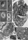
FIGURE 1
FIGURE 1
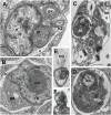
Light (A–D) and electron (E–H) microscopic images of Develorapax marinus. (A,C) Trophont, (B) swimming cell, (D) two daughter cells still connected by thin cytoplasmic bridge (arrowhead). (A,B) DIC, (C,D) phase contrast. (E) General view at longitudinal section, (F) some organelles and portion of nucleus at higher magnification (arrow shows an axial filament in the crista), (G) structure of vacuoles and dense bodies, (H) dense body at higher magnification. Scale bar: (A–D) 5 μm; (E,G) 500 nm; (F) 300 nm; (H) 125 nm. Abbreviations: af, anterior flagellum; b, bacteria; db, dense bodies; fv, food vacuole; ga, Golgi apparatus; kp, kinetoplast of the prey; k1, kinetosome of posterior flagellum; k2, kinetosome of anterior flagellum; l, lipid globule; m, mitochondrion; mp, mitochondrion of the prey; n, nucleus; nu, nucleolus; pf, posterior flagellum; pr, prey; ps, pseudopodium; r1–r4, microtubular flagellar roots; smt, secondary microtubules of r3; tm, tubular mastigonemes; tp, transversal plate; v, vacuole.
FIGURE 2
FIGURE 2
Ultrastructure of Develorapax marinus. (A,B) Nucleus and complex of associated organelles. (C) Food vacuole with partly digested kinetoplastid, (D) food vacuole at higher magnification with remained kinetoplast and mitochondrion, (E) unilateral tubular mastigonemes (tm), (F) tangential section of flagellar transition zone with six gyres of transitional helix. Scale bars: (A,B) 500 nm; (C) 1.5 μm; (D–F) 200 nm.
FIGURE 3
FIGURE 3
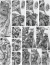
Structure of kinetid in Develorapax marinus. (A) Root 2 at longitudinal section. (B–E) Series of consecutive transverse sections of kinetosome 2. Arrowheads on (C) show two connectives between kinetosomes. (F–M) Series of consecutive longitudinal sections of kinetid. Arrows on (E,F) show the fibrillar strand of k1 becoming the core of r2. Scale bar: 400 nm.
FIGURE 4
FIGURE 4
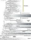
Heterokont ribosomal RNA gene phylogeny with focus on the Gyrista clade. Concatenated alignment of 18S and 28S rRNA genes with cumulative length of 4,606 positions was used for the analysis. The tree was reconstructed using the Bayesian Inference approach implemented by PhyloBayes under the CAT+GTR model with eight discrete gamma distributed rate categories. Nodes with Bayesian posterior probability of more than 0.9 and bootstrap support above 90% (RAxML) are marked with thicker lines. Resections mark branches shortened to half of their actual length.

References

    1. Acosta F., Ngugi D. K., Stingl U. (2013). Diversity of picoeukaryotes at an oligotrophic site off the Northeastern Red Sea Coast. Aquat. Biosyst. 9:16 10.1186/2046-9063-9-16 - DOI - PMC - PubMed
    1. Al-Qassab S., Lee W. J., Murray S., Simpson A. G. B., Patterson D. J. (2002). Flagellates from stromatolites and surrounding sediments in Shark Bay, Western Australia. Acta Protozool. 41 91–144.
    1. Archibald J. M. (2015). Genomic perspectives on the birth and spread of plastids. Proc. Natl. Acad. Sci. U.S.A. 112 10147–10153. 10.1073/pnas.1421374112 - DOI - PMC - PubMed
    1. Beakes G. W., Glockling S. L., Sekimoto S. (2012). The evolutionary phylogeny of the oomycete “fungi.” Protoplasma 249 3–19. 10.1007/s00709-011-0269-2 - DOI - PubMed
    1. Ben Ali A., De Baere R., De Wachter R., Van de Peer Y. (2002). Evolutionary relationships among heterokont algae (the autotrophic stramenopiles) based on combined analyses of small and large subunit ribosomal RNA. Protist 153 123–132. 10.1078/1434-4610-00091 - DOI - PubMed

LinkOut - more resources