

Multidisciplinary Oncoplastic Approach Reduces Infection in Chest Wall Resection and Reconstruction for Malignant Chest Wall Tumors
- PMID: 27536488
- PMCID: PMC4977137
- DOI: 10.1097/GOX.0000000000000751
Multidisciplinary Oncoplastic Approach Reduces Infection in Chest Wall Resection and Reconstruction for Malignant Chest Wall Tumors
Abstract
Background: Management of complex thoracic defects post tumor extipiration is challenging because of the nature of pathology, the radical approach, and the insertion of prosthetic material required for biomechanical stability. Wound complications pose a significant problem that can have detrimental effect on patient outcome. The authors outline an institutional experience of a multidisciplinary thoracic oncoplastic approach to improve outcomes.
Methods: Prospectively collected data from 71 consecutive patients treated with chest wall resection and reconstruction were analyzed (2009-2015). The demographic data, comorbidities, operative details, and outcomes with special focus on wound infection were recorded. All patients were managed in a multidisciplinary approach to optimize perioperative surgical planning.
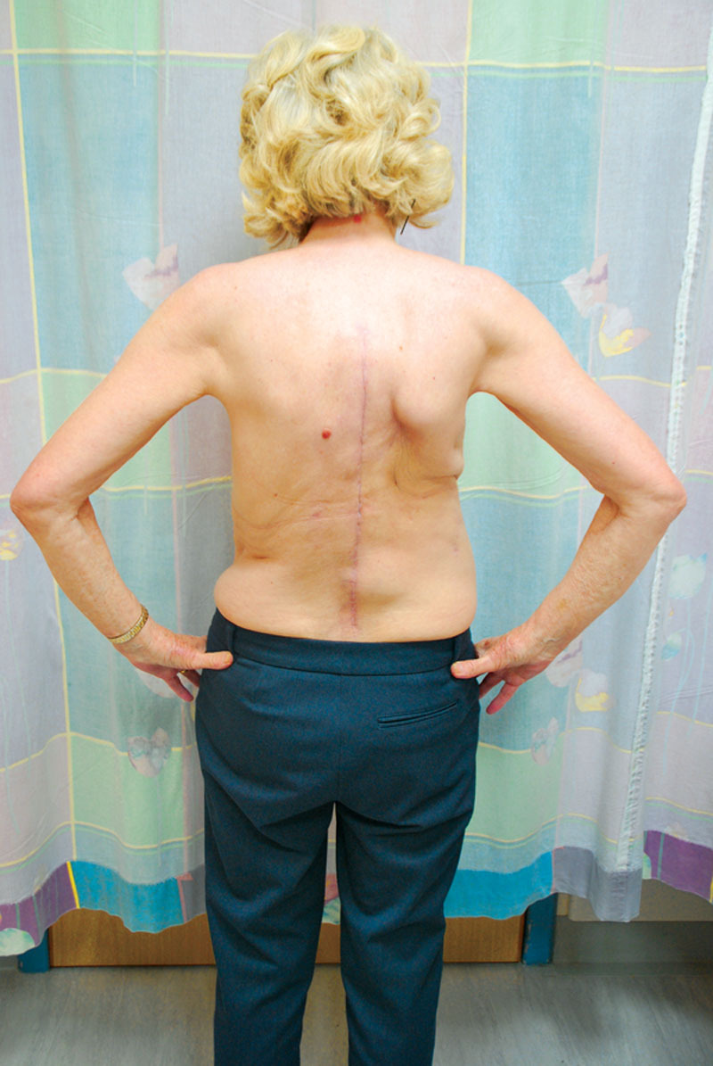
Results: Pathology included sarcoma (78%), locally advanced breast cancer (15%), and desmoids (6%), with age ranging from 17 to 82 years (median, 42 years) and preponderance of female patients (n = 44). Chest wall defects were located anterior and anterolateral (77.5%), posterior (8.4%), and apical axillary (10%) with skeletal defect size ranging from 56 to 600 cm(2) (mean, 154 cm(2)). Bony reconstruction was performed using polyprolene mesh, methyl methacrylate prosthesis, and titanium plates. Soft tissue reconstructions depended on size, location, and flap availability and were achieved using regional, distant, and free tissue flaps. The postoperative follow-up ranged from 5 to 70 months (median, 32 months). All flaps survived with good functional and aesthetic outcome, whereas 2 patients experienced surgical site infection (2.8%).
Conclusions: Multidisciplinary thoracic oncoplastic maximizes outcome for patients with large resection of chest wall tumors with reduction in surgical site infection and wound complications particularly in association with rigid skeletal chest wall reconstruction.
Figures


Similar articles
-
Chest Wall Reconstruction with Porcine Acellular Dermal Matrix (Strattice) and Autologous Tissue Transfer for High Risk Patients with Chest Wall Tumors.Plast Reconstr Surg Glob Open. 2018 May 18;6(5):e1703. doi: 10.1097/GOX.0000000000001703. eCollection 2018 May. Plast Reconstr Surg Glob Open. 2018. PMID: 29922541 Free PMC article.
-
[Plastic surgical reconstruction of extensive thoracic wall defects after oncologic resection].Chirurg. 2008 Feb;79(2):164-74. doi: 10.1007/s00104-007-1382-9. Chirurg. 2008. PMID: 17786394 German.
-
Reconstruction of large full thickness chest wall defects following resection of malignant tumors.J Egypt Natl Canc Inst. 2010 Mar;22(1):19-27. J Egypt Natl Canc Inst. 2010. PMID: 21503003
-
Materials and techniques in chest wall reconstruction: a review.J Vis Surg. 2017 Jul 26;3:95. doi: 10.21037/jovs.2017.06.10. eCollection 2017. J Vis Surg. 2017. PMID: 29078657 Free PMC article. Review.
-
Thoracic Wall Reconstruction after Tumor Resection.Front Oncol. 2015 Oct 29;5:247. doi: 10.3389/fonc.2015.00247. eCollection 2015. Front Oncol. 2015. PMID: 26579499 Free PMC article. Review.
Cited by
-
Functional and Aesthetic Thorax Reconstruction after Desmoid Tumor Resection.Plast Reconstr Surg Glob Open. 2017 Feb 22;5(2):e1248. doi: 10.1097/GOX.0000000000001248. eCollection 2017 Feb. Plast Reconstr Surg Glob Open. 2017. PMID: 28280682 Free PMC article.
-
Preoperative Long-term Therapeutic Subcutaneous Heparin Administration into Abdomen: Possible Cause for Nonobstructive Microvascular Flap Failure.Plast Reconstr Surg Glob Open. 2021 Feb 17;9(2):e3400. doi: 10.1097/GOX.0000000000003400. eCollection 2021 Feb. Plast Reconstr Surg Glob Open. 2021. PMID: 33680653 Free PMC article.
-
Contributing factors to the outcome of primary malignant chest wall tumors.J Thorac Dis. 2017 Dec;9(12):5184-5193. doi: 10.21037/jtd.2017.11.61. J Thorac Dis. 2017. PMID: 29312725 Free PMC article.
-
Chest Wall Reconstruction with Porcine Acellular Dermal Matrix (Strattice) and Autologous Tissue Transfer for High Risk Patients with Chest Wall Tumors.Plast Reconstr Surg Glob Open. 2018 May 18;6(5):e1703. doi: 10.1097/GOX.0000000000001703. eCollection 2018 May. Plast Reconstr Surg Glob Open. 2018. PMID: 29922541 Free PMC article.
-
Biologic versus synthetic prosthesis for chest wall reconstruction: a matched analysis.Eur J Cardiothorac Surg. 2023 Dec 1;64(6):ezad348. doi: 10.1093/ejcts/ezad348. Eur J Cardiothorac Surg. 2023. PMID: 37846030 Free PMC article.
References
-
- Berthet JP, Wihlm JM, Canaud L, et al. The combination of polytetrafluoroethylene mesh and titanium rib implants: an innovative process for reconstructing large full thickness chest wall defects. Eur J Cardiothorac Surg. 2012;42:444–453. - PubMed
-
- Berthet JP, Solovei L, Tiffet O, et al. Chest-wall reconstruction in case of infection of the operative site: is there any interest in titanium rib osteosynthesis? Eur J Cardiothorac Surg. 2013;44:866–874. - PubMed
-
- Gonfiotti A, Santini PF, Campanacci D, et al. Malignant primary chest-wall tumours: techniques of reconstruction and survival. Eur J Cardiothorac Surg. 2010;38:39–45. - PubMed
-
- Arnold PG, Pairolero PC. Chest-wall reconstruction: an account of 500 consecutive patients. Plast Reconstr Surg. 1996;98:804–810. - PubMed
-
- Mansour KA, Thourani VH, Losken A, et al. Chest wall resections and reconstruction: a 25-year experience. Ann Thorac Surg. 2002;73:1720–1725. discussion 1725–1726. - PubMed
LinkOut - more resources
Full Text Sources
Other Literature Sources