. 2016 Aug 4;1(12):e88529.
doi: 10.1172/jci.insight.88529.

HSV-2 ΔgD elicits FcγR-effector antibodies that protect against clinical isolates

Affiliations

HSV-2 ΔgD elicits FcγR-effector antibodies that protect against clinical isolates

Christopher D Petro et al. JCI Insight. .

Abstract

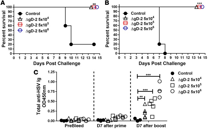
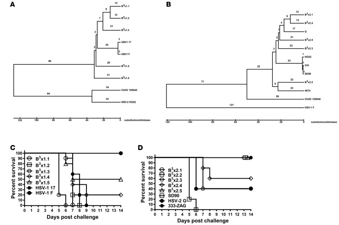
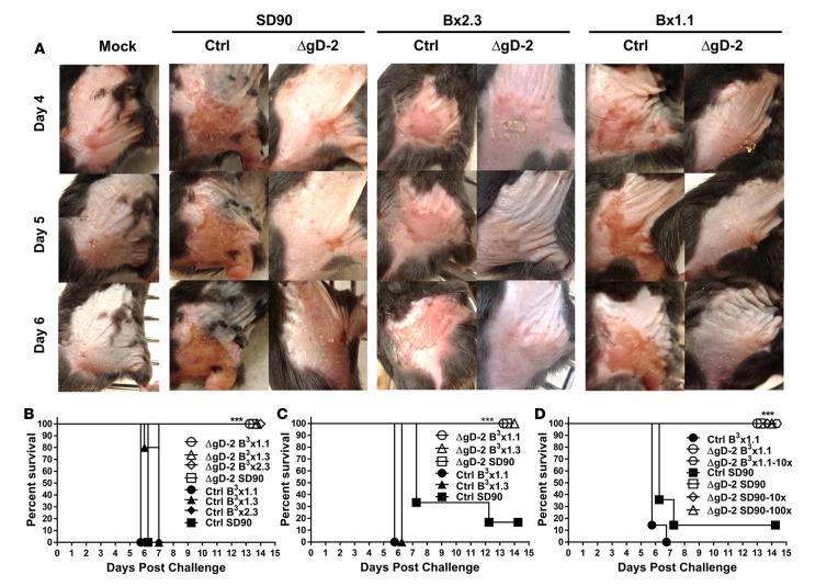
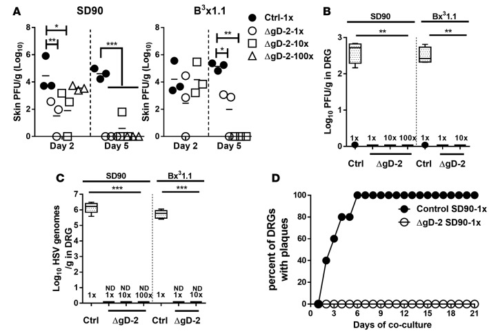
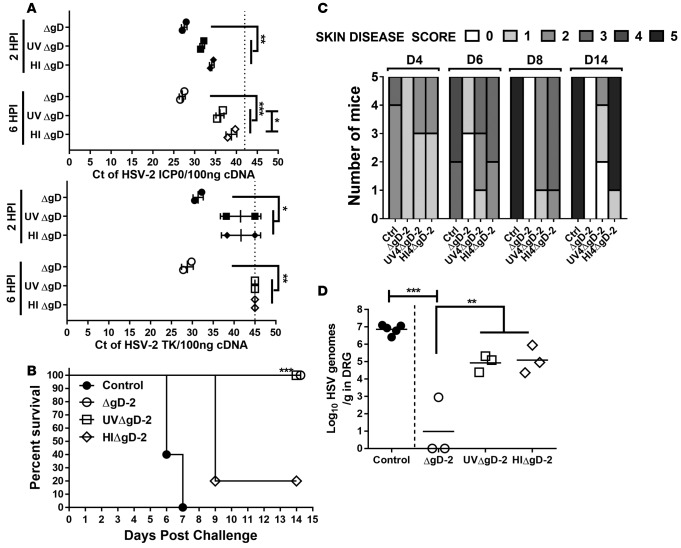
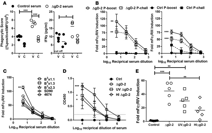
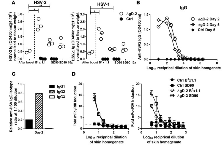
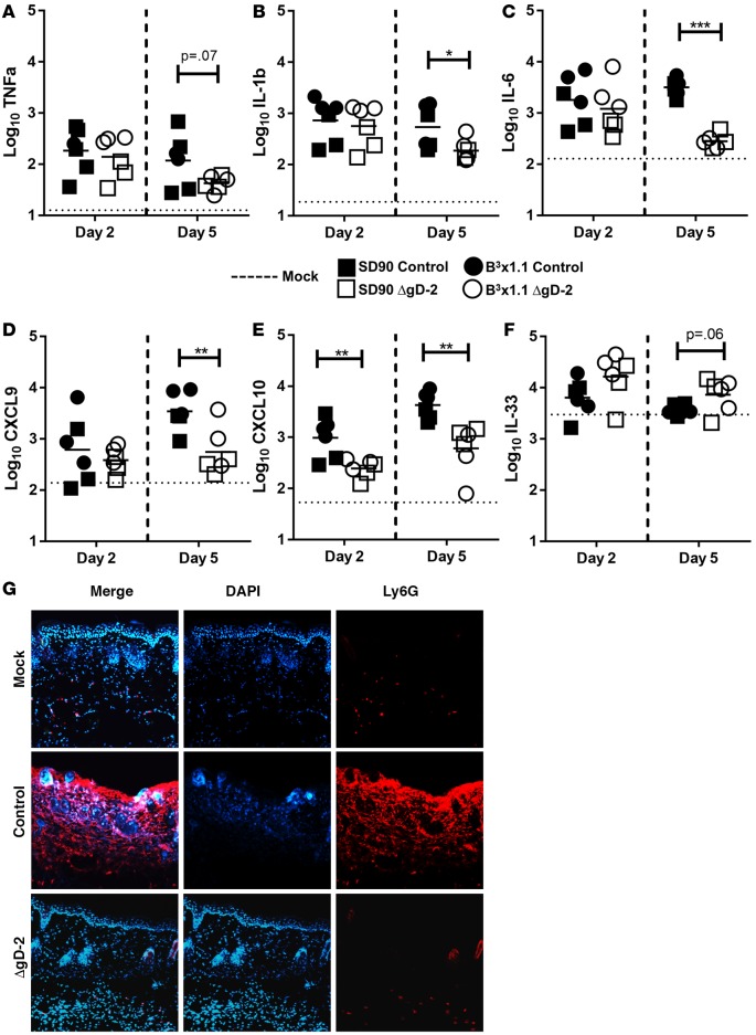
A single-cycle herpes simplex virus (HSV) deleted in glycoprotein D (ΔgD-2) elicited high titer HSV-specific antibodies (Abs) that (i) were rapidly transported into the vaginal mucosa; (ii) elicited antibody-dependent cell-mediated cytotoxicity but little neutralization; (iii) provided complete protection against lethal intravaginal challenge; and (iv) prevented establishment of latency in mice. However, clinical isolates may differ antigenically and impact vaccine efficacy. To determine the breadth and further define mechanisms of protection of this vaccine candidate, we tested ΔgD-2 against a panel of clinical isolates in a murine skin challenge model. The isolates were genetically diverse, as evidenced by genomic sequencing and in vivo virulence. Prime and boost immunization (s.c.) with live but not heat- or UV-inactivated ΔgD-2 completely protected mice from challenge with the most virulent HSV-1 and HSV-2 isolates. Furthermore, mice were completely protected against 100 times the lethal dose that typically kills 90% of animals (LD90) of a South African isolate (SD90), and no latent virus was detected in dorsal root ganglia. Immunization was associated with rapid recruitment of HSV-specific FcγRIII- and FcγRIV-activating IgG2 Abs into the skin, resolution of local cytokine and cellular inflammatory responses, and viral clearance by day 5 after challenge. Rapid clearance and the absence of latent virus suggest that ΔgD-2 elicits sterilizing immunity.


Figures

Figure 1
Figure 1. HSV-2ΔgD-2 provides complete protection following intravaginal or skin challenge with vaccine doses as low as 5 × 10⁴ PFU.
C57BL/6 mice were primed and, 21 days later, were boosted s.c. with 5 × 10⁴ PFU, 5 × 10⁵ PFU, or 5 × 10⁶ PFU of HSV-2 ΔgD-2 or VD60 lysates (control). Mice were subsequently challenged 21 days after boost with an LD90 of HSV-2(4674) either (A) intravaginally or (B) via skin scarification and followed for survival (n = 5 mice/group). (C) Serum was assessed for HSV-2 antibodies by ELISA before (PreBleed), day 7 after prime, and day 7 after boost; each symbol represents optical densitometry units (OD) for an individual mouse (n = 5/group); lines represent mean of each group. ΔgD-2– and control-vaccinated groups were compared for survival by Kaplan-Meier and for Ab responses by 2-way ANOVA; **P < 0.01, ***P < 0.001.
Figure 2
Figure 2. Clinical HSV-1 and HSV-2 isolates are genotypically and phenotypically diverse.
(A) Herpes simplex virus type-1 (HSV-1) and (B) HSV-2 phylogenic trees were constructed from whole genome alignments of de novo–assembled HSV clinical isolate and lab strain sequences by the UPGMA method in MEGA6. The branches are labeled with genetic distance in nucleotide substitutions/kilobase. All branches in HSV-1 tree showed 100% confidence; those on the HSV-2 tree were also 100% except the branches at the level of 4674, which were 92%, and the branch bearing B3 × 2.3, which was 99%. Published data for Chimpanzee α-1 herpesvirus (ChHV), HSV-1(F), or HSV-2(HG52) were used as outgroups for each analysis. (C) In vivo virulence was assessed by challenging BALB/C mice with 1 × 10⁵ PFU/mouse of HSV-1 (D) or 5 × 10⁴ PFU/mouse of HSV-2 by skin scarification. Survival curves are shown for each isolate; open symbols represent clinical isolates, and closed symbols represent laboratory strains.
Figure 3
Figure 3. HSV-2 ΔgD-2 protects mice from clinical isolates of HSV-1 and HSV-2.
C57BL/6 (n = 7 mice/group) or BALB/C (n = 5 mice/group) mice were immunized with ΔgD-2 or VD60 cell lysates (control) and subsequently challenged by skin scarification with an LD90 dose of the most virulent isolates and monitored daily. (A) Representative images from C57BL/6 mice on days 4, 5, and 6 after challenge (magnification 1.2×); (B) survival curves for C57BL/6 mice, and (C) survival curves for BALB/C mice. (D) Additional C57BL/6 mice were challenged with 10 and 100 times (10x and 100x) the LD90 dose of SD90 and 10 times the LD90 of B3 × 1.1. ΔgD-2 and control-vaccinated groups were compared for survival by Kaplan-Meier; ***P < 0.001.
Figure 4
Figure 4. Virus is rapidly cleared and no latent virus is detected in dorsal root ganglia isolated from HSV-2 ΔgD-2–vaccinated mice.
Mice were immunized with ΔgD-2 or VD60 cell lysates (control) and subsequently challenged by skin scarification with 1, 10, or 100 times the LD90 of HSV-2(SD90) or with 1 or 10 times the LD90 of HSV-1(B3 × 1.1) (n = 5 mice per group). (A) Skin biopsies were obtained on day 2 and day 5 after challenge and assayed for viral load by plaque assay on Vero cells (n = 3 samples/group, lines represent mean). (B) Replicating or (C) latent HSV in dorsal root ganglia (DRG) tissue obtained from ΔgD-2–vaccinated (day 14 after challenge) or control-vaccinated (time of euthanasia) mice were assessed by plaque assay and qPCR, respectively (n = 5 mice/group). (D) Latency was further evaluated by coculturing Vero cells with DRG isolated at day 5 after challenge from ΔgD-2– and control–immunized mice that were challenged with a 1× LD90 of HSV-2 SD90 (n = 5/group). Data in B and C are presented as box and whisker plots, with the bounds of the box representing the 25th and 75th percentile, the line representing the median, the whiskers representing the 10th and 90th percentile, and black dots indicating outliers. The HSV-2 ΔgD-2–vaccinated group and control–vaccinated groups were compared by student’s t test; *P < 0.05; **P < 0.01; ***P < 0.001.
Figure 5
Figure 5. Inactivation of ΔgD-2 leads to reduction in vaccine efficacy.
(A) Vero cells were infected with an MOI of 5 PFU/cell of ΔgD-2 or an equivalent amount of UV-inactivated (UVΔgD-2) or heat-inactivated (HIΔgD-2) virus and viral gene expression for infected cell protein-0 (ICP0, upper panel) and thymidine kinase (TK, lower panel) assessed at 2 hours (closed symbols) and 6 hours (open symbols) after infection (HPI). Data are presented as threshold cycle (Ct). Each point represents Ct values from individual experiment; lines equals mean ±SD from replicate experiments, with the dotted line indicating the Ct values for mock-infected cells. The asterisks indicate results for 2-way ANOVA comparing UVΔgD-2, HIΔgD-2, and ΔgD-2 Ct values at each time after infection (*P < 0.05, **P < 0.01, ***P < 0.001). (B and C) Mice were prime-boost vaccinated with 5 × 10⁶ PFU of live or equivalent concentrations of UVΔgD-2, HIΔgD-2, ΔgD-2, or VD60 lysates (control) and subsequently challenged with an LD90 of HSV-2(SD90) on the skin (n = 5/group). Mice were monitored for (B) survival and (C) skin disease scores. Kaplan Meier analysis was used for survival curves of ΔgD-2–, UVΔgD-2–, HIΔgD-2–, and control-vaccinated mice. (D) At day 5 after challenge, mice were euthanized and DRGs were extracted for qPCR analysis of HSV DNA (n = 3/group for UV-, HI-, or ΔgD-2–immunized mice or n = 5 for control-treated mice; lines represent the mean). **P < 0.01, ***P < 0.001 by 2-way ANOVA, ΔgD-2–vaccinated groups vs. control-vaccinated group.
Figure 6
Figure 6. HSV-2 ΔgD-2 elicits cross-reactive HSV-specific FcγR effector antibodies.
Mice were vaccinated with ΔgD-2 or VD60 lysates (control) and subsequently challenged with HSV-2(4674) on the skin (n = 5/group). (A) Antibody-dependent cellular phagocytosis (ADCP) activity of serum from control or HSV-2 ΔgD-2–vaccinated mice 7 days after boost was quantified using THP-1 monocytic cell line and beads coated with HSV-2–infected cell lysates (v) or uninfected cell lysates (c) (left panel). The %ADCP is calculated as percent of cells positive for beads multiplied by the MFI of positive cells divided by 1 × 10⁶. IFN-γ levels were measured in the supernatants 8 hours after incubation of THP-1 cells with the beads (right panel) (n = 4/group, line represents the mean). (B) Serum from 7 days postboost or 7 days postchallenge ΔgD-2– or control-immunized mice were further assessed for mFcγRIV (left panel) and mFcγRIII (right panel) activation via a luciferase effector cell reporter assay using HSV-2(4674)–infected target cells (data represented as mean ±SD values of 5 mice/group). (C) ΔgD-2 boost serum was pooled day 7 after boost and was assessed for mFcγRIV activation against cells infected with 5 different clinical isolates (n = 5 mice/pool; data represented as mean, SD obtained from replicates). (D) Day 7 postboost serum from mice vaccinated with ΔgD-2, UVΔgD-2, HIΔgD-2, or VD60 lysates (control) were evaluated for anti-HSV antibodies by ELISA using an HSV-2–infected cell lysate (data represented as mean ±SD values of 5 mice/group) and (E) mFcγRIV activation against HSV-2(4674)–infected cells (lines represent means). Dashed lines represent values from mock-infected mouse serum. For A and E, *P < 0.05, **P < 0.01, ***P < 0.001, ΔgD-2 vaccinated groups vs. control-vaccinated group via 2-way ANOVA. For B, C, and D, *P < 0.05, **P < 0.01, ***P < 0.001; AUCs were generated for each group and then analyzed via one-way ANOVA comparing ΔgD-2, UVΔgD-2, or HIΔgD-2 groups with control values (B and D) or against each other (C).
Figure 7
Figure 7. FcγR-activating HSV-specific IgG2 antibodies are rapidly recruited into the skin of ΔgD-2–vaccinated mice following viral challenge.
Mice were immunized with ΔgD-2 or VD60 cell lysates (control) and subsequently challenged with HSV-1(B3 × 1.1) and HSV-2(SD90) clinical isolates on the skin. (A) Skin biopsies were obtained 21 days after boost and day 2 after challenge, homogenized, and evaluated for the presence of anti-HSV antibodies by ELISA using an HSV-2–infected (left) or HSV-1–infected (right) cell lysate as the antigen (n = 3 mice per group, lines represent mean). Differences in HSV-specific Abs in ΔgD-2–vaccinated vs. control-vaccinated mice were compared by students t test; *P < 0.05; **P < 0.01. (B) Pools (6 mice per pool) of skin homogenates (at day 2 or day 5 after challenge) were serially diluted and assayed in an HSV-2 ELISA (results are mean ±SD obtained from testing pools in duplicate). (C) The relative proportion of different IgG subtypes in the day 2 postchallenge skin homogenate pool was determined using subtype-specific secondary antibodies in the ELISA. Results shown are with the 1:100 dilution of the pooled skin homogenates. (D) mFcγRIII (left panel) and mFcγRIV (right panel) activation was also assessed in pools of serially diluted skin homogenates (n = 3 mice/pool, mean ±SD obtained from testing pools in duplicate). Dashed lines represent mock-infected skin homogenate activation.
Figure 8
Figure 8. ΔgD-2 immunization is associated with decreased inflammatory response in the skin by day 5 after challenge compared with control-vaccinated mice.
Biopsies of skin from mice immunized with ΔgD-2 or VD60 lysates (control) at day 2 or day 5 after challenge with either SD90 or B3 × 1.1 (n = 3 mice per group) or unimmunized, uninfected controls (n = 2) were homogenized and evaluated for concentration of TNFα (A), IL-1β (B), IL-6 (C), CXCL9 (D), CXCL10 (IP-10) (E), and IL-33 (F). Results are expressed as log10 pg of analyte per gram of tissue. Each dot represents a single animal, and the lines represent mean combining the SD90 and B3 × 1.1 challenges for each vaccine. *P < 0.05, **P < 0.01, ***P < 0.001, student’s t test comparing ΔgD-2–vaccinated vs. control-vaccinated groups. The dashed lines represent mean for unimmunized, mock-infected animals. (G) Additionally, skin sections of unimmunized mock-infected mice or mice immunized with HSV-2 ΔgD-2 or VD60 lysates (control) and infected with HSV-1(B3 × 1.1) virus were harvested on day 5 after challenge and stained for neutrophils using Ly6G (red). Nuclei are stained blue with DAPI. Images were microphotographed at 20× magnification.

Similar articles

Cited by

References

    1. Bernstein DI, et al. Epidemiology, clinical presentation, and antibody response to primary infection with herpes simplex virus type 1 and type 2 in young women. Clin Infect Dis. 2013;56(3):344–351. doi: 10.1093/cid/cis891. - DOI - PMC - PubMed
    1. Gnann JW, et al. Herpes Simplex Encephalitis: Lack of Clinical Benefit of Long-term Valacyclovir Therapy. Clin Infect Dis. 2015;61(5):683–691. doi: 10.1093/cid/civ369. - DOI - PMC - PubMed
    1. Looker KJ, et al. Global estimates of prevalent and incident herpes simplex virus type 2 infections in 2012. PLoS One. 2015;10(1):e88529. doi: 10.1371/journal.pone.0114989. - DOI - PMC - PubMed
    1. et al. High seroprevalence of human herpesviruses in HIV-infected individuals attending primary healthcare facilities in rural South Africa. PLoS ONE. 2014;9(6):e88529. doi: 10.1371/journal.pone.0099243. - DOI - PMC - PubMed
    1. Whitley RJ. Herpes simplex viruses. Clin Infect Dis. 1998;26(3):541–553. doi: 10.1086/514600. - DOI - PubMed