

Rate of lumbar paravertebral muscle fat infiltration versus spinal degeneration in asymptomatic populations: an age-aggregated cross-sectional simulation study
- PMID: 27536737
- PMCID: PMC4975884
- DOI: 10.1186/s13013-016-0080-0
Rate of lumbar paravertebral muscle fat infiltration versus spinal degeneration in asymptomatic populations: an age-aggregated cross-sectional simulation study
Abstract
Background: The spinal column including its vertebrae and disks has been well examined and extensively reported in relation to age-aggregated degeneration. In contrast, paravertebral muscles are poorly represented in describing normative degeneration. Increasing evidence points to the importance of paravertebral muscle quality in low back health, and their potential as a modifiable factor in low back pain (LBP). Studies examining normative decline of paravertebral muscles are needed to advance the field's etiological understanding. With a novel approach and based on published data, we establish and compare decline rates of imaging features for degeneration of lumbar vertebrae and disks, versus fatty infiltration in paravertebral muscles in asymptomatic adults.
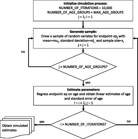
Methods: Our cross-sectional simulation study examined age-aggregated data from three published studies who reported on asymptomatic adults spanning 18-60 years. Prevalence rates of imaging degenerative features of the spinal column were examined via logistic regression and compared with percentage fatty infiltration in erector spinae, multifidus and psoas using synthetic data and Monte Carlo simulation with 10,000 endpoint-specific regression iterations. General linear regression models were employed to estimate marginal effects of age reported as a one-year change rate (with 95 % confidence intervals) for comparisons between all reported spinal features.
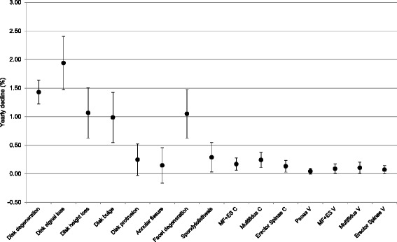
Results: Declines in multifidus (0.24 & 0.11 %/year), erector spinae (0.13 & 0.07 %/year), and psoas (0.04 %/year) occur at similarly slow rates to disk protrusion (0.25 %/year), annular fissure (0.15 %/year), and spondylolisthesis (0.29 %/year). Multifidus showed a trend for faster decline than erector spinae, particularly in men. Of the features examined, disk signal loss declined fastest, and psoas muscle the slowest.
Conclusions: Degeneration of lumbar paravertebral muscles occurs slowly in asymptomatic adults, with a tendency to be most pronounced in multifidus. Rate of decline of spinal structures represents a novel variable that warrants inclusion as a known feature of the expected degenerative cascade, and to provide a basis for comparison to diseases of the spine in research and clinical practice. Concurrent examination of spinal features using advanced imaging to improve muscle analysis would be a strong addition to the field.
Keywords: Ageing; Asymptomatic adults; Disc degeneration; Erector spinae; Fatty infiltration; Lumbar spine; Magnetic resonance imaging; Multifidus; Paravertebral muscles; Psoas.
Figures
Similar articles
-
Change in fatty infiltration of lumbar multifidus, erector spinae, and psoas muscles in asymptomatic adults of Asian or Caucasian ethnicities.Eur Spine J. 2017 Dec;26(12):3059-3067. doi: 10.1007/s00586-017-5212-6. Epub 2017 Jul 11. Eur Spine J. 2017. PMID: 28698963
-
[Imaging study of paravertebral muscle degeneration in degenerative lumbar instability].Zhonghua Wai Ke Za Zhi. 2014 Aug;52(8):571-5. Zhonghua Wai Ke Za Zhi. 2014. PMID: 25370754 Chinese.
-
Severe Lumbar Intervertebral Disc Degeneration Is Associated with Modic Changes and Fatty Infiltration in the Paraspinal Muscles at all Lumbar Levels, Except for L1-L2: A Cross-Sectional Analysis of 50 Symptomatic Women and 50 Age-Matched Symptomatic Men.World Neurosurg. 2019 Feb;122:e1069-e1077. doi: 10.1016/j.wneu.2018.10.229. Epub 2018 Nov 9. World Neurosurg. 2019. PMID: 30415054
-
Age-related degeneration of the lumbar paravertebral muscles: Systematic review and three-level meta-regression.Exp Gerontol. 2020 May;133:110856. doi: 10.1016/j.exger.2020.110856. Epub 2020 Jan 30. Exp Gerontol. 2020. PMID: 32006635
-
The Association between Imaging Parameters of the Paraspinal Muscles, Spinal Degeneration, and Low Back Pain.Biomed Res Int. 2017;2017:2562957. doi: 10.1155/2017/2562957. Epub 2017 Mar 20. Biomed Res Int. 2017. PMID: 28409152 Free PMC article. Review.
Cited by
-
Relation of bone mineral density with fat infiltration of paraspinal muscles: The Goutallier classification.Osteoporos Sarcopenia. 2024 Jun;10(2):84-88. doi: 10.1016/j.afos.2024.04.002. Epub 2024 Apr 24. Osteoporos Sarcopenia. 2024. PMID: 39035231 Free PMC article.
-
Influence of patient-specific factors when comparing multifidus fat infiltration between chronic low back pain patients and asymptomatic controls.JOR Spine. 2022 Jul 14;5(4):e1217. doi: 10.1002/jsp2.1217. eCollection 2022 Dec. JOR Spine. 2022. PMID: 36601370 Free PMC article.
-
Reduced Cervical Muscle Fat Infiltrate Is Associated with Self-Reported Recovery from Chronic Idiopathic Neck Pain Over Six Months: A Magnetic Resonance Imaging Longitudinal Cohort Study.J Clin Med. 2024 Jul 31;13(15):4485. doi: 10.3390/jcm13154485. J Clin Med. 2024. PMID: 39124753 Free PMC article.
-
Use of two-point and six-point Dixon MRI for fat fraction analysis in the lumbar vertebral bodies and paraspinal muscles in healthy dogs: comparison with magnetic resonance spectroscopy.Front Vet Sci. 2024 Sep 25;11:1412552. doi: 10.3389/fvets.2024.1412552. eCollection 2024. Front Vet Sci. 2024. PMID: 39386243 Free PMC article.
-
Incidence and Predictive Factors of New Onset Postoperative Sacroiliac Joint Pain After Posterior Lumbar Fusion Surgery for Degenerative Lumbar Disease.J Pain Res. 2023 Dec 14;16:4291-4299. doi: 10.2147/JPR.S431197. eCollection 2023. J Pain Res. 2023. PMID: 38111748 Free PMC article.
References
-
- Brinjikji W, Luetmer PH, Comstock B, Bresnahan BW, Chen LE, Deyo RA, Halabi S, Turner JA, Avins AL, James K, et al. Systematic literature review of imaging features of spinal degeneration in asymptomatic populations. Am J Neuroradiol. 2015;36(4):811–816. doi: 10.3174/ajnr.A4173. - DOI - PMC - PubMed
-
- Fischer MA, Nanz D, Shimakawa A, Schirmer T, Guggenberger R, Chhabra A, Carrino JA, Andreisek G. Quantification of muscle fat in patients with low back pain: comparison of multi-echo MR imaging with single-voxel MR spectroscopy. Radiology. 2013;266(2):555–563. doi: 10.1148/radiol.12120399. - DOI - PubMed
LinkOut - more resources
Full Text Sources
Other Literature Sources
Miscellaneous