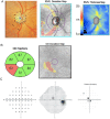
Macular Ganglion Cell Inner Plexiform Layer Thickness in Glaucomatous Eyes with Localized Retinal Nerve Fiber Layer Defects
- PMID: 27537107
- PMCID: PMC4990273
- DOI: 10.1371/journal.pone.0160549
Macular Ganglion Cell Inner Plexiform Layer Thickness in Glaucomatous Eyes with Localized Retinal Nerve Fiber Layer Defects
Abstract
Purpose: To investigate macular ganglion cell-inner plexiform layer (mGCIPL) thickness in glaucomatous eyes with visible localized retinal nerve fiber layer (RNFL) defects on stereophotographs.
Methods: 112 healthy and 149 glaucomatous eyes from the Diagnostic Innovations in Glaucoma Study (DIGS) and the African Descent and Glaucoma Evaluation Study (ADAGES) subjects had standard automated perimetry (SAP), optical coherence tomography (OCT) imaging of the macula and optic nerve head, and stereoscopic optic disc photography. Masked observers identified localized RNFL defects by grading of stereophotographs.
Result: 47 eyes had visible localized RNFL defects on stereophotographs. Eyes with visible localized RNFL defects had significantly thinner mGCIPL thickness compared to healthy eyes (68.3 ± 11.4 μm versus 79.2 ± 6.6 μm respectively, P<0.001) and similar mGCIPL thickness to glaucomatous eyes without localized RNFL defects (68.6 ± 11.2 μm, P = 1.000). The average mGCIPL thickness in eyes with RNFL defects was 14% less than similarly aged healthy controls. For 29 eyes with a visible RNFL defect in just one hemiretina (superior or inferior) mGCIPL was thinnest in the same hemiretina in 26 eyes (90%). Eyes with inferior-temporal RNFL defects also had significantly thinner inferior-temporal mGCIPL (P<0.001) and inferior mGCIPL (P = 0.030) compared to glaucomatous eyes without a visible RNFL defect.
Conclusion: The current study indicates that presence of a localized RNFL defect is likely to indicate significant macular damage, particularly in the region of the macular that topographically corresponds to the location of the RNFL defect.
Conflict of interest statement
Figures


Similar articles
-
Relationship between ganglion cell layer thickness and estimated retinal ganglion cell counts in the glaucomatous macula.Ophthalmology. 2014 Dec;121(12):2371-9. doi: 10.1016/j.ophtha.2014.06.047. Epub 2014 Aug 20. Ophthalmology. 2014. PMID: 25148790 Free PMC article.
-
Estimated retinal ganglion cell counts in glaucomatous eyes with localized retinal nerve fiber layer defects.Am J Ophthalmol. 2013 Sep;156(3):578-587.e1. doi: 10.1016/j.ajo.2013.04.015. Epub 2013 Jun 7. Am J Ophthalmol. 2013. PMID: 23746612 Free PMC article.
-
Temporal Relation between Macular Ganglion Cell-Inner Plexiform Layer Loss and Peripapillary Retinal Nerve Fiber Layer Loss in Glaucoma.Ophthalmology. 2017 Jul;124(7):1056-1064. doi: 10.1016/j.ophtha.2017.03.014. Epub 2017 Apr 10. Ophthalmology. 2017. PMID: 28408038
-
Diagnostic ability of macular ganglion cell inner plexiform layer measurements in glaucoma using swept source and spectral domain optical coherence tomography.PLoS One. 2015 May 15;10(5):e0125957. doi: 10.1371/journal.pone.0125957. eCollection 2015. PLoS One. 2015. PMID: 25978420 Free PMC article.
-
Macular imaging by optical coherence tomography in the diagnosis and management of glaucoma.Br J Ophthalmol. 2018 Jun;102(6):718-724. doi: 10.1136/bjophthalmol-2017-310869. Epub 2017 Oct 21. Br J Ophthalmol. 2018. PMID: 29055905 Review.
Cited by
-
Structural changes of macular inner retinal layers in early normal-tension and high-tension glaucoma by spectral-domain optical coherence tomography.Graefes Arch Clin Exp Ophthalmol. 2018 Jul;256(7):1245-1256. doi: 10.1007/s00417-018-3944-6. Epub 2018 Mar 9. Graefes Arch Clin Exp Ophthalmol. 2018. PMID: 29523993
-
Progressive Thinning of Retinal Nerve Fiber Layer and Ganglion Cell-Inner Plexiform Layer in Glaucoma Eyes with Disc Hemorrhage.Ophthalmol Glaucoma. 2021 Sep-Oct;4(5):541-549. doi: 10.1016/j.ogla.2021.01.003. Epub 2021 Jan 30. Ophthalmol Glaucoma. 2021. PMID: 33529795 Free PMC article.
-
Changes of macular blood flow and structure in acute primary angle closure glaucoma.Int Ophthalmol. 2022 Dec;42(12):3789-3801. doi: 10.1007/s10792-022-02399-y. Epub 2022 Jul 4. Int Ophthalmol. 2022. PMID: 35781598 Free PMC article.
-
24-2 Visual Fields Miss Central Defects Shown on 10-2 Tests in Glaucoma Suspects, Ocular Hypertensives, and Early Glaucoma.Ophthalmology. 2017 Oct;124(10):1449-1456. doi: 10.1016/j.ophtha.2017.04.021. Epub 2017 May 24. Ophthalmology. 2017. PMID: 28551166 Free PMC article.
-
Macula Vessel Density and Thickness in Early Primary Open-Angle Glaucoma.Am J Ophthalmol. 2019 Mar;199:120-132. doi: 10.1016/j.ajo.2018.11.012. Epub 2018 Nov 26. Am J Ophthalmol. 2019. PMID: 30496723 Free PMC article.
References
-
- Quigley HA, Green WR. The histology of human glaucoma cupping and optic nerve damage: Clinicopathologic correlation in 21 eyes. Ophthalmology. 1979; 86:1803–1830. - PubMed
-
- Shah NN, Bowd C, Medeiros FA, Weinreb RN, Sample PA, Hoffmann EM, et al. Combining structural and functional testing for detection of glaucoma. Ophthalmology. 2006; 113:1593–1602. - PubMed
-
- Medeiros FA, Zangwill LM, Bowd C, Weinreb RN. Comparison of the gdx VCC scanning laser polarimeter, HRT II confocal scanning laser ophthalmoscope, and stratus OCT optical coherence tomograph for the detection of glaucoma. Arch Ophthalmol. 2004; 122:827–837. - PubMed
MeSH terms
Grants and funding
LinkOut - more resources
Full Text Sources
Other Literature Sources
Medical
Miscellaneous

