Somatic Variants in the Human Lens Epithelium: A Preliminary Assessment
- PMID: 27537255
- PMCID: PMC4986767
- DOI: 10.1167/iovs.16-19726
Somatic Variants in the Human Lens Epithelium: A Preliminary Assessment
Abstract
Purpose: We hypothesize that somatic mutations accumulate in cells of the human lens and may contribute to the development of cortical or posterior sub-capsular cataracts. Here, we used a Next-generation sequencing (NGS) strategy to screen for low-allelic frequency variants in DNA extracted from human lens epithelial samples.
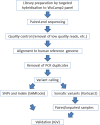
Methods: Next-Generation sequencing of 151 cancer-related genes (WUCaMP2 panel) was performed on DNA extracted from post-mortem or surgical specimens obtained from 24 individuals. Usually, pairwise comparisons were made between two or more ocular samples from the same individual, allowing putative somatic variants detected in lens samples to be differentiated from germline variants.
Results: Use of a targeted hybridization approach enabled high sequence coverage (>1000-fold) of the WUCaMP2 genes. In addition to high-frequency variants (corresponding to homozygous or heterozygous SNPs and Indels), somatic variants with allelic frequencies of 1-4% were detected in the lens epithelial samples. The presence of one such variant, a T > C point substitution at position 32907082 in BRCA2, was verified subsequently using droplet digital PCR.
Conclusions: Low-allelic fraction variants are present in the human lens epithelium, at frequencies consistent with the presence of millimeter-sized clones.
Figures


Similar articles
-
Germ-line and somatic EPHA2 coding variants in lens aging and cataract.PLoS One. 2017 Dec 21;12(12):e0189881. doi: 10.1371/journal.pone.0189881. eCollection 2017. PLoS One. 2017. PMID: 29267365 Free PMC article.
-
Molecular characterization of mouse lens epithelial cell lines and their suitability to study RNA granules and cataract associated genes.Exp Eye Res. 2015 Feb;131:42-55. doi: 10.1016/j.exer.2014.12.011. Epub 2014 Dec 19. Exp Eye Res. 2015. PMID: 25530357 Free PMC article.
-
Lim2(To3) transgenic mice establish a causative relationship between the mutation identified in the lim2 gene and cataractogenesis in the To3 mouse mutant.Mol Vis. 2000 Jun 2;6:85-94. Mol Vis. 2000. PMID: 10851259
-
Structural function of MIP/aquaporin 0 in the eye lens; genetic defects lead to congenital inherited cataracts.Handb Exp Pharmacol. 2009;(190):265-97. doi: 10.1007/978-3-540-79885-9_14. Handb Exp Pharmacol. 2009. PMID: 19096783 Review.
-
Congenital hereditary cataracts.Int J Dev Biol. 2004;48(8-9):1031-44. doi: 10.1387/ijdb.041854jg. Int J Dev Biol. 2004. PMID: 15558493 Review.
Cited by
-
A full lifespan model of vertebrate lens growth.R Soc Open Sci. 2017 Jan 18;4(1):160695. doi: 10.1098/rsos.160695. eCollection 2017 Jan. R Soc Open Sci. 2017. PMID: 28280571 Free PMC article.
-
The aging mouse lens transcriptome.Exp Eye Res. 2021 Aug;209:108663. doi: 10.1016/j.exer.2021.108663. Epub 2021 Jun 11. Exp Eye Res. 2021. PMID: 34119483 Free PMC article.
-
A comprehensive spatial-temporal transcriptomic analysis of differentiating nascent mouse lens epithelial and fiber cells.Exp Eye Res. 2018 Oct;175:56-72. doi: 10.1016/j.exer.2018.06.004. Epub 2018 Jun 5. Exp Eye Res. 2018. PMID: 29883638 Free PMC article.
-
Germ-line and somatic EPHA2 coding variants in lens aging and cataract.PLoS One. 2017 Dec 21;12(12):e0189881. doi: 10.1371/journal.pone.0189881. eCollection 2017. PLoS One. 2017. PMID: 29267365 Free PMC article.
-
The lens growth process.Prog Retin Eye Res. 2017 Sep;60:181-200. doi: 10.1016/j.preteyeres.2017.04.001. Epub 2017 Apr 11. Prog Retin Eye Res. 2017. PMID: 28411123 Free PMC article. Review.
References
-
- Bourne RR,, Stevens GA,, White RA,, et al. Causes of vision loss worldwide, 1990-2010: a systematic analysis. Lancet Glob Health. 2013; 1: e339–e349. - PubMed
-
- Klein BE,, Klein R,, Linton KL. Prevalence of age-related lens opacities in a population. The Beaver Dam Eye Study. Ophthalmology. 1992; 99: 546–552. - PubMed
-
- Stewart FA Akleyev AV, Hauer-Jensen M, et al. ; et al. ICRP publication 118: ICRP statement on tissue reactions and early and late effects of radiation in normal tissues and organs--threshold doses for tissue reactions in a radiation protection context. Ann ICRP. 2012; 41: 1–322. - PubMed
Publication types
MeSH terms
Substances
Grants and funding
LinkOut - more resources
Full Text Sources
Other Literature Sources
Medical
Miscellaneous

