

Review
doi: 10.1167/iovs.16-19933.
Clinical Utility of Optical Coherence Tomography in Glaucoma
Affiliations
- PMID: 27537415
- PMCID: PMC4991023
- DOI: 10.1167/iovs.16-19933
Item in Clipboard
Review
Clinical Utility of Optical Coherence Tomography in Glaucoma
Invest Ophthalmol Vis Sci.
.
Abstract
Optical coherence tomography (OCT) has established itself as the dominant imaging modality in the management of glaucoma and retinal diseases, providing high-resolution visualization of ocular microstructures and objective quantification of tissue thickness and change. This article reviews the history of OCT imaging with a specific focus on glaucoma. We examine the clinical utility of OCT with respect to diagnosis and progression monitoring, with additional emphasis on advances in OCT technology that continue to facilitate glaucoma research and inform clinical management strategies.
Figures
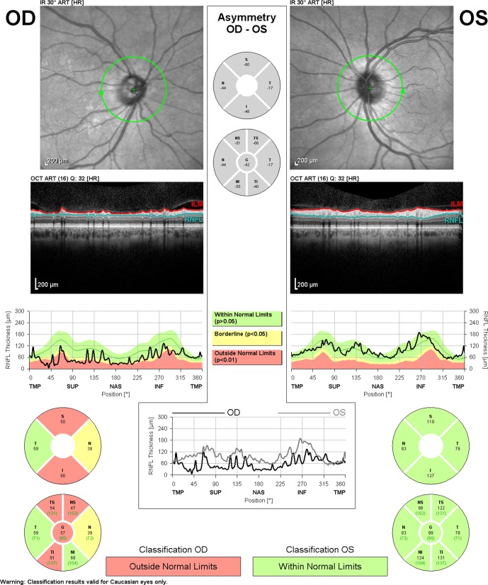
Retinal nerve fiber layer analysis from Spectralis-OCT (Heidelberg Engineering, Heidelberg, Germany) demonstrating glaucomatous damage. The RNFL evaluation in the right eye (OD) shows abnormalities in the superior and inferior quadrants.
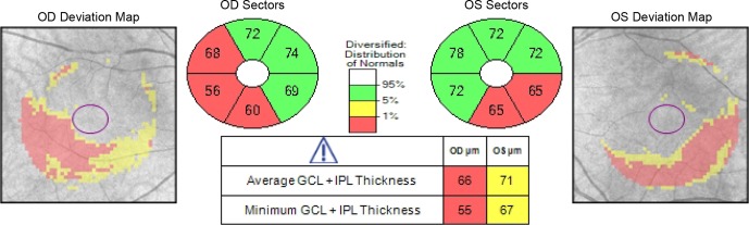
Ganglion cell analysis from Cirrus-OCT (Carl-Zeiss Meditec, Dublin, CA, USA), which includes the combination of ganglion cell and inner plexiform layers (GCIPL), shows thinning in the inferior and inferotemporal perifoveal regions of both eyes.
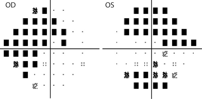
Perimetric pattern deviation maps from the same patient shown in Figure 2. Defects correspond with the locations of thinning observed in ganglion cell analysis.
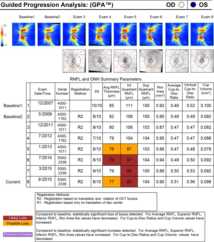
Guided progression analysis from Cirrus-OCT (Carl-Zeiss Meditec) demonstrating early glaucomatous progression. Compared with baseline examinations from December 2007, the patient has statistically significant focal RNFL loss in the inferior quadrant in the left eye. More commonly, the corresponding RNFL thickness map to “Exam 6” may show a red wedge-shaped defect in the inferior quadrant of the ONH.
References
-
- Garcia-Valenzuela E,, Shareef S,, Walsh J,, Sharma SC. Programmed cell death of retinal ganglion cells during experimental glaucoma. Exp Eye Res. 1995. ; 61: 33–44. - PubMed
-
- Quigley HA,, Dunkelberger GR,, Green WR. Retinal ganglion cell atrophy correlated with automated perimetry in human eyes with glaucoma. Am J Ophthalmol. 1989. ; 107: 453 –4 - PubMed
-
- Quigley HA,, Nickells RW,, Kerrigan LA,, Pease ME,, Thibault DJ,, Zack DJ. Retinal ganglion cell death in experimental glaucoma and after axotomy occurs by apoptosis. Invest Ophthalmol Vis Sci. 1995. ; 36: 774 –7 - PubMed
-
- Sommer A,, Miller NR,, Pollack I,, Maumenee AE,, George T. The nerve fiber layer in the diagnosis of glaucoma. Arch Ophthalmol. 1977. ; 95: 2149 –21 - PubMed
Publication types
MeSH terms
Grants and funding
LinkOut - more resources
Full Text Sources
Other Literature Sources
Medical
