Review
. 2016 Aug 18;12(9):662-8.
doi: 10.1038/nchembio.2149.

Insights into newly discovered marks and readers of epigenetic information

Affiliations
Review

Insights into newly discovered marks and readers of epigenetic information

Forest H Andrews et al. Nat Chem Biol. .

Abstract

The field of chromatin biology has been advancing at an accelerated pace. Recent discoveries of previously uncharacterized sites and types of post-translational modifications (PTMs) and the identification of new sets of proteins responsible for the deposition, removal, and reading of these marks continue raising the complexity of an already exceedingly complicated biological phenomenon. In this Perspective article we examine the biological importance of new types and sites of histone PTMs and summarize the molecular mechanisms of chromatin engagement by newly discovered epigenetic readers. We also highlight the imperative role of structural insights in understanding PTM-reader interactions and discuss future directions to enhance the knowledge of PTM readout.


Figures

Figure 1
Figure 1. Recognition of epigenetic marks by histone readers
(a) A reader domain (orange circle) binds to its target PTM (blue circle) in the histone tail, tethering the host protein to chromatin. (b) Multivalent engagement with chromatin through interactions of multiple readers, in the same protein or in different proteins (assembled in the complex), to enhance or regulate overall binding affinity and specificity. Complexes often contain proteins or subunits with catalytic domains (writers, erasers, or ATPase remodelers) and scaffolding domains necessary for the complex assembly.
Figure 2
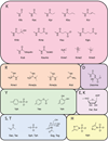
Figure 2. Modifications identified in histone proteins
fo, formylation; ma, malonylation; su, succinylation; glu, glutarylation; ub, ubiquitination; cit, citrullination; oh, hydroxylation; ar, ADP ribosylation; og, O-GlcNAcylation.
Figure 3
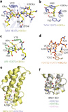
Figure 3. Novel acyllysine readers and their binding mechanisms
(a) Structure of the Taf14 YEATS domain (blue) in complex with H3K9cr peptide (yellow stick). Red dashed lines represent hydrogen bonds; spheres represent water molecules. (b) The π–π–π stacking mechanism involving the alkene moiety of Kcr in Taf14 YEATS–H3K9cr (PDB 5IOK). (c) AF9 YEATS domain (green) in complex with H3K9cr (yellow; PDB 5HJB). (d) The H3K27cr-binding site of the YEATS2 YEATS domain (beige; PDB 5IQL). H3K27cr is shown in orange. (e) The structure of BD of BRD4 (yellow) in complex with the H3K23pr (light blue; PDB 3MUK). (f) Overlay of the structures of the BRD4 BD1 in complex with H3K23pr (light blue), H3K14bu (light green; PDB 3MUL), and H3K14ac (pink; PDB 3JVK), with water shells shown as red, light green, and pink spheres, respectively.
Figure 4
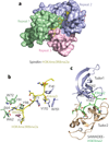
Figure 4. New methyllysine readers
(a) The structure of Spindlin of the protein Spindlin1 in complex with H3K4me3R8me2a peptide (PDB 4MZF). (b) Close-up view of the two aromatic cages of Spindlin. (c) The structure of the SHH1 SAWADEE–H3K9me2 complex (PDB 4IUT).
Figure 5
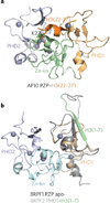
Figure 5. Novel readers of unmodified histone H3
(a) The structure of the AF10 PZP–H3(22–27) complex (PDB 5DAH). Zn-kn, zinc knuckle. (b) Superimposed structures of the BRPF1 PZP domain (brown, cyan, and blue) and the PHD1 finger (gray) of orthologous BRPF2 fused to a sequence corresponding to histone H3(1–7) tail (light green) (PDB 5ERC and 2L43). Spheres represent zinc ions.
Figure 6
Figure 6. Crosstalk of PTMs and paired readers
(a) The structure of the ZMYND11 BD–ZnF–PWWP region in complex with H3.3K36me3 peptide (yellow) (PDB 4N4I). (b) The structure of ORC1 BAH–PHD in complex with H3(1–8) (yellow) (PDB 5HH7). (c) Superimposed structures of two complexes of the ZMET2 BAH–CD–methyltransferase region bound to different H3K9me2 peptides (red) (PDB 4FT2 and 4FT4). MTase, methyltransferase.

References

    1. Strahl BD, Allis CD. The language of covalent histone modifications. Nature. 2000;403:41–45. - PubMed
    1. Jenuwein T, Allis CD. Translating the histone code. Science. 2001;293:1074–1080. - PubMed
    1. Kouzarides T. Chromatin modifications and their function. Cell. 2007;128:693–705. - PubMed
    1. Chi P, Allis CD, Wang GG. Covalent histone modification—miswritten, misinterpreted and mis-erased in human cancers. Nat. Rev. Cancer. 2010;10:457–469. - PMC - PubMed
    1. Polak P, et al. Cell-of-origin chromatin organization shapes the mutational landscape of cancer. Nature. 2015;518:360–364. - PMC - PubMed

MeSH terms

LinkOut - more resources