. 2016 Oct;35(10):1295-304.
doi: 10.1097/ICO.0000000000000966.

Superficial Keratectomy and Conjunctival Advancement Hood Flap (SKCAHF) for the Management of Bullous Keratopathy: Validation in Dogs With Spontaneous Disease

Affiliations

Superficial Keratectomy and Conjunctival Advancement Hood Flap (SKCAHF) for the Management of Bullous Keratopathy: Validation in Dogs With Spontaneous Disease

Taemi Horikawa et al. Cornea. 2016 Oct.

Abstract

Purpose: To evaluate the efficacy of superficial keratectomy and conjunctival advancement hood flap (SKCAHF) for the treatment of bullous keratopathy in canine patients.

Methods: Nine dogs (12 eyes) diagnosed with progressive corneal edema underwent superficial keratectomy followed by placement of conjunctival advancement hood flaps. The canine patients were examined pre- and postoperatively using in vivo confocal microscopy, ultrasonic pachymetry (USP), and Fourier-domain optical coherence tomography (FD-OCT). All owners responded to a survey regarding treatment outcomes.

Results: Mean central corneal thickness (CCT) as measured by FD-OCT was 1163 ± 290 μm preoperatively and significantly decreased postoperatively to 795 ± 197 μm (P = 0.001), 869 ± 190 μm (P = 0.005), and 969 ± 162 μm (P = 0.033) at median postoperative evaluations occurring at 2.2, 6.8, and 12.3 months, respectively. Owners reported significant improvement (P < 0.05) in vision and corneal cloudiness at 6.8 and 12.3 months postoperatively. The percentage of cornea covered by the conjunctival flap was correlated (P = 0.0159) with a reduction in CCT by USP at 12.3 months postoperatively. All canine patients were comfortable pre- and postoperatively.

Conclusions: SKCAHF results in a reduction of corneal thickness in canine patients with bullous keratopathy. The increase in corneal thickness over time, after performing SKCAHF, is likely because of progressive endothelial decompensation. This surgery is a potentially effective intervention for progressive corneal edema in dogs that may have value in treatment of human patients with bullous keratopathy.


Conflict of interest statement

The authors have no conflicts of interest to disclose.

Figures

Figure 1
Figure 1
External photographs of SKCAHF showing different locations and percentage of coverage by conjunctival flaps. The left (A) and right (B) eyes of a 10.8-year-old spayed female Shih Tzu (case 6) 3 days after SKCAHF with the conjunctival flaps covering 36% of cornea in each eye. The right eye (C) of a 10.5-year-old neutered male Cocker Spaniel (case 9) 13 days after SKCAHF with the conjunctival flap covering 36% of the cornea. The right eye (D) of a 8.1-year-old spayed female Boston Terrier (case 7) 9 days after SKCAHF with the conjunctival flap covering 46% of the cornea.
Figure 2
Figure 2
Representative IVCM images of corneal endothelium from a normal canine eye (A) and eyes affected with canine CED (B, C). A, The right (nonoperated) eye of an 11.7-year-old spayed female Dalmatian (case 2) with normal endothelial density of 2474.2 cells/mm2. B, The left and (C) right eyes of an 8-year-old spayed female Boston Terrier (case 7) showing marked polymegathism and pleomorphism and decreased cell densities (left 1576.3 cells/mm2; right 1099.6 cells/mm2). Only the right eye (C) of this patient was clinically affected with corneal edema and received SKCAHF.
Figure 3
Figure 3
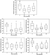
Corneal thickness measurements by USP (Pachette 3) showed significant decrease in central, superior, and temporal regions throughout the postoperative period. Inferior corneal thickness decreased at 2.2 months, but no significant decrease was found at 6.8 and 12.3 months compared with preoperative thickness. Nasal corneal thickness did not significantly change after surgery. Box plots depict median (solid line), mean (dashed line), and 25th and 75th percentiles, and whisker plots show 10th and 90th percentiles; black circles indicate outliers. The P values were determined by a Student–Newman–Keuls post hoc test (central, inferior, temporal, and nasal) or Kruskal–Wallis test (superior). *P < 0.05; P < 0.05; and P < 0.05.
Figure 4
Figure 4
The area of conjunctival flap coverage and change in CCT by USP at 12.3 months showed a significant direct linear relationship (P = 0.0159) between area covered by the flap and subsequent corneal thinning in 12 eyes of 9 patients receiving a SKCAHF surgery.
Figure 5
Figure 5
Central corneal full and stromal thickness significantly decreased throughout the postoperative period after SKCAHF as measured by FD-OCT. Although corneas thinned significantly postoperatively, no significant additional changes were found with increasing time from surgery. The change in corneal thickness was attributable to changes in the stroma. Epithelial and DM–endothelial complex thickness did not significantly change at any time point. Box plots depict median (solid line), mean (dashed line), and 25th and 75th percentiles, and whisker plots show 10th and 90th percentiles; black circles indicate outliers. The P values were determined by a Student–Newman–Keuls post hoc test (full, stromal, and epithelial) or Kruskal–Wallis test (DM–endothelial complex). *P ≤ 0.001; P < 0.01; and P < 0.05.
Figure 6
Figure 6
FD-OCT images of central cornea of both eyes from a 10.8-year-old spayed female Shih Tzu (case 6) before and after SKCAHF at 2.4, 6.9, and 12.1 months. CCT measurements are indicated on each image. Preoperatively, the stroma was markedly thickened, with stromal edema, and loss of the orderly arrangement of the collagen fibrils was noted. Corneal thickness and stromal edema continued to decrease up to 6.9 months. Collagen fibrils seemed more orderly at 6.9 months. At 12.1 months, the corneal thickness had increased but remained less than the preoperative value. Undulated anterior surface in the images from the left eye represent the conjunctival flaps. Stromal fibrosis, which seems as homogenous hyperreflective layer in the anterior stroma, was more evident postoperatively.
Figure 7
Figure 7
External photographs before and at approximately 2, 6, and 12 months after SKCAHF. A, The right eye of an 11.7-year-old neutered male Cocker Spaniel and Poodle mix (case 4). CCT measurements by USP were 952, 683, 751, and 923 μm before and at 2.2, 5.5, and 12.1 months, respectively. The conjunctival flaps are located temporally and inferionasally and became partially pigmented. Percentage of cornea covered with conjunctival flap was 32%. B, The right eye of an 8-year-old spayed female Boston Terrier (case 7). CCT measurements by USP were 1897, 719, 1000, and 1395 mm before and at 2.6, 6.9, and 13.0 months, respectively. The conjunctival flap was located temporally and became partially pigmented. Percentage of cornea covered by conjunctival flap was 46%. She also developed an arc-shaped subepithelial mineral deposit in the nasal paraxial cornea, presumably secondary to chronic topical ophthalmic steroid administration. These deposits were most notable at the 6.9-month recheck and became less obvious at the 13month recheck.

References

    1. Anshu A, Price MO, Tan DT, et al. Endothelial keratoplasty: a revolution in evolution. Surv Ophthalmol. 2012;57:236–252. - PubMed
    1. Koenig SB, Covert DJ. Early results of small-incision Descemet's stripping and automated endothelial keratoplasty. Ophthalmology. 2007;114:221–226. - PubMed
    1. Ing JJ, Ing HH, Nelson LR, et al. Ten-year postoperative results of penetrating keratoplasty. Ophthalmology. 1998;105:1855–1865. - PubMed
    1. Knezović I, Dekaris I, Gabrić N, et al. Therapeutic efficacy of 5% NaCl hypertonic solution in patients with bullous keratopathy. Coll Antropol. 2006;30:405–408. - PubMed
    1. Gasset AR, Kaufman HE. Bandage lenses in the treatment of bullous keratopathy. Am J Ophthalmol. 1971;72:376–380. - PubMed

Publication types

MeSH terms