. 2016 Aug 19:6:31695.
doi: 10.1038/srep31695.

Curcumin suppresses NTHi-induced CXCL5 expression via inhibition of positive IKKβ pathway and up-regulation of negative MKP-1 pathway

Affiliations

Curcumin suppresses NTHi-induced CXCL5 expression via inhibition of positive IKKβ pathway and up-regulation of negative MKP-1 pathway

Anuhya S Konduru et al. Sci Rep. .

Abstract

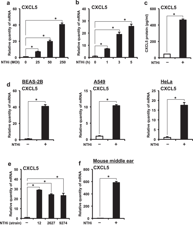
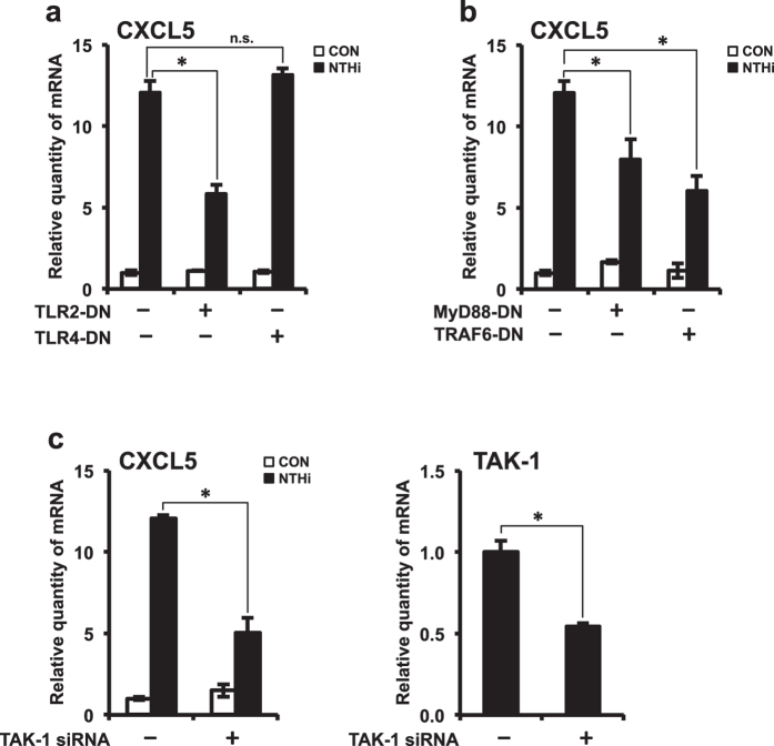
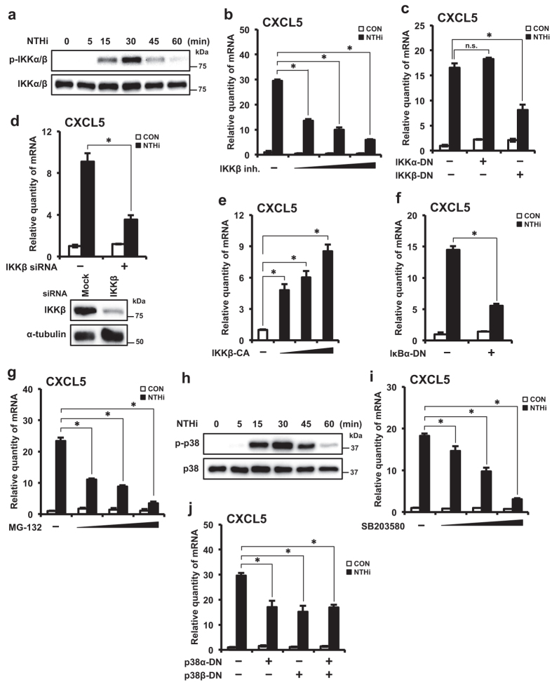
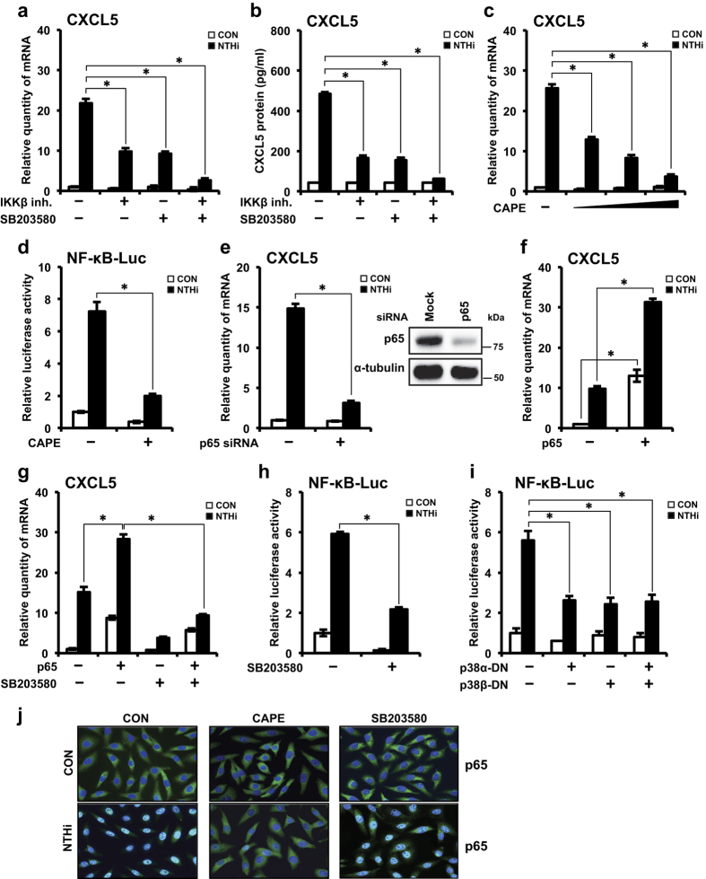
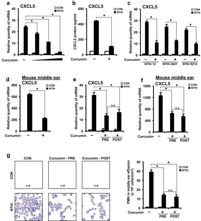
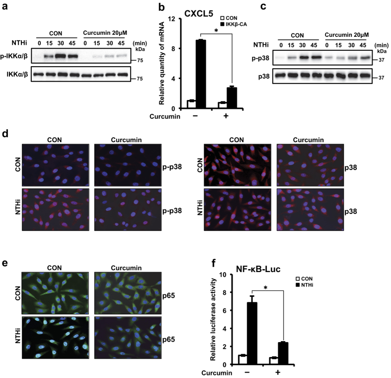
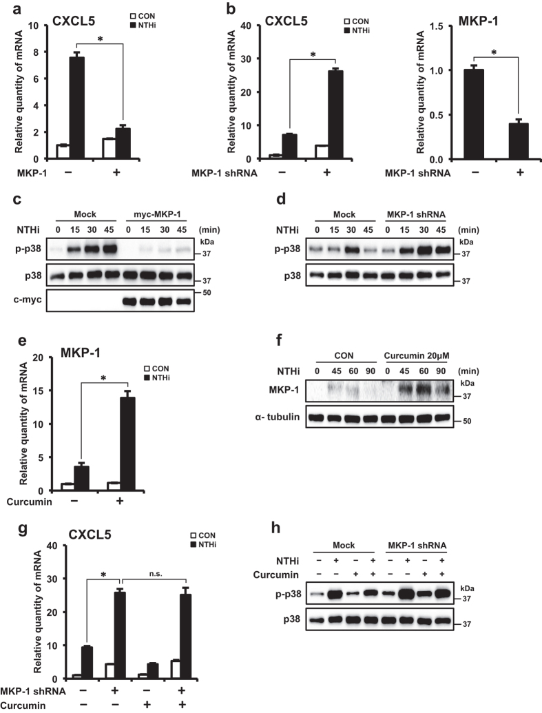
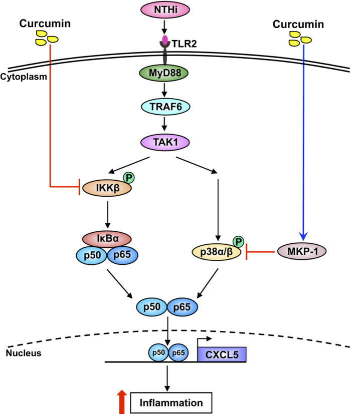
Otitis media (OM) is the most common childhood bacterial infection, and leading cause of conductive hearing loss. Nontypeable Haemophilus influenzae (NTHi) is a major bacterial pathogen for OM. OM characterized by the presence of overactive inflammatory responses is due to the aberrant production of inflammatory mediators including C-X-C motif chemokine ligand 5 (CXCL5). The molecular mechanism underlying induction of CXCL5 by NTHi is unknown. Here we show that NTHi up-regulates CXCL5 expression by activating IKKβ-IκBα and p38 MAPK pathways via NF-κB nuclear translocation-dependent and -independent mechanism in middle ear epithelial cells. Current therapies for OM are ineffective due to the emergence of antibiotic-resistant NTHi strains and risk of side effects with prolonged use of immunosuppressant drugs. In this study, we show that curcumin, derived from Curcuma longa plant, long known for its medicinal properties, inhibited NTHi-induced CXCL5 expression in vitro and in vivo. Curcumin suppressed CXCL5 expression by direct inhibition of IKKβ phosphorylation, and inhibition of p38 MAPK via induction of negative regulator MKP-1. Thus, identification of curcumin as a potential therapeutic for treating OM is of particular translational significance due to the attractiveness of targeting overactive inflammation without significant adverse effects.


Figures

Figure 1
Figure 1. NTHi up-regulates CXCL5 expression in middle ear epithelial cells in vitro and in vivo.
(a) HMEECs were stimulated with NTHi (MOI of 25, 50 or 250) for 5 h, and CXCL5 mRNA expression was measured by Q-PCR. (b) HMEECs were stimulated with NTHi (MOI of 50) for 1, 3 or 5 h, and CXCL5 mRNA expression was measured by Q-PCR. (c) HMEECs were stimulated with NTHi for 12 h, and CXCL5 protein levels in cell culture supernatants was measured by ELISA. (d) Airway epithelial BEAS-2B cells, lung epithelial A549 cells and cervical epithelial HeLa cells were stimulated with NTHi for 5 h, and CXCL5 mRNA expression was measured by Q-PCR. (e) HMEECs were stimulated with NTHi strains 12, 2627 or 9274 for 5 h, and CXCL5 mRNA expression was measured by Q-PCR. (f) Mice were trans-tympanically inoculated with NTHi (6 × 107 CFU) for 6 h, and CXCL5 mRNA expression in middle ear was measured by Q-PCR. Data are mean ± s.d. (n = 3). (a,b,e) *p < 0.05, ANOVA (Tukey’s post-hoc). (c,d,f) *p < 0.05, t-test. Data are representative of three or more independent experiments.
Figure 2
Figure 2. TLR2-MyD88-TRAF6-TAK1 signaling axis is required for NTHi-induced CXCL5 expression.
(a,b) HMEECs were transfected with Mock, TLR2-DN, TLR4-DN, MyD88-DN or TRAF6-DN plasmid. Cells were stimulated with NTHi for 5 h, and CXCL5 mRNA expression was measured. (c) HMEECs were transfected with control siRNA or TAK1 siRNA. Cells were stimulated with NTHi for 5 h, and CXCL5 mRNA expression was measured. Knockdown of TAK1 by siRNA was confirmed by Q-PCR. Data are mean ± s.d. (n = 3). (a,b) *p < 0.05, ANOVA (Tukey’s post-hoc). (c) *p < 0.05, t-test. Data are representative of three or more independent experiments.
Figure 3
Figure 3. Activation of IKKβ-IκBα and p38 signaling pathways are required for NTHi-induced CXCL5 expression.
(a) HMEECs were stimulated with NTHi for various time intervals as indicated in the figure. Phospho-IKKα/β, total IKKα/β protein levels were visualized by western blot. (b) HMEECs were pre-treated with IKKβ inhibitor (0.25, 0.5 or 1.0 μM) for 1 h, followed by stimulation with NTHi for 5 h, and CXCL5 mRNA expression was measured. (c) HMEECs were transfected with Mock, IKKα-DN or IKKβ-DN plasmid. Cells were stimulated with NTHi for 5 h, and CXCL5 mRNA expression was measured. (d) HMEECs were transfected with control siRNA or IKKβ siRNA. Cells were stimulated with NTHi for 5 h, and CXCL5 mRNA expression was measured. Knockdown of IKKβ protein by siRNA was confirmed by western blot. (e) HMEECs were transfected with Mock, IKKβ–CA (0.25, 0.5 or 1 μg) plasmid. CXCL5 mRNA expression was measured. (f) HMEECs were transfected with Mock or IκBα (S32A/S36A) plasmid. Cells were stimulated with NTHi for 5 h, and CXCL5 mRNA expression was measured. (g) HMEECs were pre-treated with MG-132 (5, 10 or 20 μM) for 1 h, followed by stimulation with NTHi for 5 h, and CXCL5 mRNA expression was measured. (h) HMEECs were stimulated with NTHi for various time intervals as indicated in the figure. Phospho-p38, total p38 protein levels were visualized by western blot. (i) HMEECs were pre-treated with SB203580 (5, 10 or 20 μM) for 1 h, followed by stimulation with NTHi for 5 h, and CXCL5 mRNA expression was measured. (j) HMEECs were transfected with Mock, p38α–DN, p38β–DN or both (p38α–DN and p38β–DN) plasmids. Cells were stimulated with NTHi for 5 h, and CXCL5 mRNA expression was measured. Data are mean ± s.d. (n = 3). (b,c,e,g,i,j) *p < 0.05, ANOVA (Tukey’s post-hoc). (d,f) *p < 0.05, t-test. n.s., not significant. Displayed immunoblots are cropped images from full-length blots, presented in Supplementary Fig. S1. Data are representative of three or more independent experiments.
Figure 4
Figure 4. IKKβ-IκBα and p38 signaling axes mediate CXCL5 induction via p65 nuclear translocation–dependent and –independent mechanism, respectively.
(a,b) HMEECs were treated with IKKβ inhibitor (0.5 μM), SB203580 (10 μM) or both for 1 h, followed by stimulation with NTHi for (a) 5 h, and CXCL5 mRNA expression was measured, (b) 12 h, and CXCL5 protein levels in cell culture supernatants was measured by ELISA. (c) HMEECs were pre-treated with CAPE (5, 10 or 25 μg/ml) for 1 h, followed by stimulation with NTHi for 5 h, and CXCL5 mRNA expression was measured. (d) HMEECs were transfected with NF-κB luciferase vector. Cells were pre-treated with CAPE (25 μg/ml) for 1 h, followed by NTHi stimulation for 5 h. NF-κB promoter activity was measured by luciferase assay. (e) HMEECs were transfected with control siRNA or p65 siRNA. Cells were stimulated with NTHi for 5 h, and CXCL5 mRNA expression was measured. Knockdown of p65 protein by siRNA was confirmed by western blot. (f,g) HMEECs were transfected with Mock or p65. Cells were (f) stimulated with NTHi for 5 h, (g) pre-treated with SB203580 (20 μM) for 1 h, followed by stimulation with NTHi for 5 h; and CXCL5 mRNA expression was measured. (h,i) HMEECs were transfected with (h) NF-κB luciferase vector alone, (i) NF-κB luciferase vector and Mock, p38α–DN, p38β–DN or both (p38α–DN and p38β–DN) plasmids. Cells were (h) pre-treated with SB203580 (20 μM) for 1 h and (h,i) stimulated with NTHi for 5 h. NF-κB promoter activity was measured by luciferase assay. (j) HMEECs were pre-treated with CAPE (25 μg/ml) or SB203580 (20 μM) for 1 h, followed by NTHi stimulation for 1 h. p65 translocation was visualized by immunofluorescence by FITC staining. DAPI, nuclear stain. Magnification: 400x. Data are mean ± s.d. (n = 3). (ac,f,g,i) *p < 0.05, ANOVA (Tukey’s post-hoc). (d,e,h) *p < 0.05, t-test. Displayed immunoblots are cropped images from full-length blots, presented in Supplementary Fig. S2. Data are representative of three or more independent experiments.
Figure 5
Figure 5. Curcumin suppresses NTHi-induced CXCL5 expression in vitro and in vivo.
(a) HMEECs were pre-treated with curcumin (10, 20 or 50 μM) for 1 h, followed by stimulation with NTHi for 5 h, and CXCL5 mRNA expression was measured. (b) HMEECs were pre-treated with curcumin (20 μM) for 1 h, followed by stimulation with NTHi for 12 h, and CXCL5 protein levels in cell culture supernatants was measured by ELISA. (c) HMEECs were pre-treated with curcumin (20 μM) for 1 h, followed by stimulation with NTHi strains 12, 2627 or 9274 for 5 h, and CXCL5 mRNA expression was measured. (d) Mice were pretreated with curcumin (50 mg/kg) (i.p) for 1 h, followed by trans-tympanic inoculation with NTHi (5 × 107 CFU) for 6 h, CXCL5 mRNA expression in dissected middle ear was measured. (e) HMEECs were pre-treated with curcumin (20 μM) 1 h prior NTHi stimulation or post-treated with curcumin (20 μM) 1 h after NTHi stimulation. 5 h after NTHi stimulation CXCL5 mRNA expression was measured. (f) Mice were pre-treated with curcumin (50 mg/kg) (i.p) for 1 h, followed by trans-tympanic inoculation with NTHi or post-treated with curcumin (50 mg/kg) (i.p) 1 h after NTHi inoculation. 6 h after NTHi inoculation CXCL5 mRNA expression in dissected middle ear was measured. (g) Mice were pre-treated with curcumin (50 mg/kg) (i.p) for 1 h, followed by trans-tympanic inoculation with NTHi or post-treated with curcumin (50 mg/kg) (i.p) 1 h after NTHi inoculation. Middle ear effusion was harvested 9 h after NTHi inoculation. Following cytocentrifugation, cells were stained with Diff-Quik staining kit. n.d., not detected. Magnification: 400x. PMN cell count in middle ear effusion was determined using a hemocytometer under the microscope. Data are mean ± s.d. (n = 3). (a,e–g) *p < 0.05, ANOVA (Tukey’s post-hoc). (b–d) *p < 0.05, t-test. n.s., not significant. n.d., not detected. Data are representative of three or more independent experiments.
Figure 6
Figure 6. Curcumin suppresses NTHi-induced CXCL5 expression via inhibition of IKKβ and p38 pathways.
(a) HMEECs were treated with curcumin (20 μM) for 1 h, followed by stimulation with NTHi for various time intervals as indicated in the figure. Phospho-IKKβ, total IKKβ protein levels were visualized by western blot. (b) HMEECs were transfected with Mock or IKKβ–CA plasmid. Cells were treated with curcumin (20 μM) for 1 h, and CXCL5 mRNA expression was measured. (c) HMEECs were treated with curcumin (20 μM) for 1 h, followed by stimulation with NTHi for various time intervals as indicated in the figure. Phospho-p38, total p38 protein levels were visualized by western blot. (d,e) HMEECs were pre-treated with curcumin (20 μM) for 1 h, followed by NTHi stimulation for (d) 30 min, (e) 1 h. (d) Phospho-p38, total p38 protein levels (Rhodamine stain), (e) p65 translocation (FITC stain) was visualized by immunofluorescence. DAPI, nuclear stain. Magnification: 400x. (f) HMEECs were transfected with NF-κB luciferase vector. Cells were pre-treated with curcumin (20 μM) for 1 h, followed by stimulated with NTHi for 5 h. NF-κB promoter activity was measured by luciferase assay. Data are mean ± s.d. (n = 3). (b,f) *p < 0.05, t-test. Displayed immunoblots are cropped images from full-length blots, presented in Supplementary Fig. S3. Data are representative of three or more independent experiments.
Figure 7
Figure 7. Curcumin suppresses NTHi-induced CXCL5 expression via up-regulation of negative regulator MKP-1.
(a,b) HMEECs were transfected with (a) Mock or myc-MKP-1, (b) Mock or MKP-1 shRNA. Cells were stimulated with NTHi for 5 h, and CXCL5 mRNA expression was measured. Knockdown of MKP-1 protein by shRNA was confirmed by Q-PCR. (c,d) HMEECs were transfected with (c) Mock or myc-MKP-1 plasmid, (d) Mock or shMKP-1 shRNA. Cells were stimulated with NTHi for times indicated. Phospho-p38, total p38, MKP-1 protein levels were visualized by western blot. (e) HMEECs were pre-treated with curcumin (20 μM) for 1 h, followed by stimulation with NTHi for 1 h, and MKP-1 mRNA expression was measured. (f) HMEECs were pre-treated with curcumin (20 μM) for 1 h, followed by stimulation with NTHi for various time intervals as indicated in the figure. MKP-1 and α-tubulin protein levels were visualized by western blot. (g) HMEECs were transfected with Mock or MKP-1 shRNA. Cells were pre-treated with curcumin (20 μM) for 1 h, followed by NTHi stimulation for 5 h, and CXCL5 mRNA expression was measured. (h) HMEECs were transfected with Mock or MKP-1 shRNA. Cells were pre-treated with curcumin (20 μM) for 1 h, followed by NTHi stimulation for 30 min. Phospho-p38, total p38 protein levels were visualized by western blot. Data are mean ± s.d. (n = 3). (a,b,e) *p < 0.05, t-test. (g) *p < 0.05, ANOVA (Tukey’s post-hoc). n.s., not significant. Displayed immunoblots are cropped images from full-length blots, presented in Supplementary Fig. S4. Data are representative of three or more independent experiments.
Figure 8
Figure 8. Schematic representation of NTHi-induced CXCL5 expression and curcumin-mediated suppression of CXCL5.

Similar articles

Cited by

References

    1. Li J. D. et al.. Panel 4: Recent advances in otitis media in molecular biology, biochemistry, genetics, and animal models. Otolaryngol Head Neck Surg 148, E52–E63 (2013). - PMC - PubMed
    1. Monasta L. et al.. Burden of disease caused by otitis media: systematic review and global estimates. PLoS One 7, e36226 (2012). - PMC - PubMed
    1. Giebink G. S. Immunoprophylaxis of otitis media. Adv Exp Med Biol 303, 149–158 (1991). - PubMed
    1. Arguedas A., Kvaerner K., Liese J., Schilder A. G. & Pelton S. I. Otitis media across nine countries: disease burden and management. Int J Pediatr Otorhinolaryngol 74, 1419–1424 (2010). - PubMed
    1. Ito M. et al.. Clonal spread of beta-lactamase-producing amoxicillin-clavulanate-resistant (BLPACR) strains of non-typeable Haemophilus influenzae among young children attending a day care in Japan. Int J Pediatr Otorhinolaryngol 74, 901–906 (2010). - PubMed

Publication types

MeSH terms