. 2016 Aug 18;16(1):188.
doi: 10.1186/s12866-016-0810-8.

Antibacterial effects of Lactobacillus and bacteriocin PLNC8 αβ on the periodontal pathogen Porphyromonas gingivalis

Affiliations

Antibacterial effects of Lactobacillus and bacteriocin PLNC8 αβ on the periodontal pathogen Porphyromonas gingivalis

Hazem Khalaf et al. BMC Microbiol. .

Abstract

Background: The complications in healthcare systems associated with antibiotic-resistant microorganisms have resulted in an intense search for new effective antimicrobials. Attractive substances from which novel antibiotics may be developed are the bacteriocins. These naturally occurring peptides are generally considered to be safe and efficient at eliminating pathogenic bacteria. Among specific keystone pathogens in periodontitis, Porphyromonas gingivalis is considered to be the most important pathogen in the development and progression of chronic inflammatory disease. The aim of the present study was to investigate the antimicrobial effects of different Lactobacillus species and the two-peptide bacteriocin PLNC8 αβ on P. gingivalis.

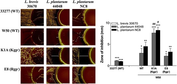
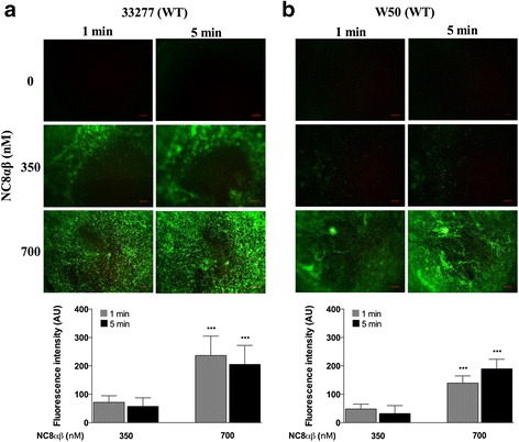
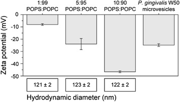
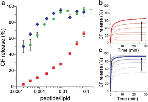
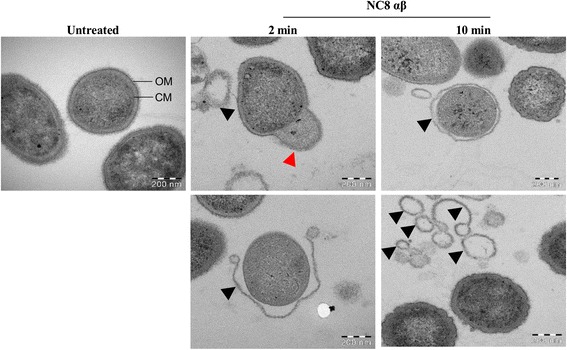
Results: Growth inhibition of P. gingivalis was obtained by viable Lactobacillus and culture media from L. plantarum NC8 and 44048, but not L. brevis 30670. The two-peptide bacteriocin from L. plantarum NC8 (PLNC8 αβ) was found to be efficient against P. gingivalis through binding followed by permeabilization of the membranes, using Surface plasmon resonance analysis and DNA staining with Sytox Green. Liposomal systems were acquired to verify membrane permeabilization by PLNC8 αβ. The antimicrobial activity of PLNC8 αβ was found to be rapid (1 min) and visualized by TEM to cause cellular distortion through detachment of the outer membrane and bacterial lysis.

Conclusion: Soluble or immobilized PLNC8 αβ bacteriocins may be used to prevent P. gingivalis colonization and subsequent pathogenicity, and thus supplement the host immune system against invading pathogens associated with periodontitis.

Keywords: Bacteriocin; Lactobacillus; P. gingivalis; PLNC8; Periodontitis.


Figures

Fig. 1
Fig. 1
Lactobacillus suppresses P. gingivalis growth. Images were acquired after coculture of P. gingivalis with Lactobacillus for 4 days, using Olympus SZX9 at 10× magnification. The zone of inhibition was measured using ImageJ, n = 3. White lines indicate the inhibition zone. Prevention of P. gingivalis growth by Lactobacillus was shown to be species dependent. */#p < 0.05; **/##p < 0.01; ***/###p < 0.001, *- significance from L. brevis 30670 within each P. gingivalis strain, #- significance from WT W50 between the same Lactobacillus strain
Fig. 2
Fig. 2
Bacteriocin PLNC8 αβ from L. plantarum NC8 is efficient against P. gingivalis. The antimicrobial activity of PLNC8 αβ on wild type (WT) P. gingivalis ATCC 33277 (a) and W50 (b), respectively, was visualized using the fluorescent dye Sytox® Green. Images were acquired using Olympus BX41 at 40× magnification. The antimicrobial effect of PLNC8 αβ was rapid and a significant number of P. gingivalis cells could fluoresce already after 1 min, indicating damaged membranes. Representative images and quantitative data of at least three independent experiments are shown. Quantitative data were normalized and the controls at each time point were set to 1. ***p < 0.001, *- significance from the control at each time point. Scale bar = 300 μm
Fig. 3
Fig. 3
Zeta potential and size of liposomes and W50 microvesicles. The zeta potential and size of liposomes with different lipid composition was measured to identify the best match with microvesicles from P. gingivalis W50. All measurements are done in triplicates
Fig. 4
Fig. 4
Dose- and time dependent release of CF in response to bacteriocin PLNC8 αβ. a Release of 5 (6)-carboxyfluorescein (CF) from liposomes (5:95 POPS: POPC) after 30 min with addition of PLNC8 α (red), β (blue) and αβ 1:2 (green). The interaction kinetics was recorded every minute for PLNC8 α (b) and PLNC8 β (c) with the liposomes and is displayed in increasing bacteriocin concentration (0.005–2 μM) indicated by the arrow. The total lipid concentration was kept constant at 25 μM. CF release without addition of bacteriocins were < 1 % and all data points are average of n = 3. The thick red and blue lines in B and C indicate the highest peptide concentration
Fig. 5
Fig. 5
Secondary structure change of PLNC8 α and β. CD spectra of 100 μM PLNC8 α (a) and PLNC8 β (b) in solution (dashed lines) and with 1 mM (total lipid concentration) 5:95 POPS:POPC liposomes (solid lines) in 10 mM PB pH 7, 20 °C
Fig. 6
Fig. 6
PLNC8 αβ binds to P. gingivalis in a dose-dependent manner. Binding of P. gingivalis to PLNC8 αβ and to anti- P.gingivalis antibodies was analyzed by SPR. Both P. gingivalis ATCC 33277 (a) and W50 (b) were found to bind to immobilized PLNC8 αβ (280 nM). The binding was verified by pre-incubating the bacteria with different concentrations of soluble PLNC8 αβ prior to analysis, which resulted in a significantly reduced binding to the immobilized bacteriocins. Pre-incubation of P. gingivalis ATCC 33277 (c) and W50 (d) with increasing concentrations of soluble PLNC8 αβ prior to analysis reduced the bacterial binding to anti-P. gingivalis antibodies in a dose-dependent manner. Results are presented from three independent experiments. *p < 0.05; **p < 0.01; ***p < 0.001
Fig. 7
Fig. 7
The bacteriocin PLNC8 αβ damages the membrane of P. gingivalis. Bacterial ultrastructure was examined using a Hitachi HT 7700 transmission electron microscope and showed typical coccobacillus shapes of untreated P. gingivalis, where the outer membrane (OM) and cytoplasmic membrane (CM) could be clearly distinguished. Treatment with PLNC8 αβ (1:2) efficiently damaged ccells by causing rupture of the bacterial membrane and leakage of intracellular content (red arrow head), eventually resulting in completely detached outer membrane (black arrow heads). Scale bar = 200 nm

References

    1. Bush K, Courvalin P, Dantas G, Davies J, Eisenstein B, Huovinen P, Jacoby GA, Kishony R, Kreiswirth BN, Kutter E, et al. Tackling antibiotic resistance. Nat Rev Microbiol. 2011;9(12):894–896. doi: 10.1038/nrmicro2693. - DOI - PMC - PubMed
    1. Cotter PD, Ross RP, Hill C. Bacteriocins - a viable alternative to antibiotics? Nat Rev Microbiol. 2013;11(2):95–105. doi: 10.1038/nrmicro2937. - DOI - PubMed
    1. Brodala N, Merricks EP, Bellinger DA, Damrongsri D, Offenbacher S, Beck J, Madianos P, Sotres D, Chang YL, Koch G, et al. Porphyromonas gingivalis bacteremia induces coronary and aortic atherosclerosis in normocholesterolemic and hypercholesterolemic pigs. Arterioscler Thromb Vasc Biol. 2005;25(7):1446–1451. doi: 10.1161/01.ATV.0000167525.69400.9c. - DOI - PubMed
    1. Lamont RJ, Chan A, Belton CM, Izutsu KT, Vasel D, Weinberg A. Porphyromonas gingivalis invasion of gingival epithelial cells. Infect Immun. 1995;63(10):3878–3885. - PMC - PubMed
    1. Deshpande RG, Khan MB, Genco CA. Invasion of aortic and heart endothelial cells by Porphyromonas gingivalis. Infect Immun. 1998;66(11):5337–5343. - PMC - PubMed

Publication types

MeSH terms