. 2016 Oct;36(5):e00388.
doi: 10.1042/BSR20160195. Epub 2016 Aug 18.

ApoE deficiency promotes colon inflammation and enhances inflammatory potential oxidized-LDL and TNF-α in colon epithelial cells

Affiliations

ApoE deficiency promotes colon inflammation and enhances inflammatory potential oxidized-LDL and TNF-α in colon epithelial cells

Ali El-Bahrawy et al. Biosci Rep. 2016 Oct.

Erratum in

Abstract

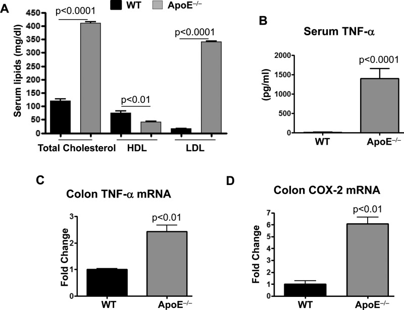
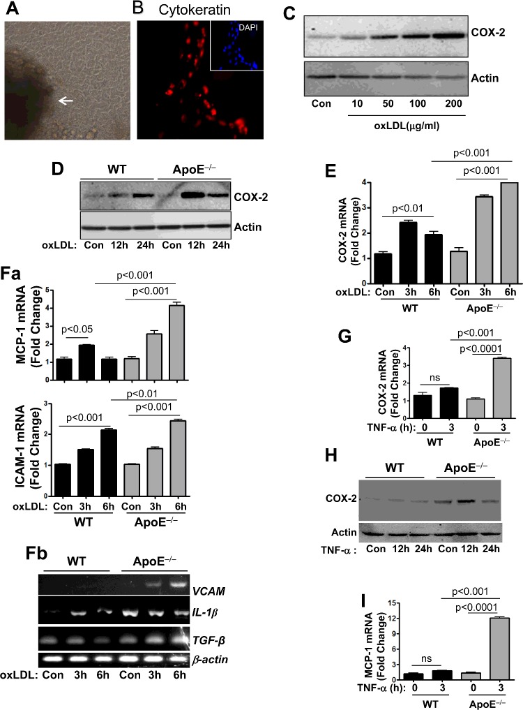
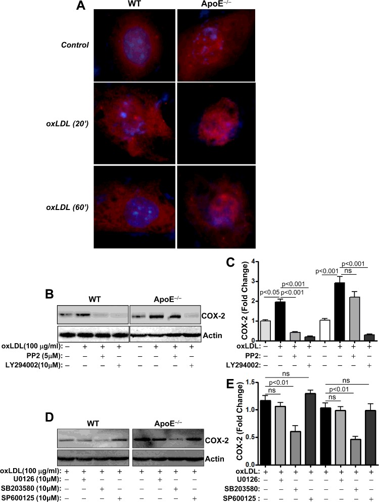
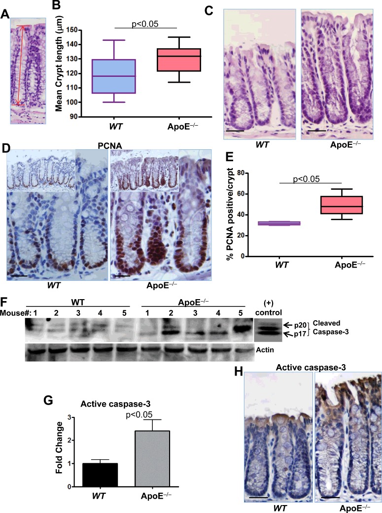
Although deficiency in Apolipoprotein E (ApoE) is linked to many diseases, its effect on colon homeostasis remains unknown. ApoE appears to control inflammation by regulating NF-kB. This study was designed to examine whether ApoE deficiency affects factors of colon integrity in vivo and given the likelihood that ApoE deficiency increases oxidized lipids and TNF-α, this study also examined whether such deficiency enhances the inflammatory potential of oxidized-LDL (oxLDL) and TNF-α, in colon epithelial cells in vitro Here we show that ApoE deficiency is associated with chronic inflammation systemically and in colonic tissues as assessed by TNF-α levels. Increased colon TNF-α mRNA coincided with a substantial increase in cyclooxygenase (COX)-2. ApoE deficiency enhanced the potential of oxLDL and TNF-a to induce COX-2 expression as well as several other inflammatory factors in primary colon epithelial cells. Interestingly, oxLDL enhanced TGF-β expression only in ApoE-/-, but not in wild-type, epithelial cells. ApoE deficiency appears to promote COX-2 expression enhancement through a mechanism that involves persistent NF-κB nuclear localization, PI3 and p38 MAP kinases but independently of Src. In mice, ApoE deficiency promoted a moderate increase in crypt length, which was associated with opposing effects of an increase in cell proliferation and apoptosis at the bottom and top of the crypt, respectively. : Our results support the notion that ApoE plays a central role in colon homeostasis and that ApoE deficiency may constitute a risk factor for colon pathologies.

Keywords: ApoE; colon; inflammation; nuclear factor kappaB; tumour necrosis factors.


Figures

Figure 1
Figure 1. Effect of ApoE deficiency on systemic and colon inflammation in mice
(A) Sera lipid profile (mg/dl) of WT and ApoE−/− mice fed a regular diet. *, difference from WT mice. (B) Sera levels of TNF-α as assessed by a Bioplex assay. (C and D) Total RNA isolated from colon of WT or ApoE−/− mice was subjected to reverse transcription followed by quantitative PCR with primers to mouse TNF-α (C), COX-2 (D) or β-actin and is expressed as fold change over levels detected in WT tissues. *, difference from WT mice.
Figure 2
Figure 2. ApoE deficiency enhances the potential of oxLDL to induce expression of COX-2 and other inflammatory and growth modulatory factors
CECs prepared from colon of WT or ApoE−/− mice using a differential attachment method. (A) Cells were visualized by bright field microscopy; white arrow indicates the colonic crypt from which the cells originated. (B) Cells cultured in chamber slides were fixed with formalin and subjected to immunofluorescence with antibodies to pan-cytokeratin (red) or staining with DAPI (blue; insert). (C) CECs isolated from WT mice were treated with 10, 50, 100 or 200 μg/ml oxLDL for 24 h. Protein extracts were subjected to immunoblot analysis with antibodies to mouse COX-2 or actin. (D) WT or ApoE−/− CECs were treated with 100 μg/ml oxLDL for 12 or 24 h. Protein extracts were subjected to immunoblot analysis with antibodies to mouse COX-2 or actin. (E) WT or ApoE−/− CECs were treated with 100 μg/ml oxLDL for 3 or 6 h. Total RNA was reversed transcribed and the resulting cDNA was amplified by quantitative PCR with primers specific to mouse COX-2 or β-actin. (Fa and Fb) Cells were treated as in (E) and the cDNA generated from the isolated RNA was amplified by quantitative PCR (Fa) with primers to mouse MCP-1 or ICAM-1 or by conventional PCR (Fb) with primers to IL-1β, VCAM-1, TGF-β or β-actin. (G) WT or ApoE−/− CECs were treated with 10 ng/ml TNF-α for 3 h. cDNA generated from the isolated RNA was amplified by quantitative PCR with primers to mouse COX-2 or β-actin. (H) WT or ApoE−/− CECs were treated with 10 ng/ml TNF-α for 12 or 24 h. Protein extracts were subjected to immunoblot analysis with antibodies to mouse COX-2 or actin. (I) The same cDNA from (G) was amplified by quantitative PCR with primers to mouse MCP-1 or β-actin.
Figure 3
Figure 3. Effects of ApoE deficiency on oxLDL-induced NF-κB nuclear translocation and sensitivity of COX-2 protein expression to Src. PI3K, MEK, p38 MAPK or JNK inhibition
(A) WT or ApoE−/− CECs cultured in chamber slides were treated with 100 μg/ml oxLDL for 20 or 60 min. Cells were then fixed with formalin and subjected to immunofluorescence with antibodies to p65 NF-κB and counterstained with DAPI. (B) WT or ApoE−/− CECs were pretreated with the Src inhibitor PP2 (5 μM) or the PI3K inhibitor LY294002 (10 μM) prior to treatment with 100 μg/ml oxLDL for 24 h. Protein extracts were subjected to immunoblot analysis with antibodies to COX-2 or actin. (C) The blots in (B) were quantified using ImageJ and normalized to actin levels. (D) WT or ApoE−/− CECs were treated with the MEK inhibitor U0126, the p38MAPK inhibitor SB203580 or the JNK inhibitor SP600125 prior to treatment with oxLDL. Protein extracts were subjected to immunoblot analysis with antibodies to mouse COX-2 or actin. (E) The blots in (D) were quantified using ImageJ and normalized to actin levels.
Figure 4
Figure 4. Effect of ApoE deficiency on crypt length, cell proliferation and apoptosis
Colonic sections from WT or ApoE−/− mice were stained with H&E and crypts length was measured as displayed in (A). (B) Average of at least 40 crypts per colon in each group. (C) Representative pictures of WT and ApoE−/− colonic tissues. (D) Colonic sections from WT or ApoE−/− mice were subjected to immunohistochemistry with antibodies to PCNA. (E) PCNA-positive cells were counted along the crypts and expressed as percent of total cells per crypt. (F) Protein extracts prepared from colons of WT or ApoE−/− mice were subjected to immunoblot analysis with antibodies to the active form of caspase-3 (p17 and p20) or actin; the rightmost panel represents immunoblot of purified caspase-3 as a positive control. (G) The immunoblots were quantified using ImageJ and normalized to actin levels. Colonic sections from WT or ApoE−/− mice were subjected to immunohistochemistry with antibodies to active caspase-3. Bars in C, D and H=30 μm.

References

    1. Mahley R.W., Huang Y., Weisgraber K.H. Putting cholesterol in its place: apoE and reverse cholesterol transport. J. Clin. Invest. 2006;116:1226–1229. doi: 10.1172/JCI28632. - DOI - PMC - PubMed
    1. Matsuura E., Kobayashi K., Tabuchi M., Lopez L.R. Oxidative modification of low-density lipoprotein and immune regulation of atherosclerosis. Prog. Lipid. Res. 2006;45:466–486. doi: 10.1016/j.plipres.2006.05.001. - DOI - PubMed
    1. Wu C.L., Zhao S.P., Yu B.L. Intracellular role of exchangeable apolipoproteins in energy homeostasis, obesity and non-alcoholic fatty liver disease. Biol. Rev. Camb. Philos. Soc. 2015;90:367–376. doi: 10.1111/brv.12116. - DOI - PubMed
    1. Zhou S., Wu H., Zeng C., Xiong X., Tang S., Tang Z., Sun X. Apolipoprotein E protects astrocytes from hypoxia and glutamate-induced apoptosis. FEBS Lett. 2013;587:254–258. doi: 10.1016/j.febslet.2012.12.003. - DOI - PubMed
    1. Wen M., Segerer S., Dantas M., Brown P.A., Hudkins K.L., Goodpaster T., Kirk E., LeBoeuf R.C., Alpers C.E. Renal injury in apolipoprotein E-deficient mice. Lab. Invest. 2002;82:999–1006. doi: 10.1097/01.LAB.0000022222.03120.D4. - DOI - PubMed