

Demodex musculi Infestation in Genetically Immunomodulated Mice
- PMID: 27538858
- PMCID: PMC4983169
Demodex musculi Infestation in Genetically Immunomodulated Mice
Abstract
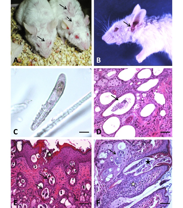
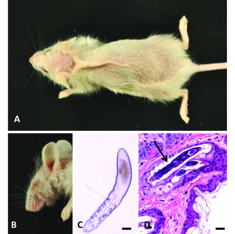
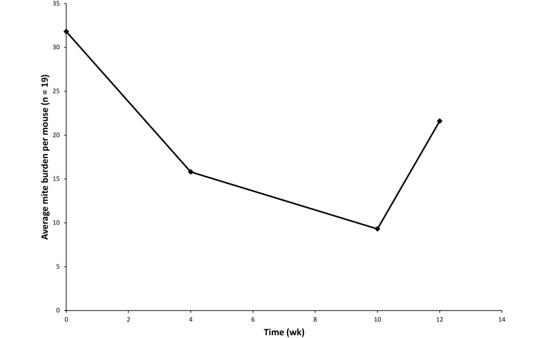
Demodex musculi, a prostigmatid mite that has been reported infrequently in laboratory mice, has been identified with increasing frequency in contemporary colonies of immunodeficient mice. Here we describe 2 episodes of D. musculi infestation with associated clinical signs in various genetically engineered mouse strains, as well as treatment strategies and an investigation into transmissibility and host susceptibility. The first case involved D. musculi associated with clinical signs and pathologic lesions in BALB/c-Tg(DO11.10)Il13(tm) mice, which have a defect in type 2 helper T cell (Th2) immunity. Subsequent investigation revealed mite transmission to both parental strains (BALB/c-Tg[DO11.10] and BALB/c-Il13(tm)), BALB/c-Il13/Il4(tm), and wild-type BALB/c. All Tg(DO11.10)Il13(tm) mice remained infested throughout the investigation, and D. musculi were recovered from all strains when they were cohoused with BALB/c-Tg(DO11.10)Il13(tm) index mice. However, only Il13(tm) and Il13/Il4(tm) mice demonstrated persistent infestation after index mice were removed. Only BALB/c-Tg(DO11.10)Il13(tm) showed clinical signs, suggesting that the phenotypic dysfunction of Th2 immunity is sufficient for persistent infestation, whereas clinical disease associated with D. musculi appears to be genotype-specific. This pattern was further exemplified in the second case, which involved NOD.Cg-Prkdc(scid)Il2r(tm1Wjl)/SzJ (NSG) and C;129S4 Rag2(tm1.1Flv) Il2rg(tm1.1Flv)/J mice with varying degrees of blepharitis, conjunctivitis, and facial pruritis. Topical amitraz decreased mite burden but did not eliminate infestation or markedly ameliorate clinical signs. Furthermore, mite burden began to increase by 1 mo posttreatment, suggesting that topical amitraz is an ineffective treatment for D. musculi. These experiences illustrate the need for vigilance regarding opportunistic and uncommon pathogens in rodent colonies, especially among mice with immunologic deficits.
Figures
Similar articles
-
Mite Burden and Immunophenotypic Response to Demodex musculi in Swiss Webster, BALB/c, C57BL/6, and NSG Mice.Comp Med. 2020 Aug 1;70(4):336-348. doi: 10.30802/AALAS-CM-19-000097. Epub 2020 Jul 16. Comp Med. 2020. PMID: 32605691 Free PMC article.
-
Characterization of Demodex musculi Infestation, Associated Comorbidities, and Topographic Distribution in a Mouse Strain with Defective Adaptive Immunity.Comp Med. 2017 Aug 1;67(4):315-329. Comp Med. 2017. PMID: 28830578 Free PMC article.
-
Demodex musculi in the Skin of Transgenic Mice.Contemp Top Lab Anim Sci. 1999 Nov;38(6):13-18. Contemp Top Lab Anim Sci. 1999. PMID: 12086441
-
Demodex folliculorum and Demodex brevis as a cause of chronic marginal blepharitis.Ann Acad Med Stetin. 2007;53(1):63-7; discussion 67. Ann Acad Med Stetin. 2007. PMID: 18561612 Review.
-
Tropical rat mites (Ornithonyssus bacoti) - serious ectoparasites.J Dtsch Dermatol Ges. 2009 Aug;7(8):667-70. doi: 10.1111/j.1610-0387.2009.07140.x. Epub 2009 Jun 8. J Dtsch Dermatol Ges. 2009. PMID: 19508683 Review. English, German.
Cited by
-
Detection of Myocoptes musculinus in Fur Swab and Fecal Samples by Using PCR Analysis.J Am Assoc Lab Anim Sci. 2019 Nov 1;58(6):796-801. doi: 10.30802/AALAS-JAALAS-19-000046. Epub 2019 Oct 29. J Am Assoc Lab Anim Sci. 2019. PMID: 31662161 Free PMC article.
-
Research-Relevant Conditions and Pathology of Laboratory Mice, Rats, Gerbils, Guinea Pigs, Hamsters, Naked Mole Rats, and Rabbits.ILAR J. 2021 Dec 31;62(1-2):77-132. doi: 10.1093/ilar/ilab022. ILAR J. 2021. PMID: 34979559 Free PMC article. Review.
-
Identification and Treatment of Fur Mites (Radfordia lemnina) in California Deer Mice (Peromyscus californicus) Using Selamectin.J Am Assoc Lab Anim Sci. 2024 Nov 1;63(6):694-700. doi: 10.30802/AALAS-JAALAS-24-055. J Am Assoc Lab Anim Sci. 2024. PMID: 39242175 Free PMC article.
-
Scleritis and anterior uveitis may herald the development of an epibulbar tumor in patients with extranodal Rosai-Dorfman disease: a case report.BMC Ophthalmol. 2019 Jul 10;19(1):144. doi: 10.1186/s12886-019-1158-2. BMC Ophthalmol. 2019. PMID: 31291929 Free PMC article.
-
Ivermectin-compounded Feed Compared with Topical Moxidectin-Imidacloprid for Eradication of Demodex musculi in Laboratory Mice.J Am Assoc Lab Anim Sci. 2018 Sep 1;57(5):483-497. doi: 10.30802/AALAS-JAALAS-18-000003. Epub 2018 Sep 5. J Am Assoc Lab Anim Sci. 2018. PMID: 30185284 Free PMC article.
References
-
- Akilov OE, Mumcuoglu KY. 2004. Immune response in demodicosis. J Eur Acad Dermatol Venereol 18:440–444. - PubMed
-
- Badescu AC, Iancu LS, Statescu L. 2013. Demodex: commensal or pathogen? Rev Med Chir Soc Med Nat Iasi 117:189–193. - PubMed
-
- Baker DG.2007. Arthropods, p 568–569. In: Fox J, Barthold SW, Davisson MT, Newcmer CE, Quimby FW, Smith AL. The mouse in biomedical research, 2nd ed: vol. 2 DiseasesNew York (NY):Elsevier.
-
- Baker DG. 2007. Parasites of rats and mice, p356–357 In: Flynn RJ, Baker DG, Flynn RJ. Flynn's parasites of laboratory animals, second ed Ames (IA): Blackwell Publishing.
MeSH terms
Substances
LinkOut - more resources
Full Text Sources
Other Literature Sources
Miscellaneous