

Mouse models of Down syndrome: gene content and consequences
- PMID: 27538963
- PMCID: PMC5471624
- DOI: 10.1007/s00335-016-9661-8
Mouse models of Down syndrome: gene content and consequences
Abstract
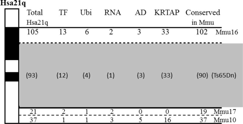
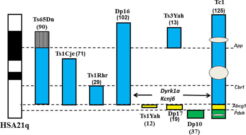
Down syndrome (DS), trisomy of human chromosome 21 (Hsa21), is challenging to model in mice. Not only is it a contiguous gene syndrome spanning 35 Mb of the long arm of Hsa21, but orthologs of Hsa21 genes map to segments of three mouse chromosomes, Mmu16, Mmu17, and Mmu10. The Ts65Dn was the first viable segmental trisomy mouse model for DS; it is a partial trisomy currently popular in preclinical evaluations of drugs for cognition in DS. Limitations of the Ts65Dn are as follows: (i) it is trisomic for 125 human protein-coding orthologs, but only 90 of these are Hsa21 orthologs and (ii) it lacks trisomy for ~75 Hsa21 orthologs. In recent years, several additional mouse models of DS have been generated, each trisomic for a different subset of Hsa21 genes or their orthologs. To best exploit these models and interpret the results obtained with them, prior to proposing clinical trials, an understanding of their trisomic gene content, relative to full trisomy 21, is necessary. Here we first review the functional information on Hsa21 protein-coding genes and the more recent annotation of a large number of functional RNA genes. We then discuss the conservation and genomic distribution of Hsa21 orthologs in the mouse genome and the distribution of mouse-specific genes. Lastly, we consider the strengths and weaknesses of mouse models of DS based on the number and nature of the Hsa21 orthologs that are, and are not, trisomic in each, and discuss their validity for use in preclinical evaluations of drug responses.
Figures
Similar articles
-
The genetic background and application of Down syndrome mouse models.Yi Chuan. 2018 Mar 20;40(3):207-217. doi: 10.16288/j.yczz.17-279. Yi Chuan. 2018. PMID: 29576544 Review.
-
The Impact of Mmu17 Non-Hsa21 Orthologous Genes in the Ts65Dn Mouse Model of Down Syndrome: The Gold Standard Refuted.Biol Psychiatry. 2023 Jul 1;94(1):84-97. doi: 10.1016/j.biopsych.2023.02.012. Epub 2023 Mar 14. Biol Psychiatry. 2023. PMID: 37074246 Free PMC article.
-
Highly penetrant myeloproliferative disease in the Ts65Dn mouse model of Down syndrome.Blood. 2008 Jan 15;111(2):767-75. doi: 10.1182/blood-2007-04-085670. Epub 2007 Sep 27. Blood. 2008. PMID: 17901249 Free PMC article.
-
Human chromosome 21 orthologous region on mouse chromosome 17 is a major determinant of Down syndrome-related developmental cognitive deficits.Hum Mol Genet. 2014 Feb 1;23(3):578-89. doi: 10.1093/hmg/ddt446. Epub 2013 Sep 16. Hum Mol Genet. 2014. PMID: 24041763 Free PMC article.
-
Mouse models of cognitive disorders in trisomy 21: a review.Behav Genet. 2006 May;36(3):387-404. doi: 10.1007/s10519-006-9056-9. Epub 2006 Mar 8. Behav Genet. 2006. PMID: 16523244 Review.
Cited by
-
A non-mosaic transchromosomic mouse model of down syndrome carrying the long arm of human chromosome 21.Elife. 2020 Jun 29;9:e56223. doi: 10.7554/eLife.56223. Elife. 2020. PMID: 32597754 Free PMC article.
-
Context Fear Conditioning in Down Syndrome Mouse Models: Effects of Trisomic Gene Content, Age, Sex and Genetic Background.Genes (Basel). 2021 Sep 28;12(10):1528. doi: 10.3390/genes12101528. Genes (Basel). 2021. PMID: 34680922 Free PMC article.
-
Influence of allelic differences in Down syndrome.Prog Brain Res. 2020;251:29-54. doi: 10.1016/bs.pbr.2019.09.001. Epub 2019 Oct 24. Prog Brain Res. 2020. PMID: 32057311 Free PMC article. Review.
-
Infantile Spasms in Pediatric Down Syndrome: Potential Mechanisms Driving Therapeutic Considerations.Children (Basel). 2024 Dec 13;11(12):1513. doi: 10.3390/children11121513. Children (Basel). 2024. PMID: 39767942 Free PMC article. Review.
-
Aberrant Oligodendrogenesis in Down Syndrome: Shift in Gliogenesis?Cells. 2019 Dec 7;8(12):1591. doi: 10.3390/cells8121591. Cells. 2019. PMID: 31817891 Free PMC article. Review.
References
-
- Ahn KJ, Jeong HK, Choi HS, Ryoo SR, Kim YJ, Goo JS, Choi SY, Han JS, Ha I, Song WJ. DYRK1A BAC transgenic mice show altered synaptic plasticity with learning and memory defects. Neurobiol Dis. 2006;22:463–472. - PubMed
-
- Andrews SJ, Rothnagel JA. Emerging evidence for functional peptides encoded by short open reading frames. Nat Rev Genet. 2014;15:193–204. - PubMed
-
- Antonarakis SE, Lyle R, Dermitzakis ET, Reymond A, Deutsch S. Chromosome 21 and down syndrome: from genomics to pathophysiology. Nat Rev Genet. 2004;5:725–38. - PubMed
Publication types
MeSH terms
Grants and funding
LinkOut - more resources
Full Text Sources
Other Literature Sources
Medical
Molecular Biology Databases
Miscellaneous
